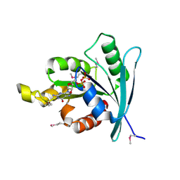
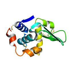
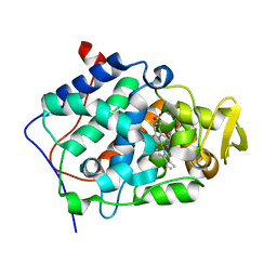
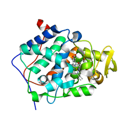
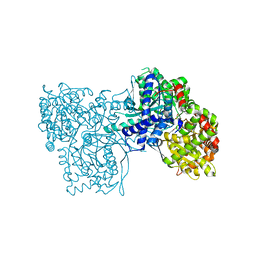
2QOH
 
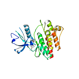 | | Crystal Structure of Abl kinase bound with PPY-A | | 分子名称: | 5-[3-(2-METHOXYPHENYL)-1H-PYRROLO[2,3-B]PYRIDIN-5-YL]-N,N-DIMETHYLPYRIDINE-3-CARBOXAMIDE, Proto-oncogene tyrosine-protein kinase ABL1 | | 著者 | Zhou, T, Dalgarno, D, Zhu, X. | | 登録日 | 2007-07-20 | | 公開日 | 2007-09-18 | | 最終更新日 | 2023-10-25 | | 実験手法 | X-RAY DIFFRACTION (1.95 Å) | | 主引用文献 | Crystal Structure of the T315I Mutant of Abl Kinase
Chem.Biol.Drug Des., 70, 2007
|
|
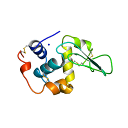
1P5X
 | | STRUCTURE OF THE D55N MUTANT OF PHOSPHOLIPASE C FROM BACILLUS CEREUS | | 分子名称: | Phospholipase C, ZINC ION | | 著者 | Antikainen, N.M, Monzingo, A.F, Franklin, C.L, Robertus, J.D, Martin, S.F. | | 登録日 | 2003-04-28 | | 公開日 | 2003-09-30 | | 最終更新日 | 2023-08-16 | | 実験手法 | X-RAY DIFFRACTION (2 Å) | | 主引用文献 | Using X-ray crystallography of the Asp55Asn mutant of the phosphatidylcholine-preferring phospholipase C from Bacillus cereus to support the mechanistic role of Asp55 as the general base.
Arch.Biochem.Biophys., 417, 2003
|
|
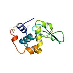
1P6E
 | | STRUCTURE OF THE D55N MUTANT OF PHOSPHOLIPASE C FROM BACILLUS CEREUS IN COMPLEX WITH 1,2-DI-N-PENTANOYL-SN-GLYCERO-3-DITHIOPHOSPHOCHOLINE | | 分子名称: | 1,2-DI-N-PENTANOYL-SN-GLYCERO-3-DITHIOPHOSPHOCHOLINE, Phospholipase C, ZINC ION | | 著者 | Antikainen, N.M, Monzingo, A.F, Franklin, C.L, Robertus, J.D, Martin, S.F. | | 登録日 | 2003-04-29 | | 公開日 | 2003-09-30 | | 最終更新日 | 2023-08-16 | | 実験手法 | X-RAY DIFFRACTION (2.3 Å) | | 主引用文献 | Using X-ray crystallography of the Asp55Asn mutant of the phosphatidylcholine-preferring phospholipase C from Bacillus cereus to support the mechanistic role of Asp55 as the general base.
Arch.Biochem.Biophys., 417, 2003
|
|
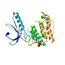
2WHL
 | | Understanding how diverse mannanases recognise heterogeneous substrates | | 分子名称: | ACETATE ION, BETA-MANNANASE, beta-D-mannopyranose-(1-4)-beta-D-mannopyranose-(1-4)-alpha-D-mannopyranose | | 著者 | Tailford, L.E, Ducros, V.M.A, Flint, J.E, Roberts, S.M, Morland, C, Zechel, D.L, Smith, N, Bjornvad, M.E, Borchert, T.V, Wilson, K.S, Davies, G.J, Gilbert, H.J. | | 登録日 | 2009-05-05 | | 公開日 | 2009-05-26 | | 最終更新日 | 2023-12-13 | | 実験手法 | X-RAY DIFFRACTION (1.4 Å) | | 主引用文献 | Understanding How Diverse -Mannanases Recognise Heterogeneous Substrates.
Biochemistry, 48, 2009
|
|
2WJJ
 | | Structure and function of the FeoB G-domain from Methanococcus jannaschii | | 分子名称: | FERROUS IRON TRANSPORT PROTEIN B HOMOLOG, GUANOSINE-5'-DIPHOSPHATE | | 著者 | Koester, S, Wehner, M, Herrmann, C, Kuehlbrandt, W, Yildiz, O. | | 登録日 | 2009-05-26 | | 公開日 | 2009-07-28 | | 最終更新日 | 2011-07-13 | | 実験手法 | X-RAY DIFFRACTION (2.405 Å) | | 主引用文献 | Structure and Function of the Feob G-Domain from Methanococcus Jannaschii
J.Mol.Biol., 392, 2009
|
|
1OUC
 | | CONTRIBUTION OF HYDROPHOBIC RESIDUES TO THE STABILITY OF HUMAN LYSOZYME: X-RAY STRUCTURE OF THE V110A MUTANT | | 分子名称: | LYSOZYME, SODIUM ION | | 著者 | Takano, K, Yamagata, Y, Fujii, S, Yutani, K. | | 登録日 | 1996-08-23 | | 公開日 | 1997-02-12 | | 最終更新日 | 2021-11-03 | | 実験手法 | X-RAY DIFFRACTION (1.8 Å) | | 主引用文献 | Contribution of the hydrophobic effect to the stability of human lysozyme: calorimetric studies and X-ray structural analyses of the nine valine to alanine mutants.
Biochemistry, 36, 1997
|
|
1OUJ
 | | CONTRIBUTION OF HYDROPHOBIC RESIDUES TO THE STABILITY OF HUMAN LYSOZYME: X-RAY STRUCTURE OF THE V99A MUTANT | | 分子名称: | LYSOZYME, SODIUM ION | | 著者 | Takano, K, Yamagata, Y, Fujii, S, Yutani, K. | | 登録日 | 1996-08-23 | | 公開日 | 1997-02-12 | | 最終更新日 | 2021-11-03 | | 実験手法 | X-RAY DIFFRACTION (1.8 Å) | | 主引用文献 | Contribution of the hydrophobic effect to the stability of human lysozyme: calorimetric studies and X-ray structural analyses of the nine valine to alanine mutants.
Biochemistry, 36, 1997
|
|
1OUI
 | | CONTRIBUTION OF HYDROPHOBIC RESIDUES TO THE STABILITY OF HUMAN LYSOZYME: X-RAY STRUCTURE OF THE V93A MUTANT | | 分子名称: | LYSOZYME, SODIUM ION | | 著者 | Takano, K, Yamagata, Y, Fujii, S, Yutani, K. | | 登録日 | 1996-08-23 | | 公開日 | 1997-02-12 | | 最終更新日 | 2021-11-03 | | 実験手法 | X-RAY DIFFRACTION (1.8 Å) | | 主引用文献 | Contribution of the hydrophobic effect to the stability of human lysozyme: calorimetric studies and X-ray structural analyses of the nine valine to alanine mutants.
Biochemistry, 36, 1997
|
|
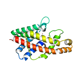
2WQO
 | | STRUCTURE OF NEK2 BOUND TO THE AMINOPYRIDINE CCT241950 | | 分子名称: | 4-[2-AMINO-5-(3,4,5-TRIMETHOXYPHENYL)PYRIDIN-3-YL]BENZOIC ACID, CHLORIDE ION, SERINE/THREONINE-PROTEIN KINASE NEK2 | | 著者 | Mas-Droux, C, Bayliss, R. | | 登録日 | 2009-08-24 | | 公開日 | 2009-12-08 | | 最終更新日 | 2024-05-08 | | 実験手法 | X-RAY DIFFRACTION (2.167 Å) | | 主引用文献 | An Autoinhibitory Tyrosine Motif in the Cell-Cycle- Regulated Nek7 Kinase is Released Through Binding of Nek9.
Mol.Cell, 36, 2009
|
|
1TAY
 | |
1TY9
 | | X-RAY CRYSTAL STRUCTURE OF PHZG FROM PSEUDOMONAS FLUORESCENS | | 分子名称: | FLAVIN MONONUCLEOTIDE, Phenazine biosynthesis protein phzG, SULFATE ION | | 著者 | Parsons, J.F, Eisenstein, E, Ladner, J.E. | | 登録日 | 2004-07-07 | | 公開日 | 2004-11-23 | | 最終更新日 | 2023-08-23 | | 実験手法 | X-RAY DIFFRACTION (1.8 Å) | | 主引用文献 | Structure of the phenazine biosynthesis enzyme PhzG.
Acta Crystallogr.,Sect.D, 60, 2004
|
|
1U67
 | | Crystal Structure of Arachidonic Acid Bound to a Mutant of Prostagladin H Synthase-1 that Forms Predominantly 11-HPETE. | | 分子名称: | 2-acetamido-2-deoxy-alpha-D-glucopyranose-(1-4)-2-acetamido-2-deoxy-beta-D-glucopyranose, ARACHIDONIC ACID, PROTOPORPHYRIN IX CONTAINING CO, ... | | 著者 | Harman, C.A, Rieke, C.J, Garavito, R.M, Smith, W.L. | | 登録日 | 2004-07-29 | | 公開日 | 2004-09-07 | | 最終更新日 | 2023-08-23 | | 実験手法 | X-RAY DIFFRACTION (3.1 Å) | | 主引用文献 | Crystal structure of arachidonic Acid bound to a mutant of prostaglandin endoperoxide h synthase-1 that forms predominantly 11-hydroperoxyeicosatetraenoic Acid.
J.Biol.Chem., 279, 2004
|
|
3BOK
 | |
3BTN
 | | Crystal structure of antizyme inhibitor, an ornithine decarboxylase homologous protein | | 分子名称: | Antizyme inhibitor 1 | | 著者 | Dym, O, Unger, T, Albeck, S, Kahana, C, Israel Structural Proteomics Center (ISPC) | | 登録日 | 2007-12-30 | | 公開日 | 2008-04-15 | | 最終更新日 | 2024-02-21 | | 実験手法 | X-RAY DIFFRACTION (2.05 Å) | | 主引用文献 | Crystallographic and biochemical studies revealing the structural basis for antizyme inhibitor function.
Protein Sci., 17, 2008
|
|
2E2F
 | |
2END
 | | CRYSTAL STRUCTURE OF A PYRIMIDINE DIMER SPECIFIC EXCISION REPAIR ENZYME FROM BACTERIOPHAGE T4: REFINEMENT AT 1.45 ANGSTROMS AND X-RAY ANALYSIS OF THE THREE ACTIVE SITE MUTANTS | | 分子名称: | ENDONUCLEASE V | | 著者 | Vassylyev, D.G, Ariyoshi, M, Matsumoto, O, Katayanagi, K, Ohtsuka, E, Morikawa, K. | | 登録日 | 1994-08-08 | | 公開日 | 1994-10-15 | | 最終更新日 | 2024-02-14 | | 実験手法 | X-RAY DIFFRACTION (1.45 Å) | | 主引用文献 | Crystal structure of a pyrimidine dimer-specific excision repair enzyme from bacteriophage T4: refinement at 1.45 A and X-ray analysis of the three active site mutants.
J.Mol.Biol., 249, 1995
|
|
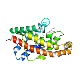
2EUN
 | | Cytochrome c peroxidase (CCP) in complex with 2,4-diaminopyrimidine | | 分子名称: | PROTOPORPHYRIN IX CONTAINING FE, PYRIMIDINE-2,4-DIAMINE, cytochrome c peroxidase | | 著者 | Brenk, R, Vetter, S.W, Boyce, S.E, Goodin, D.B, Shoichet, B.K. | | 登録日 | 2005-10-29 | | 公開日 | 2006-04-11 | | 最終更新日 | 2024-02-14 | | 実験手法 | X-RAY DIFFRACTION (1.7 Å) | | 主引用文献 | Probing molecular docking in a charged model binding site.
J.Mol.Biol., 357, 2006
|
|
2EUT
 | | Cytochrome c peroxidase (CCP) in complex with 2-amino-4-picoline | | 分子名称: | 4-METHYLPYRIDIN-2-AMINE, PROTOPORPHYRIN IX CONTAINING FE, cytochrome c peroxidase | | 著者 | Brenk, R, Vetter, S.W, Boyce, S.E, Goodin, D.B, Shoichet, B.K. | | 登録日 | 2005-10-29 | | 公開日 | 2006-04-11 | | 最終更新日 | 2023-08-23 | | 実験手法 | X-RAY DIFFRACTION (1.12 Å) | | 主引用文献 | Probing molecular docking in a charged model binding site.
J.Mol.Biol., 357, 2006
|
|
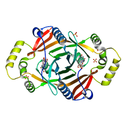
2F90
 | | Crystal structure of bisphosphoglycerate mutase in complex with 3-phosphoglycerate and AlF4- | | 分子名称: | 3-PHOSPHOGLYCERIC ACID, Bisphosphoglycerate mutase, TETRAFLUOROALUMINATE ION | | 著者 | Wang, Y, Liu, L, Wei, Z, Gong, W. | | 登録日 | 2005-12-05 | | 公開日 | 2006-10-24 | | 最終更新日 | 2024-03-13 | | 実験手法 | X-RAY DIFFRACTION (2 Å) | | 主引用文献 | Seeing the process of histidine phosphorylation in human bisphosphoglycerate mutase
J.Biol.Chem., 281, 2006
|
|
2F2C
 | |
2EUP
 | | Cytochrome c peroxidase (CCP) in complex with 2-amino-5-picoline | | 分子名称: | 5-METHYLPYRIDIN-2-AMINE, PROTOPORPHYRIN IX CONTAINING FE, cytochrome c peroxidase | | 著者 | Brenk, R, Vetter, S.W, Boyce, S.E, Goodin, D.B, Shoichet, B.K. | | 登録日 | 2005-10-29 | | 公開日 | 2006-04-11 | | 最終更新日 | 2023-08-23 | | 実験手法 | X-RAY DIFFRACTION (1.4 Å) | | 主引用文献 | Probing molecular docking in a charged model binding site.
J.Mol.Biol., 357, 2006
|
|
2F3Q
 | |
2EUF
 | |
2EUU
 | | Cytochrome c peroxidase (CCP) in complex with 1H-imidazol-2-ylmethanol | | 分子名称: | 1H-IMIDAZOL-2-YLMETHANOL, PROTOPORPHYRIN IX CONTAINING FE, cytochrome c peroxidase | | 著者 | Brenk, R, Vetter, S.W, Boyce, S.E, Goodin, D.B, Shoichet, B.K. | | 登録日 | 2005-10-29 | | 公開日 | 2006-04-11 | | 最終更新日 | 2023-08-23 | | 実験手法 | X-RAY DIFFRACTION (1.45 Å) | | 主引用文献 | Probing molecular docking in a charged model binding site.
J.Mol.Biol., 357, 2006
|
|
1QHC
 | |


