8VAL
 
 | |
8VAQ
 | |
2INT
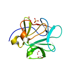
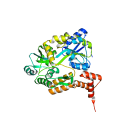
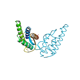 | | CRYSTAL STRUCTURE OF RECOMBINANT HUMAN INTERLEUKIN-4 | | 分子名称: | INTERLEUKIN-4 | | 著者 | Walter, M.R, Cook, W.J, Zhao, B.G, Cameron Junior, R, Ealick, S.E, Walter Junior, R.L, Reichert, P, Nagabhushan, T.L, Trotta, P.P, Bugg, C.E. | | 登録日 | 1993-07-22 | | 公開日 | 1994-01-31 | | 最終更新日 | 2019-08-14 | | 実験手法 | X-RAY DIFFRACTION (2.35 Å) | | 主引用文献 | Crystal structure of recombinant human interleukin-4.
J.Biol.Chem., 267, 1992
|
|
8VAP
 | |
8VAM
 | |
8VAS
 | |
1NS3
 | | STRUCTURE OF HCV PROTEASE (BK STRAIN) | | 分子名称: | NS3 PROTEASE, NS4A PEPTIDE, ZINC ION | | 著者 | Yan, Y, Munshi, S, Chen, Z. | | 登録日 | 1997-04-05 | | 公開日 | 1998-04-08 | | 最終更新日 | 2024-02-14 | | 実験手法 | X-RAY DIFFRACTION (2.8 Å) | | 主引用文献 | Complex of NS3 protease and NS4A peptide of BK strain hepatitis C virus: a 2.2 A resolution structure in a hexagonal crystal form.
Protein Sci., 7, 1998
|
|
1OK8
 | |
3CRI
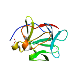
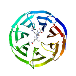
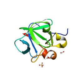 | | Crystal structure of human fibroblast growth factor-1 with mutations Glu81Ser, Glu82Asn and Lys101Ala | | 分子名称: | FORMIC ACID, Heparin-binding growth factor 1, SULFATE ION | | 著者 | Meher, A.K, Honjo, E, Kuroki, R, Lee, J, Somasundaram, T, Blaber, M. | | 登録日 | 2008-04-07 | | 公開日 | 2009-02-17 | | 最終更新日 | 2024-02-21 | | 実験手法 | X-RAY DIFFRACTION (2.1 Å) | | 主引用文献 | Engineering an improved crystal contact across a solvent-mediated interface of human fibroblast growth factor 1.
Acta Crystallogr.,Sect.F, 65, 2009
|
|
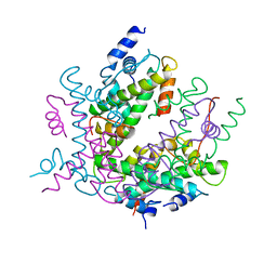
2JJK
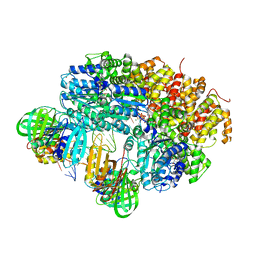
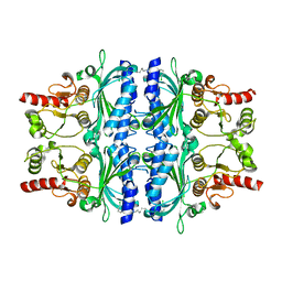 | | FRUCTOSE-1,6-BISPHOSPHATASE(D-FRUCTOSE-1,6-BISPHOSPHATE -1- PHOSPHOHYDROLASE) (E.C.3.1.3.11) COMPLEXED WITH A DUAL BINDING AMP SITE INHIBITOR | | 分子名称: | FRUCTOSE-1,6-BISPHOSPHATASE 1, N,N'-(heptane-1,7-diyldicarbamoyl)bis(3-chlorobenzenesulfonamide) | | 著者 | Ruf, A, Joseph, C, Benz, J, Fol, B, Tetaz, T, Hebeisen, P. | | 登録日 | 2008-04-09 | | 公開日 | 2008-07-22 | | 最終更新日 | 2024-05-08 | | 実験手法 | X-RAY DIFFRACTION (2 Å) | | 主引用文献 | Allosteric Fbpase Inhibitors Gain 10(5) Times in Potency When Simultaneously Binding Two Neighboring AMP Sites.
Bioorg.Med.Chem.Lett., 18, 2008
|
|
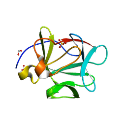
3CRH
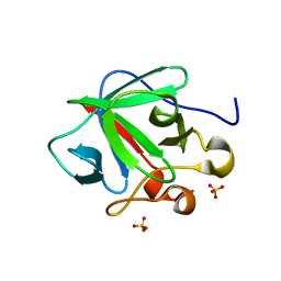 | | Crystal structure of human fibroblast growth factor-1 with mutations Glu81Ser and Lys101Ala | | 分子名称: | Heparin-binding growth factor 1, SULFATE ION | | 著者 | Meher, A.K, Honjo, E, Kuroki, R, Lee, J, Somasundaram, T, Blaber, M. | | 登録日 | 2008-04-07 | | 公開日 | 2009-02-17 | | 最終更新日 | 2024-02-21 | | 実験手法 | X-RAY DIFFRACTION (2.15 Å) | | 主引用文献 | Engineering an improved crystal contact across a solvent-mediated interface of human fibroblast growth factor 1.
Acta Crystallogr.,Sect.F, 65, 2009
|
|
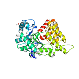
3RIB
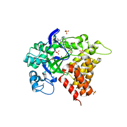 | | Human lysine methyltransferase Smyd2 in complex with AdoHcy | | 分子名称: | N-lysine methyltransferase SMYD2, S-ADENOSYL-L-HOMOCYSTEINE, SULFATE ION, ... | | 著者 | Xu, S, Zhang, T, Zhong, C, Ding, J. | | 登録日 | 2011-04-13 | | 公開日 | 2011-07-20 | | 最終更新日 | 2024-03-20 | | 実験手法 | X-RAY DIFFRACTION (2.79 Å) | | 主引用文献 | Structure of human lysine methyltransferase Smyd2 reveals insights into the substrate divergence in Smyd proteins
J Mol Cell Biol, 3, 2011
|
|
5EMM
 | |
2GLI
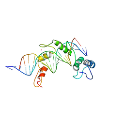 | | FIVE-FINGER GLI/DNA COMPLEX | | 分子名称: | COBALT (II) ION, DNA (5'-D(*AP*CP*GP*TP*GP*GP*AP*CP*CP*AP*CP*CP*CP*AP*AP*GP*AP*CP*GP*AP*A)-3'), DNA (5'-D(*TP*TP*TP*CP*GP*TP*CP*TP*TP*GP*GP*GP*TP*GP*GP*TP*CP*CP*AP*CP*G)-3'), ... | | 著者 | Pavletich, N.P, Pabo, C.O. | | 登録日 | 1993-11-09 | | 公開日 | 1993-11-09 | | 最終更新日 | 2024-02-14 | | 実験手法 | X-RAY DIFFRACTION (2.6 Å) | | 主引用文献 | Crystal structure of a five-finger GLI-DNA complex: new perspectives on zinc fingers.
Science, 261, 1993
|
|
3S7B
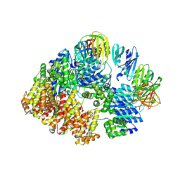
 | | Structural Basis of Substrate Methylation and Inhibition of SMYD2 | | 分子名称: | (R,R)-2,3-BUTANEDIOL, N-cyclohexyl-N~3~-[2-(3,4-dichlorophenyl)ethyl]-N-(2-{[2-(5-hydroxy-3-oxo-3,4-dihydro-2H-1,4-benzoxazin-8-yl)ethyl]amino}ethyl)-beta-alaninamide, N-lysine methyltransferase SMYD2, ... | | 著者 | Ferguson, A.D. | | 登録日 | 2011-05-26 | | 公開日 | 2011-08-10 | | 最終更新日 | 2024-02-28 | | 実験手法 | X-RAY DIFFRACTION (2.42 Å) | | 主引用文献 | Structural Basis of Substrate Methylation and Inhibition of SMYD2.
Structure, 19, 2011
|
|
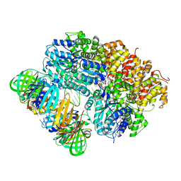
2HIO
 | | HISTONE OCTAMER (CHICKEN), CHROMOSOMAL PROTEIN | | 分子名称: | PROTEIN (HISTONE H2A), PROTEIN (HISTONE H2B), PROTEIN (HISTONE H3), ... | | 著者 | Arents, G, Moudrianakis, E.N. | | 登録日 | 1999-06-15 | | 公開日 | 2000-01-12 | | 最終更新日 | 2023-12-27 | | 実験手法 | X-RAY DIFFRACTION (3.1 Å) | | 主引用文献 | The nucleosomal core histone octamer at 3.1 A resolution: a tripartite protein assembly and a left-handed superhelix.
Proc.Natl.Acad.Sci.USA, 88, 1991
|
|
1N64
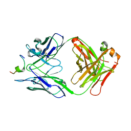 | | Crystal structure analysis of the immunodominant antigenic site on Hepatitis C virus protein bound to mAb 19D9D6 | | 分子名称: | Fab 19D9D6 heavy chain, Fab 19D9D6 light chain, Genome polyprotein Capsid protein C | | 著者 | Menez, R, Bossus, M, Muller, B, Sibai, G, Dalbon, P, Ducancel, F, Jolivet-Reynaud, C, Stura, E. | | 登録日 | 2002-11-08 | | 公開日 | 2003-02-25 | | 最終更新日 | 2013-09-18 | | 実験手法 | X-RAY DIFFRACTION (2.34 Å) | | 主引用文献 | Crystal structure of a hydrophobic immunodominant
antigenic site on hepatitis C virus core protein
complexed to monoclonal antibody 19D9D6.
J.Immunol., 170, 2003
|
|
3FJF
 | |
3FJE
 | |
3FJH
 | |
1K5M
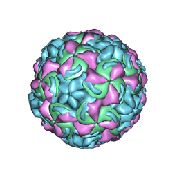 | | Crystal Structure of a Human Rhinovirus Type 14:Human Immunodeficiency Virus Type 1 V3 Loop Chimeric Virus MN-III-2 | | 分子名称: | CHIMERA OF HRV14 COAT PROTEIN VP2 (P1B) AND the V3 loop of HIV-1 gp120, COAT PROTEIN VP1 (P1D), COAT PROTEIN VP3 (P1C), ... | | 著者 | Ding, J, Smith, A.D, Geisler, S.C, Ma, X, Arnold, G.F, Arnold, E. | | 登録日 | 2001-10-11 | | 公開日 | 2002-07-17 | | 最終更新日 | 2023-08-16 | | 実験手法 | X-RAY DIFFRACTION (2.7 Å) | | 主引用文献 | Crystal Structure of a Human Rhinovirus
that Displays Part of the HIV-1 V3 Loop and
Induces Neutralizing Antibodies against
HIV-1
Structure, 10, 2002
|
|
3FJJ
 | |
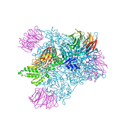
8IRS
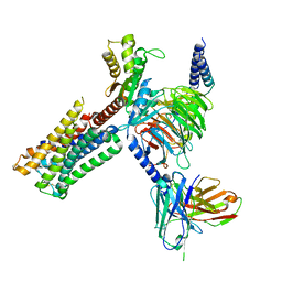 | | Dopamine Receptor D2R-Gi-Rotigotine complex | | 分子名称: | Guanine nucleotide-binding protein G(I)/G(S)/G(O) subunit gamma-2, Guanine nucleotide-binding protein G(I)/G(S)/G(T) subunit beta-1, Guanine nucleotide-binding protein G(i) subunit alpha-1, ... | | 著者 | Xu, P, Huang, S, Zhuang, Y, Mao, C, Zhang, Y, Wang, Y, Li, H, Jiang, Y, Zhang, Y, Xu, H.E. | | 登録日 | 2023-03-19 | | 公開日 | 2023-06-07 | | 最終更新日 | 2023-11-08 | | 実験手法 | ELECTRON MICROSCOPY (3 Å) | | 主引用文献 | Structural genomics of the human dopamine receptor system.
Cell Res., 33, 2023
|
|
2H13
 | |
1MH3
 | |


