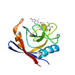
7OYM
 
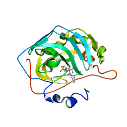 | | Carbonic anhydrase II in complex with Hit2 (MH65) | | 分子名称: | Carbonic anhydrase 2, Hit2 (MH65), ZINC ION | | 著者 | Kugler, M, Brynda, J, Rezacova, P. | | 登録日 | 2021-06-24 | | 公開日 | 2023-01-18 | | 最終更新日 | 2024-02-07 | | 実験手法 | X-RAY DIFFRACTION (0.98 Å) | | 主引用文献 | Identification of specific carbonic anhydrase inhibitors via in situ click chemistry, phage-display and synthetic peptide libraries: comparison of the methods and structural study.
Rsc Med Chem, 14, 2023
|
|
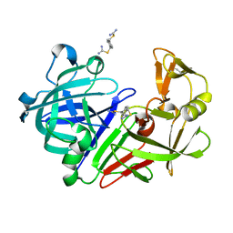
2V8T
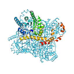
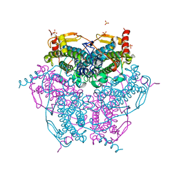 | | Crystal structure of Mn catalase from Thermus Thermophilus complexed with chloride | | 分子名称: | CHLORIDE ION, LITHIUM ION, MANGANESE (II) ION, ... | | 著者 | Antonyuk, S.V, Barynin, V.V, Vaguine, A.A, Melik-Adamyan, W.R, Popov, A.N, Lamsin, V.S, Harrison, P.M, Artymiuk, P.J. | | 登録日 | 2007-08-14 | | 公開日 | 2007-09-11 | | 最終更新日 | 2023-12-13 | | 実験手法 | X-RAY DIFFRACTION (0.98 Å) | | 主引用文献 | Three-Dimentional Structure of the Enzyme Dimanganese Catalase from Thermus Thermophilus at 1 Angstrom Resolution
Crystallogr.Rep.(Transl. Kristallografiya), 45, 2000
|
|
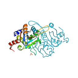
7AVE
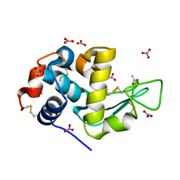 | | Perdeuterated refolded hen egg-white lysozyme at 100 K | | 分子名称: | ACETATE ION, Lysozyme C, NITRATE ION | | 著者 | Ramos, J, Laux, V, Haertlein, M, Erba Boeri, E, Forsyth, V.T, Larsen, S, Mossou, E, Langkilde, A.E. | | 登録日 | 2020-11-05 | | 公開日 | 2021-05-12 | | 最終更新日 | 2024-01-31 | | 実験手法 | X-RAY DIFFRACTION (0.98 Å) | | 主引用文献 | Structural insights into protein folding, stability and activity using in vivo perdeuteration of hen egg-white lysozyme.
Iucrj, 8, 2021
|
|
5SY4
 | |
4AWT
 | |
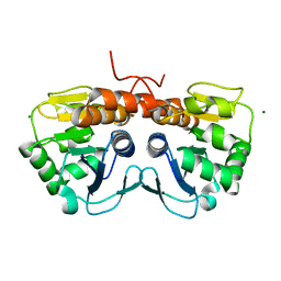
7ATV
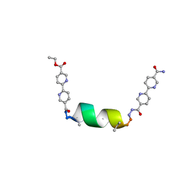
 | | Structure of protein kinase ck2 catalytic subunit (csnk2a2 gene product) in complex with the bivalent inhibitor KN2 | | 分子名称: | 1,2-ETHANEDIOL, CHLORIDE ION, Casein kinase II subunit alpha', ... | | 著者 | Lindenblatt, D, Applegate, V, Nickelsen, A, Klussmann, M, Neundorf, I, Goetz, C, Jose, J, Niefind, K. | | 登録日 | 2020-10-31 | | 公開日 | 2021-08-04 | | 最終更新日 | 2024-06-19 | | 実験手法 | X-RAY DIFFRACTION (0.98 Å) | | 主引用文献 | Molecular Plasticity of Crystalline CK2 alpha ' Leads to KN2, a Bivalent Inhibitor of Protein Kinase CK2 with Extraordinary Selectivity.
J.Med.Chem., 65, 2022
|
|
5MNM
 | |
6SG6
 | |
5MOR
 | | Joint X-ray/neutron structure of cationic trypsin in complex with benzylamine | | 分子名称: | (phenylmethyl)azanium, CALCIUM ION, Cationic trypsin, ... | | 著者 | Schiebel, J, Schrader, T.E, Ostermann, A, Heine, A, Klebe, G. | | 登録日 | 2016-12-14 | | 公開日 | 2018-02-28 | | 最終更新日 | 2024-05-01 | | 実験手法 | NEUTRON DIFFRACTION (0.98 Å), X-RAY DIFFRACTION | | 主引用文献 | Joint X-ray/neutron structure of cationic trypsin in complex with benzylamine
to be published
|
|
3WBO
 | |
5MB5
 | |
4HVW
 | |
7V2G
 | | The 0.98 angstrom structure of the human FABP3 Y19F mutant complexed with palmitic acid | | 分子名称: | Fatty acid-binding protein, heart, HEXAETHYLENE GLYCOL, ... | | 著者 | Sugiyama, S, Takahashi, J, Matsuoka, S, Tsuchikawa, H, Sonoyama, M, Inoue, Y, Hayashi, F, Murata, M. | | 登録日 | 2021-08-09 | | 公開日 | 2022-08-10 | | 最終更新日 | 2023-11-29 | | 実験手法 | X-RAY DIFFRACTION (0.98 Å) | | 主引用文献 | The 0.98 angstrom structure of the human FABP3 Y19F mutant complexed with palmitic acid
To Be Published
|
|
7PMT
 | | Human Cyclophilin D in complex with N-[(5-ethyl-4-oxo-1,2,3,4,5,6- hexahydro-1,5-benzodiazocin-8-yl)methyl]-7-methyl-2-oxo-1H,2H-pyrazolo[1,5-a]pyrimidine-6-carboxamide | | 分子名称: | DIMETHYL SULFOXIDE, Peptidyl-prolyl cis-trans isomerase F, mitochondrial, ... | | 著者 | Silva, D.O, Graedler, U, Bandeiras, T.M. | | 登録日 | 2021-09-02 | | 公開日 | 2022-09-14 | | 最終更新日 | 2024-01-31 | | 実験手法 | X-RAY DIFFRACTION (0.98 Å) | | 主引用文献 | Human Cyclophilin D in complex with N-[(4-aminophenyl)methyl]-7-methyl-2-oxo-1H,2H-pyrazolo[1,5-a]pyrimidine-6-carboxamide
To Be Published
|
|
3D1P
 | | Atomic resolution structure of uncharacterized protein from Saccharomyces cerevisiae | | 分子名称: | ACETATE ION, CHLORIDE ION, Putative thiosulfate sulfurtransferase YOR285W | | 著者 | Nocek, B, Evdokimova, E, Kudritska, M, Savchenko, A, Edwards, A.M, Joachimiak, A, Midwest Center for Structural Genomics (MCSG) | | 登録日 | 2008-05-06 | | 公開日 | 2008-07-08 | | 最終更新日 | 2011-07-13 | | 実験手法 | X-RAY DIFFRACTION (0.98 Å) | | 主引用文献 | Atomic resolution structure of uncharacterized protein from Saccharomyces cerevisiae.
To be Published
|
|
5SAR
 | | Endothiapepsin in complex with compound FU290-1 | | 分子名称: | (1,4-phenylene)bis(methylene) dicarbamimidothioate, Endothiapepsin | | 著者 | Wollenhaupt, J, Metz, A, Messini, N, Barthel, T, Klebe, G, Weiss, M.S. | | 登録日 | 2021-05-28 | | 公開日 | 2021-09-01 | | 最終更新日 | 2021-09-29 | | 実験手法 | X-RAY DIFFRACTION (0.98 Å) | | 主引用文献 | Frag4Lead: growing crystallographic fragment hits by catalog using fragment-guided template docking.
Acta Crystallogr D Struct Biol, 77, 2021
|
|
1K4I
 | | Crystal Structure of 3,4-dihydroxy-2-butanone 4-phosphate synthase in complex with two Magnesium ions | | 分子名称: | 3,4-Dihydroxy-2-Butanone 4-Phosphate Synthase, MAGNESIUM ION, SULFATE ION | | 著者 | Liao, D.-I, Zheng, Y.-J, Viitanen, P.V, Jordan, D.B. | | 登録日 | 2001-10-08 | | 公開日 | 2002-03-06 | | 最終更新日 | 2023-08-16 | | 実験手法 | X-RAY DIFFRACTION (0.98 Å) | | 主引用文献 | Structural definition of the active site and catalytic mechanism of 3,4-dihydroxy-2-butanone-4-phosphate synthase.
Biochemistry, 41, 2002
|
|
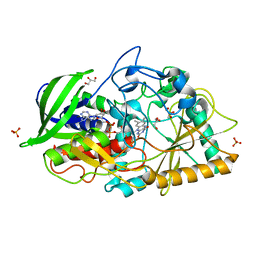
1IC6
 | | STRUCTURE OF A SERINE PROTEASE PROTEINASE K FROM TRITIRACHIUM ALBUM LIMBER AT 0.98 A RESOLUTION | | 分子名称: | CALCIUM ION, NITRATE ION, PROTEINASE K | | 著者 | Betzel, C, Gourinath, S, Kumar, P, Kaur, P, Perbandt, M, Eschenburg, S, Singh, T.P. | | 登録日 | 2001-03-30 | | 公開日 | 2001-04-11 | | 最終更新日 | 2011-07-13 | | 実験手法 | X-RAY DIFFRACTION (0.98 Å) | | 主引用文献 | Structure of a serine protease proteinase K from Tritirachium album limber at 0.98 A resolution.
Biochemistry, 40, 2001
|
|
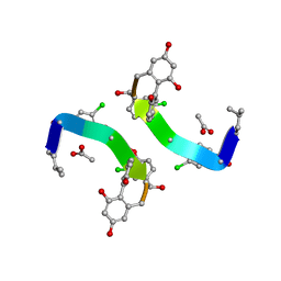
8V56
 | | UIC-1 mutant - UIC-1-B5W | | 分子名称: | UIC-1-B5W | | 著者 | Heinz-Kunert, S.L. | | 登録日 | 2023-11-30 | | 公開日 | 2024-03-06 | | 最終更新日 | 2024-03-20 | | 実験手法 | X-RAY DIFFRACTION (0.98 Å) | | 主引用文献 | Pore Restructuring of Peptide Frameworks by Mutations at Distal Packing Residues.
Biomacromolecules, 25, 2024
|
|
1S5M
 | | Xylose Isomerase in Substrate and Inhibitor Michaelis States: Atomic Resolution Studies of a Metal-Mediated Hydride Shift | | 分子名称: | MANGANESE (II) ION, SODIUM ION, Xylose isomerase, ... | | 著者 | Fenn, T.D, Ringe, D, Petsko, G.A. | | 登録日 | 2004-01-21 | | 公開日 | 2004-02-10 | | 最終更新日 | 2023-08-23 | | 実験手法 | X-RAY DIFFRACTION (0.98 Å) | | 主引用文献 | Xylose isomerase in substrate and inhibitor michaelis States: atomic resolution studies of a metal-mediated hydride shift(,).
Biochemistry, 43, 2004
|
|
3JUD
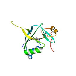 | | Human gamma-glutamylamine cyclotransferase, E82Q mutant | | 分子名称: | AIG2-like domain-containing protein 1, NITRATE ION | | 著者 | Oakley, A.J. | | 登録日 | 2009-09-15 | | 公開日 | 2010-02-09 | | 最終更新日 | 2023-11-01 | | 実験手法 | X-RAY DIFFRACTION (0.98 Å) | | 主引用文献 | Identification and characterization of {gamma}-glutamylamine cyclotransferase: An enzyme responsible for {gamma}-glutamyl-{epsilon}-lysine catabolism
J.Biol.Chem., 285, 2010
|
|
1GHG
 | | CRYSTAL STRUCTURE OF VANCOMYCIN AGLYCON | | 分子名称: | ACETIC ACID, DIMETHYL SULFOXIDE, VANCOMYCIN AGLYCON | | 著者 | Kaplan, J, Korty, B.D, Axelsen, P.H, Loll, P.J. | | 登録日 | 2000-12-13 | | 公開日 | 2001-02-12 | | 最終更新日 | 2023-12-27 | | 実験手法 | X-RAY DIFFRACTION (0.98 Å) | | 主引用文献 | The Role of Sugar Residues in Molecular Recognition by Vancomycin
J.Med.Chem., 44, 2001
|
|
3B3R
 | | Crystal structure of Streptomyces cholesterol oxidase H447Q/E361Q mutant bound to glycerol (0.98A) | | 分子名称: | Cholesterol oxidase, FLAVIN-N7 PROTONATED-ADENINE DINUCLEOTIDE, GLYCEROL, ... | | 著者 | Lyubimov, A.Y, Heard, K, Tang, H, Sampson, N.S, Vrielink, A. | | 登録日 | 2007-10-22 | | 公開日 | 2007-12-18 | | 最終更新日 | 2023-11-01 | | 実験手法 | X-RAY DIFFRACTION (0.98 Å) | | 主引用文献 | Distortion of flavin geometry is linked to ligand binding in cholesterol oxidase
Protein Sci., 16, 2007
|
|
6Q4G
 | | CDK2 in complex with FragLite37 | | 分子名称: | 2-[3-(2-azanyl-9~{H}-purin-6-yl)phenyl]ethanoic acid, Cyclin-dependent kinase 2 | | 著者 | Wood, D.J, Martin, M.P, Noble, M.E.M. | | 登録日 | 2018-12-05 | | 公開日 | 2019-03-20 | | 最終更新日 | 2024-01-24 | | 実験手法 | X-RAY DIFFRACTION (0.98 Å) | | 主引用文献 | FragLites-Minimal, Halogenated Fragments Displaying Pharmacophore Doublets. An Efficient Approach to Druggability Assessment and Hit Generation.
J.Med.Chem., 62, 2019
|
|
6T81
 | | Human Carbonic anhydrase II bound by 2-Naphthalenesulfonamide. | | 分子名称: | AZIDE ION, BICINE, SODIUM ION, ... | | 著者 | Smirnov, A, Manakova, E, Grazulis, S. | | 登録日 | 2019-10-23 | | 公開日 | 2020-10-14 | | 最終更新日 | 2024-01-24 | | 実験手法 | X-RAY DIFFRACTION (0.98 Å) | | 主引用文献 | Isoform-Selective Enzyme Inhibitors by Exploring Pocket Size According to the Lock-and-Key Principle.
Biophys.J., 119, 2020
|
|


