8TV4
 
 | |
1TXP
 | |
5MR3
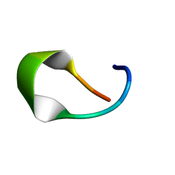
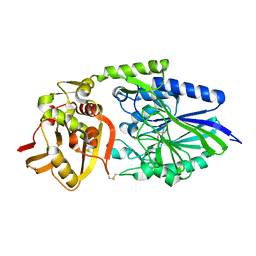
 | | Crystal structure of red abalone egg VERL repeat 2 with linker in complex with sperm lysin at 1.8 A resolution | | 分子名称: | 2-acetamido-2-deoxy-beta-D-glucopyranose, CHLORIDE ION, Egg-lysin, ... | | 著者 | Nishimura, K, Raj, I, Sadat Al-Hosseini, H, De Sanctis, D, Jovine, L. | | 登録日 | 2016-12-21 | | 公開日 | 2017-06-14 | | 最終更新日 | 2024-01-17 | | 実験手法 | X-RAY DIFFRACTION (1.8 Å) | | 主引用文献 | Structural Basis of Egg Coat-Sperm Recognition at Fertilization.
Cell, 169, 2017
|
|
4Y32
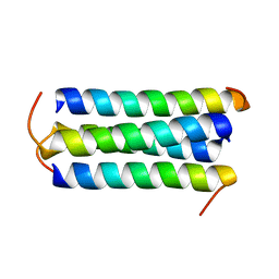
 | | Crystal structure of C-terminal modified Tau peptide-hybrid 109B with 14-3-3sigma | | 分子名称: | (2S)-2-(2-methoxyethyl)pyrrolidine, 14-3-3 protein sigma, ARG-THR-PRO-SEP-LEU-PRO-CNC(C(C)O)C(=O)N1CCCC1CCOC | | 著者 | Bartel, M, Milroy, L, Bier, D, Brunsveld, L, Ottmann, C. | | 登録日 | 2015-02-10 | | 公開日 | 2015-12-23 | | 最終更新日 | 2024-01-10 | | 実験手法 | X-RAY DIFFRACTION (1.7 Å) | | 主引用文献 | Stabilizer-Guided Inhibition of Protein-Protein Interactions.
Angew.Chem.Int.Ed.Engl., 54, 2015
|
|
5MKG
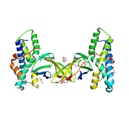
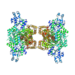
 | | PA3825-EAL Ca-CdG Structure | | 分子名称: | 9,9'-[(2R,3R,3aS,5S,7aR,9R,10R,10aS,12S,14aR)-3,5,10,12-tetrahydroxy-5,12-dioxidooctahydro-2H,7H-difuro[3,2-d:3',2'-j][1,3,7,9,2,8]tetraoxadiphosphacyclododecine-2,9-diyl]bis(2-amino-1,9-dihydro-6H-purin-6-one), CALCIUM ION, Diguanylate phosphodiesterase | | 著者 | Horrell, S, Bellini, D, Strange, R, Wagner, A, Walsh, M. | | 登録日 | 2016-12-04 | | 公開日 | 2016-12-14 | | 最終更新日 | 2024-06-19 | | 実験手法 | X-RAY DIFFRACTION (2.44 Å) | | 主引用文献 | Dimerisation induced formation of the active site and the identification of three metal sites in EAL-phosphodiesterases.
Sci Rep, 7, 2017
|
|
1OI8
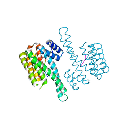
 | | 5'-Nucleotidase (E. coli) with an Engineered Disulfide Bridge (P90C, L424C) | | 分子名称: | CARBONATE ION, MANGANESE (II) ION, PROTEIN USHA, ... | | 著者 | Schultz-Heienbrok, R, Maier, T, Straeter, N. | | 登録日 | 2003-06-10 | | 公開日 | 2004-06-10 | | 最終更新日 | 2023-12-13 | | 実験手法 | X-RAY DIFFRACTION (2.1 Å) | | 主引用文献 | Trapping a 96 Degree Domain Rotation in Two Distinct Conformations by Engineered Disulfide Bridges
Protein Sci., 13, 2004
|
|
4XXD
 | | Crystal Structure of mid-region amyloid beta capture by solanezumab | | 分子名称: | Amyloid-beta fragment, Fab Heavy Chain, Fab Light Chain | | 著者 | Hermans, S.J, Crespi, G.A.N, Parker, M.W, Miles, L.A. | | 登録日 | 2015-01-30 | | 公開日 | 2015-04-29 | | 最終更新日 | 2023-09-27 | | 実験手法 | X-RAY DIFFRACTION (2.41 Å) | | 主引用文献 | Molecular basis for mid-region amyloid-beta capture by leading Alzheimer's disease immunotherapies.
Sci Rep, 5, 2015
|
|
5MFQ
 | | Crystal structure of the GluK1 ligand-binding domain in complex with kainate and BPAM-344 at 1.90 A resolution | | 分子名称: | 3-(CARBOXYMETHYL)-4-ISOPROPENYLPROLINE, 4-cyclopropyl-7-fluoro-3,4-dihydro-2H-1,2,4-benzothiadiazine 1,1-dioxide, CHLORIDE ION, ... | | 著者 | Larsen, A.P, Frydenvang, K, Kastrup, J.S. | | 登録日 | 2016-11-18 | | 公開日 | 2017-04-12 | | 最終更新日 | 2024-01-17 | | 実験手法 | X-RAY DIFFRACTION (1.9 Å) | | 主引用文献 | Identification and Structure-Function Study of Positive Allosteric Modulators of Kainate Receptors.
Mol. Pharmacol., 91, 2017
|
|
1OIE
 | |
4XZ2
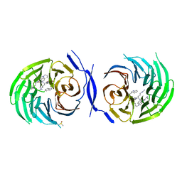
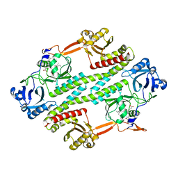
 | | Human platelet phosphofructokinase in an R-state in complex with ADP and F6P, crystal form I | | 分子名称: | 1,6-di-O-phosphono-beta-D-fructofuranose, 6-O-phosphono-beta-D-fructofuranose, ADENOSINE-5'-DIPHOSPHATE, ... | | 著者 | Kloos, M, Strater, N. | | 登録日 | 2015-02-03 | | 公開日 | 2015-06-03 | | 最終更新日 | 2024-01-10 | | 実験手法 | X-RAY DIFFRACTION (2.67 Å) | | 主引用文献 | Crystal structure of human platelet phosphofructokinase-1 locked in an activated conformation.
Biochem.J., 469, 2015
|
|
8C4C
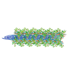
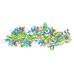 | | F-actin decorated by SipA497-669 | | 分子名称: | ADENOSINE-5'-DIPHOSPHATE, Actin, alpha skeletal muscle, ... | | 著者 | Yuan, B, Wald, J, Marlovits, T.C. | | 登録日 | 2023-01-03 | | 公開日 | 2024-01-17 | | 実験手法 | ELECTRON MICROSCOPY (2.7 Å) | | 主引用文献 | Structural basis for subversion of host cell actin cytoskeleton during Salmonella infection.
Sci Adv, 9, 2023
|
|
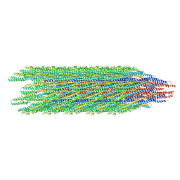
8C4E
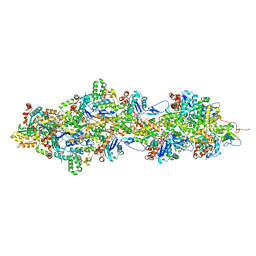 | | F-actin decorated by SipA426-685 | | 分子名称: | ADENOSINE-5'-DIPHOSPHATE, Actin, alpha skeletal muscle, ... | | 著者 | Yuan, B, Wald, J, Marlovits, T.C. | | 登録日 | 2023-01-03 | | 公開日 | 2024-01-17 | | 実験手法 | ELECTRON MICROSCOPY (2.6 Å) | | 主引用文献 | Structural basis for subversion of host cell actin cytoskeleton during Salmonella infection.
Sci Adv, 9, 2023
|
|
4Y60
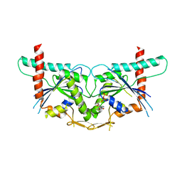
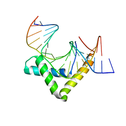 | | Structure of SOX18-HMG/PROX1-DNA | | 分子名称: | DNA (5'-D(*CP*AP*CP*TP*AP*GP*CP*AP*TP*TP*GP*TP*CP*TP*GP*GP*G)-3'), DNA (5'-D(*GP*CP*CP*CP*AP*GP*AP*CP*AP*AP*TP*GP*CP*TP*AP*GP*T)-3'), Transcription factor SOX-18 | | 著者 | Narasimhan, K, Prokoph, N, Kolatkar, P, Robinson, H, Jauch, R. | | 登録日 | 2015-02-12 | | 公開日 | 2016-03-02 | | 最終更新日 | 2024-03-20 | | 実験手法 | X-RAY DIFFRACTION (1.75 Å) | | 主引用文献 | Structure and decoy-mediated inhibition of the SOX18/Prox1-DNA interaction.
Nucleic Acids Res., 44, 2016
|
|
4Y9M
 | | PA3825-EAL Metal-Free-Apo Structure | | 分子名称: | PA3825-EAL, PHOSPHATE ION | | 著者 | Bellini, D, Horrell, S, Wagner, A, Strange, R, Walsh, M.A. | | 登録日 | 2015-02-17 | | 公開日 | 2016-03-09 | | 最終更新日 | 2024-01-10 | | 実験手法 | X-RAY DIFFRACTION (1.6 Å) | | 主引用文献 | Dimerisation induced formation of the active site and the identification of three metal sites in EAL-phosphodiesterases.
Sci Rep, 7, 2017
|
|
3ZJH
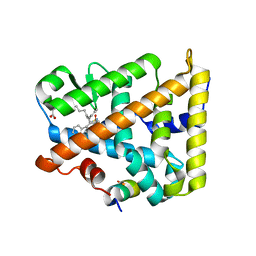
 | | Trp(60)B9Ala mutation of M.acetivorans protoglobin in complex with cyanide | | 分子名称: | CYANIDE ION, PROTOGLOBIN, PROTOPORPHYRIN IX CONTAINING FE | | 著者 | Pesce, A, Tilleman, L, Donne, J, Aste, E, Ascenzi, P, Ciaccio, C, Coletta, M, Moens, L, Viappiani, C, Dewilde, S, Bolognesi, M, Nardini, M. | | 登録日 | 2013-01-18 | | 公開日 | 2013-06-26 | | 最終更新日 | 2023-12-20 | | 実験手法 | X-RAY DIFFRACTION (1.7 Å) | | 主引用文献 | Structure and Haem-Distal Site Plasticity in Methanosarcina Acetivorans Protoglobin.
Plos One, 8, 2013
|
|
8DAF
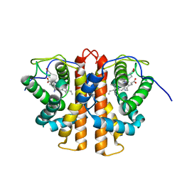
 | | Human SF-1 LBD bound to synthetic agonist 6N-10CA and bacterial phospholipid | | 分子名称: | 10-[(3aR,6S,6aR)-3-phenyl-3a-(1-phenylethenyl)-6-(sulfamoylamino)-1,3a,4,5,6,6a-hexahydropentalen-2-yl]decanoic acid (non-preferred name), DI-PALMITOYL-3-SN-PHOSPHATIDYLETHANOLAMINE, Nuclear receptor coactivator 2, ... | | 著者 | D'Agostino, E.H, Cato, M.L, Ortlund, E.A. | | 登録日 | 2022-06-13 | | 公開日 | 2023-06-28 | | 最終更新日 | 2023-10-25 | | 実験手法 | X-RAY DIFFRACTION (2.59 Å) | | 主引用文献 | Comparison of activity, structure, and dynamics of SF-1 and LRH-1 complexed with small molecule modulators.
J.Biol.Chem., 299, 2023
|
|
5MWJ
 | | Structure Enabled Discovery of a Stapled Peptide Inhibitor to Target the Oncogenic Transcriptional Repressor TLE1 | | 分子名称: | DIMETHYL SULFOXIDE, Transducin-like enhancer protein 1, pepide inhibtor | | 著者 | McGrath, S, Tortorici, M, Vidler, L, Drouin, L, Westwood, I, Gimeson, P, Van Montfort, R, Hoelder, S. | | 登録日 | 2017-01-18 | | 公開日 | 2017-04-05 | | 最終更新日 | 2024-01-17 | | 実験手法 | X-RAY DIFFRACTION (2.04 Å) | | 主引用文献 | Structure-Enabled Discovery of a Stapled Peptide Inhibitor to Target the Oncogenic Transcriptional Repressor TLE1.
Chemistry, 23, 2017
|
|
8CYE
 | |
8CWM
 | |
5MYJ
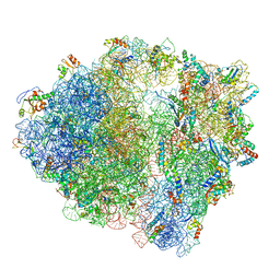 | | Structure of 70S ribosome from Lactococcus lactis | | 分子名称: | 16S ribosomal RNA, 23S ribosomal RNA, 30S ribosomal protein S10, ... | | 著者 | Franken, L.E, Oostergetel, G.T, Pijning, T, Puri, P, Boekema, E.J, Poolman, B, Guskov, A. | | 登録日 | 2017-01-26 | | 公開日 | 2017-10-11 | | 最終更新日 | 2024-05-15 | | 実験手法 | ELECTRON MICROSCOPY (5.6 Å) | | 主引用文献 | A general mechanism of ribosome dimerization revealed by single-particle cryo-electron microscopy.
Nat Commun, 8, 2017
|
|
8CXM
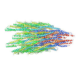
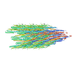
 | | Cryo-EM structure of the supercoiled E. coli K12 flagellar filament core, Normal waveform | | 分子名称: | Flagellin | | 著者 | Sonani, R.R, Kreutzberger, M.A.B, Sebastian, A.L, Scharf, B, Egelman, E.H. | | 登録日 | 2022-05-21 | | 公開日 | 2022-09-07 | | 最終更新日 | 2024-06-12 | | 実験手法 | ELECTRON MICROSCOPY (3.21 Å) | | 主引用文献 | Convergent evolution in the supercoiling of prokaryotic flagellar filaments.
Cell, 185, 2022
|
|
8CVI
 | |
4R70
 | |
3T5D
 | |
5NL4
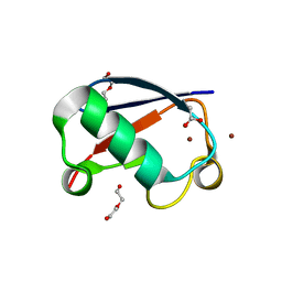 | | Crystal structure of Zn1.3-E16V human ubiquitin (hUb) mutant adduct, from a solution 35 mM zinc acetate/1.3 mM E16V hUb | | 分子名称: | 1,2-ETHANEDIOL, ACETATE ION, DI(HYDROXYETHYL)ETHER, ... | | 著者 | Fermani, S, Falini, G. | | 登録日 | 2017-04-04 | | 公開日 | 2017-04-26 | | 最終更新日 | 2024-01-17 | | 実験手法 | X-RAY DIFFRACTION (1.32 Å) | | 主引用文献 | Aggregation Pathways of Native-Like Ubiquitin Promoted by Single-Point Mutation, Metal Ion Concentration, and Dielectric Constant of the Medium.
Chemistry, 24, 2018
|
|


