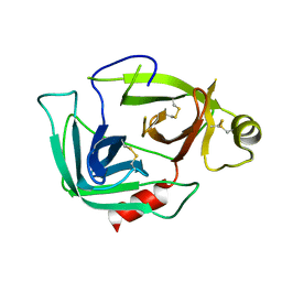
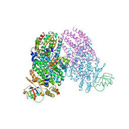
4FQ2
 
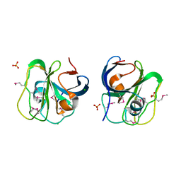
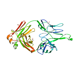 | | Crystal Structure of 10-1074 Fab | | 分子名称: | 1,2-ETHANEDIOL, Fab heavy chain, Fab light chain, ... | | 著者 | Scharf, L, Bjorkman, P.J. | | 登録日 | 2012-06-24 | | 公開日 | 2012-11-14 | | 最終更新日 | 2021-05-19 | | 実験手法 | X-RAY DIFFRACTION (1.9 Å) | | 主引用文献 | Complex-type N-glycan recognition by potent broadly neutralizing HIV antibodies.
Proc.Natl.Acad.Sci.USA, 109, 2012
|
|
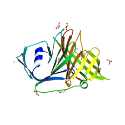
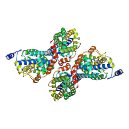
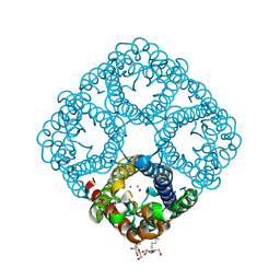
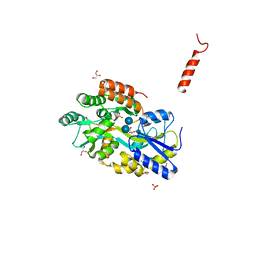
3T7J
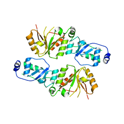 | | Crystal structure of Rtt107p (residues 820-1070) | | 分子名称: | Regulator of Ty1 transposition protein 107 | | 著者 | Li, X, Li, F, Wu, J, Shi, Y. | | 登録日 | 2011-07-30 | | 公開日 | 2012-02-15 | | 最終更新日 | 2024-03-20 | | 実験手法 | X-RAY DIFFRACTION (2.042 Å) | | 主引用文献 | Structure of C-terminal Tandem BRCT Repeats of Rtt107 Protein Reveals Critical Role in Interaction with Phosphorylated Histone H2A during DNA Damage Repair
J.Biol.Chem., 287, 2012
|
|
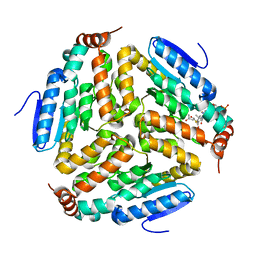
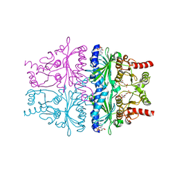
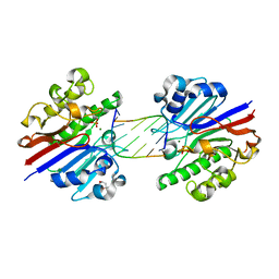
3T8N
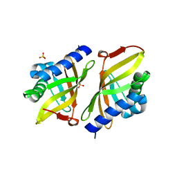 | | Crystal structure of ketosteroid isomerase Y16AD103A from Pseudomonas putida | | 分子名称: | SULFATE ION, Steroid Delta-isomerase, {[-(BIS-CARBOXYMETHYL-AMINO)-ETHYL]-CARBOXYMETHYL-AMINO}-ACETIC ACID | | 著者 | Gonzalez, A, Tsai, Y, Schwans, J, Sunden, F, Herschlag, D. | | 登録日 | 2011-08-01 | | 公開日 | 2011-11-23 | | 最終更新日 | 2023-09-13 | | 実験手法 | X-RAY DIFFRACTION (1.47 Å) | | 主引用文献 | Evaluating the catalytic contribution from the oxyanion hole in ketosteroid isomerase.
J.Am.Chem.Soc., 133, 2011
|
|
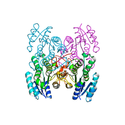
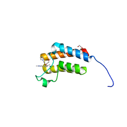
3G4T
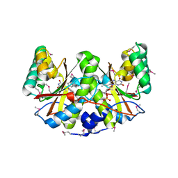
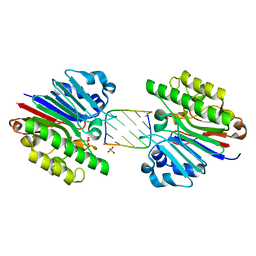 | | Mth0212 (WT) in complex with a 7bp dsDNA | | 分子名称: | 5'-D(*CP*G*TP*AP*CP*TP*AP*CP*G)-3', 5'-D(*CP*GP*TP*AP*(UPS)P*TP*AP*CP*G)-3', Exodeoxyribonuclease, ... | | 著者 | Lakomek, K, Dickmanns, A, Ficner, R. | | 登録日 | 2009-02-04 | | 公開日 | 2010-03-09 | | 最終更新日 | 2023-11-01 | | 実験手法 | X-RAY DIFFRACTION (2.64 Å) | | 主引用文献 | Crystal Structure Analysis of DNA Uridine Endonuclease Mth212 Bound to DNA
J.Mol.Biol., 399, 2010
|
|
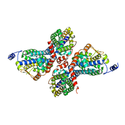
3T8I
 | | Structural analysis of thermostable S. solfataricus purine-specific nucleoside hydrolase | | 分子名称: | 1,2-ETHANEDIOL, CALCIUM ION, DI(HYDROXYETHYL)ETHER, ... | | 著者 | Minici, C, Cacciapuoti, G, De Leo, E, Porcelli, M, Degano, M. | | 登録日 | 2011-08-01 | | 公開日 | 2012-05-16 | | 最終更新日 | 2023-09-13 | | 実験手法 | X-RAY DIFFRACTION (1.8 Å) | | 主引用文献 | New Determinants in the Catalytic Mechanism of Nucleoside Hydrolases from the Structures of Two Isozymes from Sulfolobus solfataricus.
Biochemistry, 51, 2012
|
|
2O62
 | |
3PUP
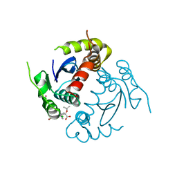
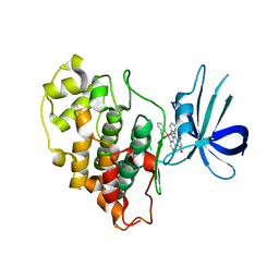 | | Structure of Glycogen Synthase Kinase 3 beta (GSK3B) in complex with a ruthenium octasporine ligand (OS1) | | 分子名称: | Glycogen synthase kinase-3 beta, Ruthenium octasporine | | 著者 | Filippakopoulos, P, Kraling, K, Essen, L.O, Meggers, E, Knapp, S. | | 登録日 | 2010-12-06 | | 公開日 | 2010-12-22 | | 最終更新日 | 2023-09-06 | | 実験手法 | X-RAY DIFFRACTION (2.99 Å) | | 主引用文献 | Structurally sophisticated octahedral metal complexes as highly selective protein kinase inhibitors.
J.Am.Chem.Soc., 133, 2011
|
|
3PVT
 | |
3PW8
 | |
3Q0J
 | |
3PWQ
 | |
3Q17
 | | Structure of a slow CLC Cl-/H+ antiporter from a cyanobacterium in Bromide | | 分子名称: | BROMIDE ION, Sll0855 protein | | 著者 | Jayaram, H, Robertson, J.L, Fang, W, Williams, C, Miller, C. | | 登録日 | 2010-12-16 | | 公開日 | 2011-01-19 | | 最終更新日 | 2023-09-13 | | 実験手法 | X-RAY DIFFRACTION (3.6 Å) | | 主引用文献 | Structure of a Slow CLC Cl(-)/H(+) Antiporter from a Cyanobacterium.
Biochemistry, 50, 2011
|
|
3PXV
 | |
3Q29
 | | Cyrstal structure of human alpha-synuclein (1-19) fused to maltose binding protein (MBP) | | 分子名称: | GLYCEROL, Maltose-binding periplasmic protein/alpha-synuclein chimeric protein, SULFATE ION, ... | | 著者 | Zhao, M, Sawaya, M.R, Cascio, D, Eisenberg, D. | | 登録日 | 2010-12-19 | | 公開日 | 2011-06-01 | | 最終更新日 | 2023-09-13 | | 実験手法 | X-RAY DIFFRACTION (2.3 Å) | | 主引用文献 | Structures of segments of alpha-synuclein fused to maltose-binding protein suggest intermediate states during amyloid formation
Protein Sci., 20, 2011
|
|
4GW6
 | |
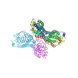
2O97
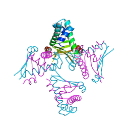 | | Crystal Structure of E. coli HU heterodimer | | 分子名称: | CHLORIDE ION, DNA-binding protein HU-alpha, DNA-binding protein HU-beta, ... | | 著者 | Guo, F, Adhya, S. | | 登録日 | 2006-12-13 | | 公開日 | 2007-03-06 | | 最終更新日 | 2023-12-27 | | 実験手法 | X-RAY DIFFRACTION (2.45 Å) | | 主引用文献 | Spiral structure of Escherichia coli HU{alpha}beta provides foundation for DNA supercoiling.
Proc.Natl.Acad.Sci.Usa, 104, 2007
|
|
3G01
 | |
2O9G
 | |
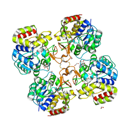
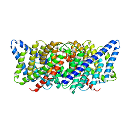
4GWS
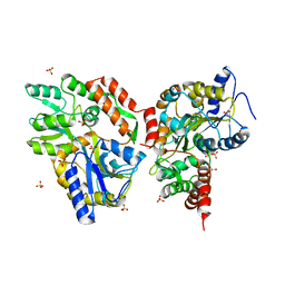
 | | Crystal Structure of AMP complexes of Porcine Liver Fructose-1,6-bisphosphatase with Filled Central Cavity | | 分子名称: | 6-O-phosphono-beta-D-fructofuranose, ADENOSINE MONOPHOSPHATE, Fructose-1,6-bisphosphatase 1, ... | | 著者 | Gao, Y, Honzatko, R.B. | | 登録日 | 2012-09-03 | | 公開日 | 2013-09-04 | | 最終更新日 | 2023-09-13 | | 実験手法 | X-RAY DIFFRACTION (2.75 Å) | | 主引用文献 | Hydrophobic Central Cavity in Fructose-1,6-bisphosphatase is Essential for the Synergism in AMP/Fructose 2,6-bisphosphate Inhibition
To be Published
|
|
3GED
 | | Fingerprint and Structural Analysis of a Apo SCOR enzyme from Clostridium thermocellum | | 分子名称: | GLYCEROL, SODIUM ION, Short-chain dehydrogenase/reductase SDR, ... | | 著者 | Huether, R, Liu, Z.J, Xu, H, Wang, B.C, Pletnev, V, Mao, Q, Umland, T, Duax, W. | | 登録日 | 2009-02-25 | | 公開日 | 2009-03-17 | | 最終更新日 | 2023-09-06 | | 実験手法 | X-RAY DIFFRACTION (1.698 Å) | | 主引用文献 | Sequence fingerprint and structural analysis of the SCOR enzyme A3DFK9 from Clostridium thermocellum.
Proteins, 78, 2010
|
|
3Q14
 | | Toluene 4 monooxygenase HD Complex with p-cresol | | 分子名称: | FE (III) ION, P-CRESOL, PENTAETHYLENE GLYCOL, ... | | 著者 | Acheson, J.F, Bailey, L.J, Fox, B.G. | | 登録日 | 2010-12-16 | | 公開日 | 2011-12-21 | | 最終更新日 | 2024-02-21 | | 実験手法 | X-RAY DIFFRACTION (1.75 Å) | | 主引用文献 | Crystallographic analysis of active site contributions to regiospecificity in the diiron enzyme toluene 4-monooxygenase.
Biochemistry, 51, 2012
|
|
3Q26
 | | Cyrstal structure of human alpha-synuclein (10-42) fused to maltose binding protein (MBP) | | 分子名称: | GLYCEROL, Maltose-binding periplasmic protein/alpha-synuclein chimeric protein, SULFATE ION, ... | | 著者 | Zhao, M, Sawaya, M.R, Cascio, D, Eisenberg, D. | | 登録日 | 2010-12-19 | | 公開日 | 2011-06-01 | | 最終更新日 | 2023-09-13 | | 実験手法 | X-RAY DIFFRACTION (1.54 Å) | | 主引用文献 | Structures of segments of alpha-synuclein fused to maltose-binding protein suggest intermediate states during amyloid formation
Protein Sci., 20, 2011
|
|
3G3C
 | | Mth0212 (WT) in complex with a 6bp dsDNA containing a single one nucleotide long 3'-overhang | | 分子名称: | (4R)-2-METHYLPENTANE-2,4-DIOL, 5'-D(*CP*GP*TP*AP*(UPS)P*TP*AP*CP*G)-3', 5'-D(*CP*GP*TP*AP*CP*TP*AP*CP*G)-3', ... | | 著者 | Lakomek, K, Dickmanns, A, Ficner, R. | | 登録日 | 2009-02-02 | | 公開日 | 2010-03-09 | | 最終更新日 | 2023-11-01 | | 実験手法 | X-RAY DIFFRACTION (3.04 Å) | | 主引用文献 | Crystal Structure Analysis of DNA Uridine Endonuclease Mth212 Bound to DNA
J.Mol.Biol., 399, 2010
|
|
3Q2F
 | | Crystal Structure of the bromodomain of human BAZ2B in complex with a triazolo ligand | | 分子名称: | 1,2-ETHANEDIOL, 4-{4-[3-(1H-imidazol-1-yl)propyl]-5-methyl-4H-1,2,4-triazol-3-yl}-1-methyl-1H-pyrazol-5-amine, Bromodomain adjacent to zinc finger domain protein 2B | | 著者 | Muniz, J.R.C, Filippakopoulos, P, Picaud, S, Felletar, I, Keates, T, von Delft, F, Arrowsmith, C.H, Edwards, A.M, Weigelt, J, Bountra, C, Knapp, S, Structural Genomics Consortium (SGC) | | 登録日 | 2010-12-20 | | 公開日 | 2011-01-26 | | 最終更新日 | 2023-11-01 | | 実験手法 | X-RAY DIFFRACTION (2.06 Å) | | 主引用文献 | Crystal Structure of the bromodomain of human BAZ2B in complex with a triazolo ligand
TO BE PUBLISHED
|
|
3PJY
 | |


