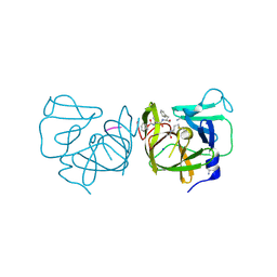
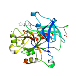
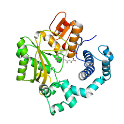
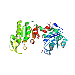
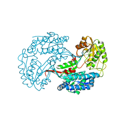
4IIE
 
 | | Crystal structure of beta-glucosidase 1 from Aspergillus aculeatus in complex with calystegine B(2) | | 分子名称: | (4R)-2-METHYLPENTANE-2,4-DIOL, 2-acetamido-2-deoxy-beta-D-glucopyranose, 2-acetamido-2-deoxy-beta-D-glucopyranose-(1-4)-2-acetamido-2-deoxy-beta-D-glucopyranose, ... | | 著者 | Suzuki, K, Sumitani, J, Kawaguchi, T, Fushinobu, S. | | 登録日 | 2012-12-20 | | 公開日 | 2013-04-10 | | 最終更新日 | 2020-07-29 | | 実験手法 | X-RAY DIFFRACTION (2 Å) | | 主引用文献 | Crystal structures of glycoside hydrolase family 3 beta-glucosidase 1 from Aspergillus aculeatus
Biochem.J., 452, 2013
|
|
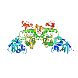
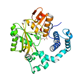
4IMQ
 | | Structural Basis of Substrate Specificity and Protease Inhibition in Norwalk Virus | | 分子名称: | 3C-like protease, PEPTIDE INHIBITOR, syc8, ... | | 著者 | Prasad, B.V.V, Muhaxhiri, Z, Deng, L, Shanker, S, Sankaran, B, Estes, M.K, Palzkill, T, Song, Y. | | 登録日 | 2013-01-03 | | 公開日 | 2013-02-20 | | 最終更新日 | 2023-11-15 | | 実験手法 | X-RAY DIFFRACTION (1.5 Å) | | 主引用文献 | Structural basis of substrate specificity and protease inhibition in norwalk virus.
J.Virol., 87, 2013
|
|
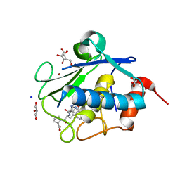
1V47
 | | Crystal structure of ATP sulfurylase from Thermus thermophillus HB8 in complex with APS | | 分子名称: | ADENOSINE-5'-PHOSPHOSULFATE, ATP sulfurylase, CHLORIDE ION, ... | | 著者 | Taguchi, Y, Sugishima, M, Fukuyama, K, RIKEN Structural Genomics/Proteomics Initiative (RSGI) | | 登録日 | 2003-11-11 | | 公開日 | 2004-04-06 | | 最終更新日 | 2023-10-25 | | 実験手法 | X-RAY DIFFRACTION (2.49 Å) | | 主引用文献 | Crystal structure of a novel zinc-binding ATP sulfurylase from Thermus thermophilus HB8
Biochemistry, 43, 2004
|
|
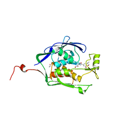
3BKM
 | | Structure of anti-amyloid-beta Fab WO2 (Form A, P212121) | | 分子名称: | SODIUM ION, WO2 IgG2a Fab fragment Heavy Chain, WO2 IgG2a Fab fragment Light Chain Kappa, ... | | 著者 | Miles, L.A, Wun, K.S, Crespi, G.A, Fodero-Tavoletti, M, Galatis, D, Bageley, C.J, Beyreuther, K, Masters, C.L, Cappai, R, McKinstry, W.J, Barnham, K.J, Parker, M.W. | | 登録日 | 2007-12-07 | | 公開日 | 2008-04-15 | | 最終更新日 | 2023-11-01 | | 実験手法 | X-RAY DIFFRACTION (1.6 Å) | | 主引用文献 | Amyloid-beta-anti-amyloid-beta complex structure reveals an extended conformation in the immunodominant B-cell epitope.
J.Mol.Biol., 377, 2008
|
|
4IHS
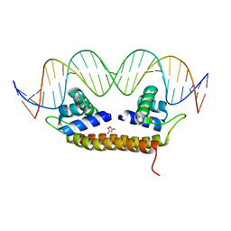 | | Crystal Structure of BenM_DBD/catB site 1 DNA Complex | | 分子名称: | HTH-type transcriptional regulator BenM, MALONATE ION, SODIUM ION, ... | | 著者 | Alanazi, A, Momany, C, Neidle, E.L. | | 登録日 | 2012-12-19 | | 公開日 | 2013-07-10 | | 最終更新日 | 2023-09-20 | | 実験手法 | X-RAY DIFFRACTION (3.1 Å) | | 主引用文献 | The DNA-binding domain of BenM reveals the structural basis for the recognition of a T-N11-A sequence motif by LysR-type transcriptional regulators.
Acta Crystallogr.,Sect.D, 69, 2013
|
|
4H1P
 | |
4HDB
 | | Crystal Structure of HIV-1 protease mutants D30N complexed with inhibitor GRL-0519 | | 分子名称: | (3R,3aS,3bR,6aS,7aS)-octahydrodifuro[2,3-b:3',2'-d]furan-3-yl [(1S,2R)-1-benzyl-2-hydroxy-3-{[(4-methoxyphenyl)sulfonyl](2-methylpropyl)amino}propyl]carbamate, CHLORIDE ION, HIV-1 Protease, ... | | 著者 | Zhang, H, Wang, Y.-F, Shen, C.H, Agniswamy, J, Weber, I.T. | | 登録日 | 2012-10-02 | | 公開日 | 2013-08-14 | | 最終更新日 | 2023-09-20 | | 実験手法 | X-RAY DIFFRACTION (1.49 Å) | | 主引用文献 | Novel P2 tris-tetrahydrofuran group in antiviral compound 1 (GRL-0519) fills the S2 binding pocket of selected mutants of HIV-1 protease.
J.Med.Chem., 56, 2013
|
|
2ZHW
 | | Exploring thrombin S3 pocket | | 分子名称: | Hirudin variant-2, N-cycloheptylglycyl-N-(4-carbamimidoylbenzyl)-L-prolinamide, SODIUM ION, ... | | 著者 | Brandt, T, Baum, B, Heine, A, Klebe, G. | | 登録日 | 2008-02-08 | | 公開日 | 2009-02-10 | | 最終更新日 | 2023-11-15 | | 実験手法 | X-RAY DIFFRACTION (2.02 Å) | | 主引用文献 | Exploring thrombin S3 pocket
To be Published
|
|
4H88
 | | Structure of POM1 FAB fragment complexed with mouse PrPc Fragment 120-230 | | 分子名称: | Major prion protein, POM1 FAB CHAIN H, POM1 FAB CHAIN L, ... | | 著者 | Baral, P.K, Wieland, B, Swayampakula, M, James, M.N. | | 登録日 | 2012-09-21 | | 公開日 | 2013-07-31 | | 最終更新日 | 2013-09-18 | | 実験手法 | X-RAY DIFFRACTION (1.9 Å) | | 主引用文献 | The toxicity of antiprion antibodies is mediated by the flexible tail of the prion protein.
Nature, 501, 2013
|
|
3HVI
 | | Rat catechol O-methyltransferase in complex with a catechol-type, N6-ethyladenine-containing bisubstrate inhibitor | | 分子名称: | (4S,5S)-1,2-DITHIANE-4,5-DIOL, CHLORIDE ION, Catechol O-methyltransferase, ... | | 著者 | Ehler, A, Schlatter, D, Stihle, M, Benz, J, Rudolph, M.G. | | 登録日 | 2009-06-16 | | 公開日 | 2009-10-13 | | 最終更新日 | 2024-03-20 | | 実験手法 | X-RAY DIFFRACTION (1.2 Å) | | 主引用文献 | Molecular recognition at the active site of catechol-o-methyltransferase: energetically favorable replacement of a water molecule imported by a bisubstrate inhibitor.
Angew.Chem.Int.Ed.Engl., 48, 2009
|
|
2ZO3
 | | Bisphenylic Thrombin Inhibitors | | 分子名称: | Hirudin variant-1, SODIUM ION, Thrombin heavy chain, ... | | 著者 | Baum, B, Heine, A, Klebe, G. | | 登録日 | 2008-05-05 | | 公開日 | 2009-04-14 | | 最終更新日 | 2023-11-15 | | 実験手法 | X-RAY DIFFRACTION (1.7 Å) | | 主引用文献 | Think twice: understanding the high potency of bis(phenyl)methane inhibitors of thrombin
J.Mol.Biol., 391, 2009
|
|
3I58
 | |
4HHD
 | |
2ZF0
 | | Exploring Thrombin S1 Pocket | | 分子名称: | D-phenylalanyl-N-(3-methylbenzyl)-L-prolinamide, Hirudin variant-1, SODIUM ION, ... | | 著者 | Baum, B, Heine, A, Klebe, G. | | 登録日 | 2007-12-18 | | 公開日 | 2008-11-04 | | 最終更新日 | 2023-11-15 | | 実験手法 | X-RAY DIFFRACTION (2.2 Å) | | 主引用文献 | Exploring Thrombin S1 pocket
To be Published
|
|
3L27
 | | Crystal structure of Zaire Ebola VP35 interferon inhibitory domain R312A mutant | | 分子名称: | CHLORIDE ION, GLYCEROL, PHOSPHATE ION, ... | | 著者 | Leung, D.W, Prins, K.C, Borek, D.M, Farahbakhsh, M, Tufariello, J.M, Ramanan, P, Nix, J.C, Helgeson, L.A, Otwinowski, Z, Honzatko, R.B, Basler, C.F, Amarasinghe, G.K. | | 登録日 | 2009-12-14 | | 公開日 | 2010-01-26 | | 最終更新日 | 2023-09-06 | | 実験手法 | X-RAY DIFFRACTION (1.95 Å) | | 主引用文献 | Structural basis for dsRNA recognition and interferon antagonism by Ebola VP35.
Nat.Struct.Mol.Biol., 17, 2010
|
|
4IQU
 | | Tdt core in complex with inhibitor (2Z,5E)-6-[4-(4-fluorobenzoyl)-1H-pyrrol-2-yl]-2-hydroxy-4-oxohexa-2,5-dienoic acid | | 分子名称: | (2Z,5E)-6-[4-(4-fluorobenzoyl)-1H-pyrrol-2-yl]-2-hydroxy-4-oxohexa-2,5-dienoic acid, DNA nucleotidylexotransferase, SODIUM ION | | 著者 | Gouge, J, Delarue, M. | | 登録日 | 2013-01-13 | | 公開日 | 2013-09-04 | | 最終更新日 | 2024-03-13 | | 実験手法 | X-RAY DIFFRACTION (2.4 Å) | | 主引用文献 | New nucleotide-competitive non-nucleoside inhibitors of terminal deoxynucleotidyl transferase: discovery, characterization, and crystal structure in complex with the target.
J.Med.Chem., 56, 2013
|
|
4IQV
 | | Tdt core in complex with inhibitor 6-[4-(3-fluorobenzoyl)-1H-pyrrol-2-yl]-2-hydroxy-4-oxohexa-2,5-dienoic acid and ssDNA | | 分子名称: | 5'-D(*GP*CP*CP*G)-3', 6-[4-(3-fluorobenzoyl)-1H-pyrrol-2-yl]-2-hydroxy-4-oxohexa-2,5-dienoic acid, DNA nucleotidylexotransferase, ... | | 著者 | Gouge, J, Delarue, M. | | 登録日 | 2013-01-13 | | 公開日 | 2013-09-04 | | 最終更新日 | 2023-09-20 | | 実験手法 | X-RAY DIFFRACTION (2.9 Å) | | 主引用文献 | New nucleotide-competitive non-nucleoside inhibitors of terminal deoxynucleotidyl transferase: discovery, characterization, and crystal structure in complex with the target.
J.Med.Chem., 56, 2013
|
|
4IQW
 | | Tdt core in complex with inhibitor (2Z,5E)-6-[4-(4-fluorobenzoyl)-1H-pyrrol-2-yl]-2-hydroxy-4-oxohexa-2,5-dienoic acid and ssDNA | | 分子名称: | (2Z,5E)-6-[4-(4-fluorobenzoyl)-1H-pyrrol-2-yl]-2-hydroxy-4-oxohexa-2,5-dienoic acid, 5'-D(*GP*CP*CP*G)-3', DNA nucleotidylexotransferase, ... | | 著者 | Gouge, J, Delarue, M. | | 登録日 | 2013-01-13 | | 公開日 | 2013-09-04 | | 最終更新日 | 2023-09-20 | | 実験手法 | X-RAY DIFFRACTION (2.6 Å) | | 主引用文献 | New nucleotide-competitive non-nucleoside inhibitors of terminal deoxynucleotidyl transferase: discovery, characterization, and crystal structure in complex with the target.
J.Med.Chem., 56, 2013
|
|
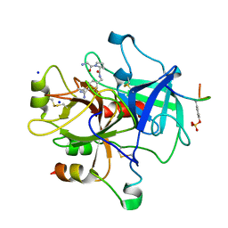
4IN9
 | | Structure of karilysin MMP-like catalytic domain in complex with inhibitory tetrapeptide SWFP | | 分子名称: | GLYCEROL, Karilysin protease, POTASSIUM ION, ... | | 著者 | Guevara, T, Ksiazek, M, Skottrup, P.D, Cerda-Costa, N, Trillo-Muyo, S, de Diego, I, Riise, E, Potempa, J, Gomis-Ruth, F.X. | | 登録日 | 2013-01-04 | | 公開日 | 2013-05-15 | | 最終更新日 | 2023-09-20 | | 実験手法 | X-RAY DIFFRACTION (1.55 Å) | | 主引用文献 | Structure of the catalytic domain of the Tannerella forsythia matrix metallopeptidase karilysin in complex with a tetrapeptidic inhibitor.
Acta Crystallogr.,Sect.F, 69, 2013
|
|
4IS9
 | | Crystal Structure of the Escherichia coli LpxC/L-161,240 complex | | 分子名称: | (4R)-2-(3,4-dimethoxy-5-propylphenyl)-N-hydroxy-4,5-dihydro-1,3-oxazole-4-carboxamide, ISOPROPYL ALCOHOL, SODIUM ION, ... | | 著者 | Lee, C.-J, Zhou, P. | | 登録日 | 2013-01-16 | | 公開日 | 2013-10-30 | | 最終更新日 | 2024-02-28 | | 実験手法 | X-RAY DIFFRACTION (2.13 Å) | | 主引用文献 | Structural Basis of the Promiscuous Inhibitor Susceptibility of Escherichia coli LpxC.
Acs Chem.Biol., 9, 2014
|
|
1V6S
 | |
4GRN
 | |
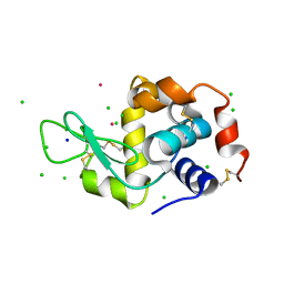
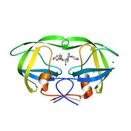
3HUP
 | | High-resolution structure of the extracellular domain of human CD69 | | 分子名称: | CHLORIDE ION, Early activation antigen CD69, SODIUM ION | | 著者 | Kolenko, P, Dohnalek, J, Skalova, T, Hasek, J, Duskova, J, Vanek, O, Bezouska, K. | | 登録日 | 2009-06-15 | | 公開日 | 2009-12-15 | | 最終更新日 | 2023-11-01 | | 実験手法 | X-RAY DIFFRACTION (1.371 Å) | | 主引用文献 | The high-resolution structure of the extracellular domain of human CD69 using a novel polymer
Acta Crystallogr.,Sect.F, 65, 2009
|
|
4GVC
 | | Crystal Structure of T-cell Lymphoma Invasion and Metastasis-1 PDZ in complex with phosphorylated Syndecan1 Peptide | | 分子名称: | 5-(DIMETHYLAMINO)-1-NAPHTHALENESULFONIC ACID(DANSYL ACID), CHLORIDE ION, SODIUM ION, ... | | 著者 | Liu, X, Shepherd, T.R, Murray, A.M, Xu, Z, Fuentes, E.J. | | 登録日 | 2012-08-30 | | 公開日 | 2013-03-13 | | 最終更新日 | 2023-12-06 | | 実験手法 | X-RAY DIFFRACTION (1.54 Å) | | 主引用文献 | The structure of the Tiam1 PDZ domain/ phospho-syndecan1 complex reveals a ligand conformation that modulates protein dynamics.
Structure, 21, 2013
|
|
3I44
 | |


