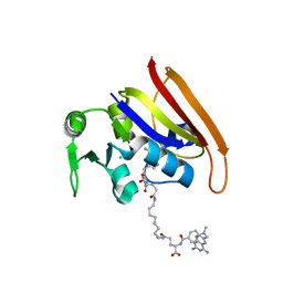
4XH2
 
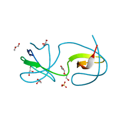
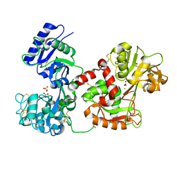
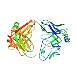 | | Crystal structure of human paxillin LD4 motif in complex with Fab fragment | | 分子名称: | ACETATE ION, ACETYL GROUP, Fab Heavy Chain, ... | | 著者 | Nocula-Lugowska, M, Lugowski, M, Salgia, R, Kossiakoff, A.A. | | 登録日 | 2015-01-04 | | 公開日 | 2015-07-01 | | 最終更新日 | 2023-09-27 | | 実験手法 | X-RAY DIFFRACTION (2 Å) | | 主引用文献 | Engineering Synthetic Antibody Inhibitors Specific for LD2 or LD4 Motifs of Paxillin.
J.Mol.Biol., 427, 2015
|
|
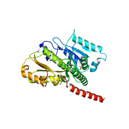
3OCR
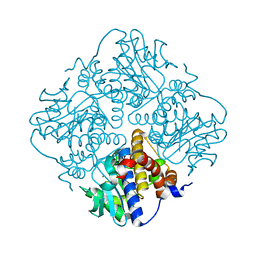
 | | Crystal structure of aldolase II superfamily protein from Pseudomonas syringae | | 分子名称: | Class II aldolase/adducin domain protein, SULFATE ION | | 著者 | Chang, C, Kagan, O, Savchenko, A, Edwards, A, Joachimiak, A, Midwest Center for Structural Genomics (MCSG) | | 登録日 | 2010-08-10 | | 公開日 | 2010-08-25 | | 最終更新日 | 2017-11-08 | | 実験手法 | X-RAY DIFFRACTION (1.95 Å) | | 主引用文献 | Crystal structure of aldolase II superfamily protein from Pseudomonas syringae
To be Published
|
|
4XXF
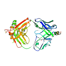
 | | L-fuculose 1-phosphate aldolase from Glaciozyma antarctica PI12 | | 分子名称: | Fuculose-1-phosphate aldolase, ZINC ION | | 著者 | Jaafar, N.R, Abu Bakar, F.D, Abdul Murad, A.M, Illias, R, Littler, D, Beddoe, T, Rossjohn, J, Mahadi, M.N. | | 登録日 | 2015-01-30 | | 公開日 | 2015-02-25 | | 最終更新日 | 2023-11-08 | | 実験手法 | X-RAY DIFFRACTION (1.34 Å) | | 主引用文献 | Crystal structure of fuculose aldolase from the Antarctic psychrophilic yeast Glaciozyma antarctica PI12.
Acta Crystallogr.,Sect.F, 72, 2016
|
|
2VRF
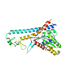
 | | CRYSTAL STRUCTURE OF THE HUMAN BETA-2-SYNTROPHIN PDZ DOMAIN | | 分子名称: | 1,2-ETHANEDIOL, BETA-2-SYNTROPHIN | | 著者 | Sun, Z, Roos, A.K, Pike, A.C.W, Pilka, E.S, Cooper, C, Elkins, J.M, Murray, J, Arrowsmith, C.H, Doyle, D, Edwards, A, von Delft, F, Bountra, C, Oppermann, U. | | 登録日 | 2008-03-31 | | 公開日 | 2008-04-22 | | 最終更新日 | 2023-12-13 | | 実験手法 | X-RAY DIFFRACTION (2 Å) | | 主引用文献 | Crystal Structure of the Human Beta-2-Syntrophin Pdz Domain
To be Published
|
|
4XGZ
 | | Crystal structure of human paxillin LD2 motif in complex with Fab fragment | | 分子名称: | 1,2-ETHANEDIOL, FAB HEAVY CHAIN, FAB LIGHT CHAIN, ... | | 著者 | Nocula-Lugowska, M, Lugowski, M, Salgia, R, Kossiakoff, A.A. | | 登録日 | 2015-01-04 | | 公開日 | 2015-07-01 | | 最終更新日 | 2023-09-27 | | 実験手法 | X-RAY DIFFRACTION (2.5 Å) | | 主引用文献 | Engineering Synthetic Antibody Inhibitors Specific for LD2 or LD4 Motifs of Paxillin.
J.Mol.Biol., 427, 2015
|
|
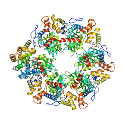
7AHO
 | | RUVBL1-RUVBL2 heterohexameric ring after binding of RNA helicase DHX34 | | 分子名称: | ADENOSINE-5'-DIPHOSPHATE, RuvB-like 1, RuvB-like 2 | | 著者 | Lopez-Perrote, A, Rodriguez, C.F, Llorca, O. | | 登録日 | 2020-09-25 | | 公開日 | 2020-11-25 | | 実験手法 | ELECTRON MICROSCOPY (4.18 Å) | | 主引用文献 | Regulation of RUVBL1-RUVBL2 AAA-ATPases by the nonsense-mediated mRNA decay factor DHX34, as evidenced by Cryo-EM.
Elife, 9, 2020
|
|
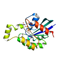
4XRE
 | | Crystal structure of Gnk2 complexed with mannose | | 分子名称: | Antifungal protein ginkbilobin-2, alpha-D-mannopyranose | | 著者 | Miyakawa, T, Hatano, K, Miyauchi, Y, Suwa, Y, Sawano, Y, Tanokura, M. | | 登録日 | 2015-01-21 | | 公開日 | 2015-02-25 | | 最終更新日 | 2023-11-08 | | 実験手法 | X-RAY DIFFRACTION (2.597 Å) | | 主引用文献 | A secreted protein with plant-specific cysteine-rich motif functions as a mannose-binding lectin that exhibits antifungal activity.
Plant Physiol., 166, 2014
|
|
4Y92
 | |
2WOZ
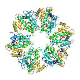
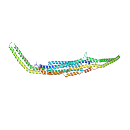
 | | The novel beta-propeller of the BTB-Kelch protein Krp1 provides the binding site for Lasp-1 that is necessary for pseudopodia extension | | 分子名称: | KELCH REPEAT AND BTB DOMAIN-CONTAINING PROTEIN 10 | | 著者 | Gray, C.H, McGarry, L.C, Spence, H.J, Riboldi-Tunnicliffe, A, Ozanne, B.W. | | 登録日 | 2009-07-31 | | 公開日 | 2009-09-01 | | 最終更新日 | 2024-05-08 | | 実験手法 | X-RAY DIFFRACTION (2 Å) | | 主引用文献 | Novel beta-propeller of the BTB-Kelch protein Krp1 provides a binding site for Lasp-1 that is necessary for pseudopodial extension.
J. Biol. Chem., 284, 2009
|
|
3Q84
 | |
6FO1
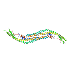
 | | Human R2TP subcomplex containing 1 RUVBL1-RUVBL2 hexamer bound to 1 RBD domain from RPAP3. | | 分子名称: | ADENOSINE-5'-DIPHOSPHATE, RNA polymerase II-associated protein 3, RuvB-like 1, ... | | 著者 | Martino, F, Munoz-Hernandez, H, Rodriguez, C.F, Pearl, L.H, Llorca, O. | | 登録日 | 2018-02-05 | | 公開日 | 2018-04-04 | | 最終更新日 | 2019-12-11 | | 実験手法 | ELECTRON MICROSCOPY (3.57 Å) | | 主引用文献 | RPAP3 provides a flexible scaffold for coupling HSP90 to the human R2TP co-chaperone complex.
Nat Commun, 9, 2018
|
|
3OCH
 | | Chemically Self-assembled Antibody Nanorings (CSANs): Design and Characterization of an Anti-CD3 IgM Biomimetic | | 分子名称: | (2S,2'S)-5,5'-(nonane-1,9-diyldiimino)bis(2-{[(4-{[(2,4-diaminopteridin-6-yl)methyl](methyl)amino}phenyl)carbonyl]amino}-5-oxopentanoic acid) (non-preferred name), CHLORIDE ION, Dihydrofolate reductase, ... | | 著者 | Cody, V. | | 登録日 | 2010-08-10 | | 公開日 | 2010-12-08 | | 最終更新日 | 2023-09-06 | | 実験手法 | X-RAY DIFFRACTION (1.79 Å) | | 主引用文献 | Chemically Self-Assembled Antibody Nanorings (CSANs): Design and Characterization of an Anti-CD3 IgM Biomimetic.
J.Am.Chem.Soc., 132, 2010
|
|
2XKB
 | |
6ZMO
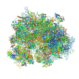 | | SARS-CoV-2 Nsp1 bound to the human LYAR-80S-eEF1a ribosome complex | | 分子名称: | 18S ribosomal RNA, 28S ribosomal RNA, 40S ribosomal protein S10, ... | | 著者 | Thoms, M, Buschauer, R, Ameismeier, M, Denk, T, Kratzat, H, Mackens-Kiani, T, Cheng, J, Berninghausen, O, Becker, T, Beckmann, R. | | 登録日 | 2020-07-03 | | 公開日 | 2020-08-19 | | 最終更新日 | 2024-05-01 | | 実験手法 | ELECTRON MICROSCOPY (3.1 Å) | | 主引用文献 | Structural basis for translational shutdown and immune evasion by the Nsp1 protein of SARS-CoV-2.
Science, 369, 2020
|
|
2GCP
 | |
2GCO
 | |
3QYQ
 | | 1.8 Angstrom resolution crystal structure of a putative deoxyribose-phosphate aldolase from Toxoplasma gondii ME49 | | 分子名称: | Deoxyribose-phosphate aldolase, putative, SULFATE ION, ... | | 著者 | Halavaty, A.S, Ruan, J, Minasov, G, Shuvalova, L, Ueno, A, Igarashi, M, Ngo, H, Anderson, W.F, Center for Structural Genomics of Infectious Diseases (CSGID) | | 登録日 | 2011-03-03 | | 公開日 | 2011-03-30 | | 最終更新日 | 2023-09-13 | | 実験手法 | X-RAY DIFFRACTION (1.8 Å) | | 主引用文献 | Structural and functional divergence of the aldolase fold in Toxoplasma gondii.
J.Mol.Biol., 427, 2015
|
|
3QRL
 | |
2GCN
 | |
2XN4
 | | Crystal structure of the kelch domain of human KLHL2 (Mayven) | | 分子名称: | 1,2-ETHANEDIOL, KELCH-LIKE PROTEIN 2, SODIUM ION, ... | | 著者 | Canning, P, Hozjan, V, Cooper, C.D.O, Ayinampudi, V, Vollmar, M, Pike, A.C.W, von Delft, F, Weigelt, J, Arrowsmith, C.H, Edwards, A.M, Bountra, C, Bullock, A.N. | | 登録日 | 2010-07-30 | | 公開日 | 2010-08-18 | | 最終更新日 | 2023-12-20 | | 実験手法 | X-RAY DIFFRACTION (1.99 Å) | | 主引用文献 | Structural Basis for Cul3 Assembly with the Btb-Kelch Family of E3 Ubiquitin Ligases.
J.Biol.Chem., 288, 2013
|
|
4ZNX
 | |
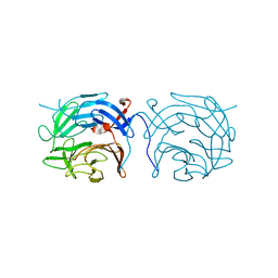
3QYT
 | | Diferric bound human serum transferrin | | 分子名称: | 2-acetamido-2-deoxy-beta-D-glucopyranose, CARBONATE ION, FE (III) ION, ... | | 著者 | Yang, N, Zhang, H, Wang, M, Hao, Q, Sun, H. | | 登録日 | 2011-03-03 | | 公開日 | 2012-03-14 | | 最終更新日 | 2023-11-01 | | 実験手法 | X-RAY DIFFRACTION (2.8 Å) | | 主引用文献 | Iron and bismuth bound human serum transferrin reveals a partially-opened conformation in the N-lobe
Sci Rep, 2, 2012
|
|
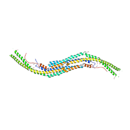
2ZET
 | | Crystal structure of the small GTPase Rab27B complexed with the Slp homology domain of Slac2-a/melanophilin | | 分子名称: | GUANOSINE-5'-TRIPHOSPHATE, MAGNESIUM ION, Melanophilin, ... | | 著者 | Kukimoto-Niino, M, Sakamoto, A, Kanno, E, Hanawa-Suetsugu, K, Terada, T, Shirouzu, M, Fukuda, M, Yokoyama, S, RIKEN Structural Genomics/Proteomics Initiative (RSGI) | | 登録日 | 2007-12-17 | | 公開日 | 2008-09-30 | | 最終更新日 | 2021-11-10 | | 実験手法 | X-RAY DIFFRACTION (3 Å) | | 主引用文献 | Structural basis for the exclusive specificity of Slac2-a/melanophilin for the Rab27 GTPases.
Structure, 16, 2008
|
|
3QNI
 | |
3Q0K
 | |


