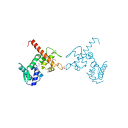
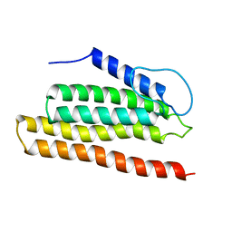
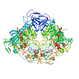
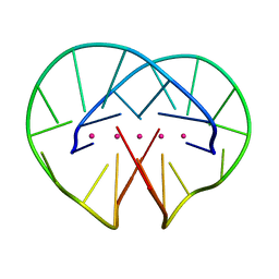
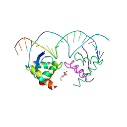
1F4K
 
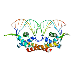 | | CRYSTAL STRUCTURE OF THE REPLICATION TERMINATOR PROTEIN/B-SITE DNA COMPLEX | | 分子名称: | 5'-D(*CP*TP*AP*TP*GP*AP*AP*CP*AP*TP*AP*AP*TP*GP*TP*TP*CP*AP*TP*AP*G)-3', 5'-D(*CP*TP*AP*TP*GP*AP*AP*CP*AP*TP*TP*AP*TP*GP*TP*TP*CP*AP*TP*AP*G)-3', REPLICATION TERMINATION PROTEIN | | 著者 | Wilce, J.A, Vivian, J.P, Hastings, A.F, Otting, G, Folmer, R.H.A, Duggin, I.G, Wake, R.G, Wilce, M.C.J. | | 登録日 | 2000-06-08 | | 公開日 | 2001-06-08 | | 最終更新日 | 2024-02-07 | | 実験手法 | X-RAY DIFFRACTION (2.5 Å) | | 主引用文献 | Structure of the RTP-DNA complex and the mechanism of polar replication fork arrest
Nat.Struct.Biol., 8, 2001
|
|
1FMQ
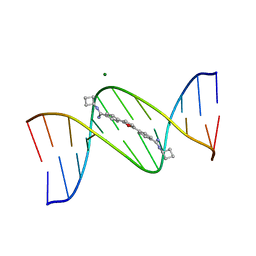 | | Cyclo-butyl-bis-furamidine complexed with dCGCGAATTCGCG | | 分子名称: | 2,5-BIS{[4-(N-CYCLOBUTYLDIAMINOMETHYL)PHENYL]}FURAN, 5'-D(*CP*GP*CP*GP*AP*AP*TP*TP*CP*GP*CP*G)-3', MAGNESIUM ION | | 著者 | Simpson, I.J, Lee, M, Kumar, A, Boykin, D.W, Neidle, S. | | 登録日 | 2000-08-18 | | 公開日 | 2000-09-11 | | 最終更新日 | 2024-02-07 | | 実験手法 | X-RAY DIFFRACTION (2 Å) | | 主引用文献 | DNA minor groove interactions and the biological activity of 2,5-bis-[4-(N-alkylamidino)phenyl] furans
Bioorg.Med.Chem.Lett., 10, 2000
|
|
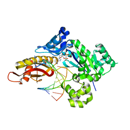
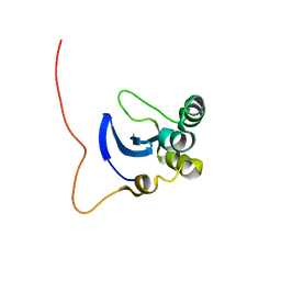
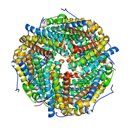
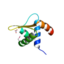
1SSO
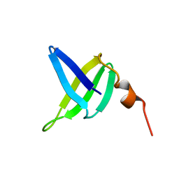 | | SOLUTION STRUCTURE AND DNA-BINDING PROPERTIES OF A THERMOSTABLE PROTEIN FROM THE ARCHAEON SULFOLOBUS SOLFATARICUS | | 分子名称: | SSO7D | | 著者 | Baumann, H, Knapp, S, Lundback, T, Ladenstein, R, Hard, T. | | 登録日 | 1995-03-31 | | 公開日 | 1995-05-08 | | 最終更新日 | 2024-05-22 | | 実験手法 | SOLUTION NMR | | 主引用文献 | Solution structure and DNA-binding properties of a thermostable protein from the archaeon Sulfolobus solfataricus.
Nat.Struct.Biol., 1, 1994
|
|
1FMS
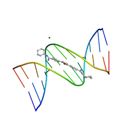 | | Structure of complex between cyclohexyl-bis-furamidine and d(CGCGAATTCGCG) | | 分子名称: | 2,5-BIS{[4-(N-CYCLOHEXYLDIAMINOMETHYL)PHENYL]}FURAN, 5'-D(*CP*GP*CP*GP*AP*AP*TP*TP*CP*GP*CP*G)-3', MAGNESIUM ION | | 著者 | Simpson, I.J, Lee, M, Kumar, A, Boykin, D.W, Neidle, S. | | 登録日 | 2000-08-18 | | 公開日 | 2000-09-11 | | 最終更新日 | 2024-02-07 | | 実験手法 | X-RAY DIFFRACTION (1.9 Å) | | 主引用文献 | DNA minor groove interactions and the biological activity of 2,5-bis-[4-(N-alkylamidino)phenyl] furans
Bioorg.Med.Chem.Lett., 10, 2000
|
|
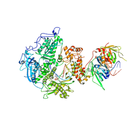
4B4C
 | | Crystal structure of the DNA-binding domain of human CHD1. | | 分子名称: | 1,2-ETHANEDIOL, CHROMODOMAIN-HELICASE-DNA-BINDING PROTEIN 1, GLYCEROL, ... | | 著者 | Allerston, C.K, Cooper, C.D.O, Shrestha, L, Vollmar, M, Burgess-Brown, N, Yue, W.W, Carpenter, E.P, Arrowsmith, C.H, Edwards, A, Bountra, C, von Delft, F, Gileadi, O. | | 登録日 | 2012-07-30 | | 公開日 | 2012-08-15 | | 最終更新日 | 2024-05-01 | | 実験手法 | X-RAY DIFFRACTION (1.62 Å) | | 主引用文献 | Crystal Structure of the DNA-Binding Domain of Human Chd1.
To be Published
|
|
8V1T
 | | Herpes simplex virus 1 polymerase holoenzyme bound to DNA and acyclovir triphosphate in closed conformation | | 分子名称: | ACYCLOVIR TRIPHOSPHATE, DNA polymerase, DNA polymerase processivity factor, ... | | 著者 | Pan, J, Abraham, J, Coen, D.M, Shankar, S, Yang, P, Hogle, J. | | 登録日 | 2023-11-21 | | 公開日 | 2024-09-04 | | 実験手法 | ELECTRON MICROSCOPY (2.8 Å) | | 主引用文献 | Viral DNA polymerase structures reveal mechanisms for drug selectivity
Cell(Cambridge,Mass.), 2024
|
|
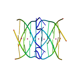
3IRR
 | | Crystal Structure of a Z-Z junction (with HEPES intercalating) | | 分子名称: | 4-(2-HYDROXYETHYL)-1-PIPERAZINE ETHANESULFONIC ACID, DNA (5'-D(*A*CP*CP*GP*CP*GP*CP*GP*AP*CP*GP*CP*GP*CP*G)-3'), DNA (5'-D(*G*TP*CP*GP*CP*GP*CP*GP*TP*CP*GP*CP*GP*CP*G)-3'), ... | | 著者 | Athanasiadis, A, de Rosa, M. | | 登録日 | 2009-08-24 | | 公開日 | 2010-05-19 | | 最終更新日 | 2023-09-06 | | 実験手法 | X-RAY DIFFRACTION (2.65 Å) | | 主引用文献 | Crystal structure of a junction between two Z-DNA helices.
Proc.Natl.Acad.Sci.USA, 107, 2010
|
|
8UJT
 | | Crystal structure of human polymerase eta with incoming dAMPnPP nucleotide opposite urea lesion | | 分子名称: | 2'-deoxy-5'-O-[(R)-hydroxy{[(R)-hydroxy(phosphonooxy)phosphoryl]amino}phosphoryl]adenosine, DNA (5'-D(*AP*GP*CP*GP*TP*CP*AP*T)-3'), DNA (5'-D(*CP*AP*TP*(XB9)P*AP*TP*GP*AP*CP*GP*CP*T)-3'), ... | | 著者 | Tomar, R, Egli, M, Stone, M.P. | | 登録日 | 2023-10-11 | | 公開日 | 2024-03-20 | | 最終更新日 | 2024-05-01 | | 実験手法 | X-RAY DIFFRACTION (2.31 Å) | | 主引用文献 | Replication Bypass of the N -(2-Deoxy-d-erythro-pentofuranosyl)-urea DNA Lesion by Human DNA Polymerase eta.
Biochemistry, 63, 2024
|
|
8UJV
 | | Crystal structure of human polymerase eta with incoming dCMPnPP nucleotide opposite urea lesion | | 分子名称: | 2'-deoxy-5'-O-[(R)-hydroxy{[(R)-hydroxy(phosphonooxy)phosphoryl]amino}phosphoryl]cytidine, DNA (5'-D(*AP*GP*CP*GP*TP*CP*AP*T)-3'), DNA (5'-D(*CP*AP*TP*(UD8)P*AP*TP*GP*AP*CP*GP*CP*T)-3'), ... | | 著者 | Tomar, R, Egli, M, Stone, M.P. | | 登録日 | 2023-10-11 | | 公開日 | 2024-03-20 | | 最終更新日 | 2024-05-01 | | 実験手法 | X-RAY DIFFRACTION (2.23 Å) | | 主引用文献 | Replication Bypass of the N -(2-Deoxy-d-erythro-pentofuranosyl)-urea DNA Lesion by Human DNA Polymerase eta.
Biochemistry, 63, 2024
|
|
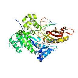
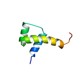
2NCJ
 | | Solution Structure of the PriC DNA replication restart protein | | 分子名称: | Uncharacterized protein | | 著者 | Cornilescu, C.C, Cornilescu, G, Wessel, S.R, Keck, J.L, Markley, J.L. | | 登録日 | 2016-04-07 | | 公開日 | 2016-07-06 | | 最終更新日 | 2024-05-15 | | 実験手法 | SOLUTION NMR | | 主引用文献 | Structure and Function of the PriC DNA Replication Restart Protein.
J.Biol.Chem., 291, 2016
|
|
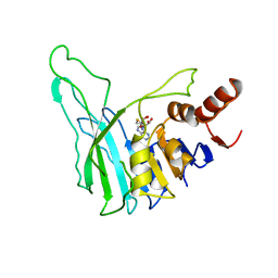
1L3G
 | | NMR Structure of the DNA-binding Domain of Cell Cycle Protein, Mbp1(2-124) from Saccharomyces cerevisiae | | 分子名称: | TRANSCRIPTION FACTOR Mbp1 | | 著者 | Nair, M, McIntosh, P.B, Frenkiel, T.A, Kelly, G, Taylor, I.A, Smerdon, S.J, Lane, A.N. | | 登録日 | 2002-02-27 | | 公開日 | 2003-02-18 | | 最終更新日 | 2024-05-22 | | 実験手法 | SOLUTION NMR | | 主引用文献 | NMR Structure of the DNA-Binding Domain of the Cell Cycle Protein Mbp1 from Saccharomyces cerevisiae
Biochemistry, 42, 2003
|
|
1SAN
 | |
1BOO
 | | PVUII DNA METHYLTRANSFERASE (CYTOSINE-N4-SPECIFIC) | | 分子名称: | PROTEIN (N-4 CYTOSINE-SPECIFIC METHYLTRANSFERASE PVU II), S-ADENOSYL-L-HOMOCYSTEINE | | 著者 | Gong, W, O'Gara, M, Blumenthal, R.M, Cheng, X. | | 登録日 | 1998-07-31 | | 公開日 | 1998-08-12 | | 最終更新日 | 2024-05-22 | | 実験手法 | X-RAY DIFFRACTION (2.8 Å) | | 主引用文献 | Structure of pvu II DNA-(cytosine N4) methyltransferase, an example of domain permutation and protein fold assignment.
Nucleic Acids Res., 25, 1997
|
|
6W8W
 | | Crystal structure of mouse DNMT1 in complex with CCG DNA | | 分子名称: | CCG DNA (5'-D(*AP*CP*TP*TP*AP*(5CM)P*GP*GP*AP*AP*GP*G)-3'), CCG DNA (5'-D(*CP*CP*TP*TP*CP*(C49)P*GP*TP*AP*AP*GP*T)-3'), DNA (cytosine-5)-methyltransferase 1, ... | | 著者 | Anteneh, H, Song, J. | | 登録日 | 2020-03-21 | | 公開日 | 2020-07-08 | | 最終更新日 | 2023-10-18 | | 実験手法 | X-RAY DIFFRACTION (3 Å) | | 主引用文献 | DNMT1 activity, base flipping mechanism and genome-wide DNA methylation are regulated by the DNA sequence context
Nat Commun, 2020
|
|
4R44
 | | Racemic crystal structure of a tetramolecular DNA G-quadruplex | | 分子名称: | 5'-D(*TP*GP*GP*GP*GP*T)-3', POTASSIUM ION, SODIUM ION | | 著者 | Mandal, P.K, Collie, G.W, Kauffmann, B, Huc, I. | | 登録日 | 2014-08-19 | | 公開日 | 2014-11-12 | | 最終更新日 | 2023-09-20 | | 実験手法 | X-RAY DIFFRACTION (2.695 Å) | | 主引用文献 | Racemic DNA crystallography.
Angew.Chem.Int.Ed.Engl., 53, 2014
|
|
8HX1
 | |
1L1T
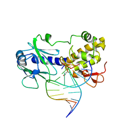 | | MutM (Fpg) Bound to Abasic-Site Containing DNA | | 分子名称: | 5'-D(*AP*G*GP*TP*AP*GP*AP*CP*CP*TP*GP*GP*AP*CP*GP*C)-3', 5'-D(*TP*GP*C*GP*TP*CP*CP*AP*(HPD)P*GP*TP*CP*TP*AP*CP*C)-3', MutM, ... | | 著者 | Fromme, J.C, Verdine, G.L. | | 登録日 | 2002-02-19 | | 公開日 | 2002-06-14 | | 最終更新日 | 2024-02-14 | | 実験手法 | X-RAY DIFFRACTION (1.8 Å) | | 主引用文献 | Structural insights into lesion recognition and repair by the bacterial 8-oxoguanine DNA glycosylase MutM.
Nat.Struct.Biol., 9, 2002
|
|
4R45
 | | Racemic crystal structure of a bimolecular DNA G-quadruplex (P-1) | | 分子名称: | 5'-D(*GP*GP*GP*GP*TP*TP*TP*TP*GP*GP*GP*G)-3', POTASSIUM ION | | 著者 | Mandal, P.K, Collie, G.W, Kauffmann, B, Huc, I. | | 登録日 | 2014-08-19 | | 公開日 | 2014-11-12 | | 最終更新日 | 2023-09-20 | | 実験手法 | X-RAY DIFFRACTION (1.9 Å) | | 主引用文献 | Racemic DNA crystallography.
Angew.Chem.Int.Ed.Engl., 53, 2014
|
|
2OBP
 | |
6WMB
 | |
6WMA
 | |
6WMC
 | |
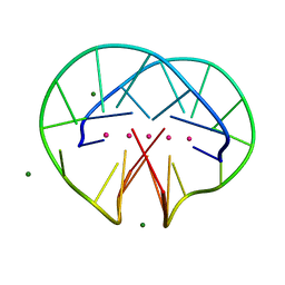
2Q2K
 | | Structure of nucleic-acid binding protein | | 分子名称: | 4-(2-HYDROXYETHYL)-1-PIPERAZINE ETHANESULFONIC ACID, DNA (5'-D(*AP*GP*TP*AP*TP*AP*(5IU)P*AP*CP*(5IU)P*AP*GP*TP*AP*TP*AP*TP*AP*CP*T)-3'), Hypothetical protein | | 著者 | Schumacher, M.A, Glover, T, Firth, N. | | 登録日 | 2007-05-28 | | 公開日 | 2008-02-05 | | 最終更新日 | 2024-02-21 | | 実験手法 | X-RAY DIFFRACTION (3 Å) | | 主引用文献 | Segrosome structure revealed by a complex of ParR with centromere DNA.
Nature, 450, 2007
|
|
4R47
 | | Racemic crystal structure of a bimolecular DNA G-quadruplex (P21/n) | | 分子名称: | 5'-D(*GP*GP*GP*GP*TP*TP*TP*TP*GP*GP*GP*G)-3', MAGNESIUM ION, POTASSIUM ION | | 著者 | Mandal, P.K, Collie, G.W, Kauffmann, B, Huc, I. | | 登録日 | 2014-08-19 | | 公開日 | 2014-11-12 | | 最終更新日 | 2023-09-20 | | 実験手法 | X-RAY DIFFRACTION (1.85 Å) | | 主引用文献 | Racemic DNA crystallography.
Angew.Chem.Int.Ed.Engl., 53, 2014
|
|
4OL8
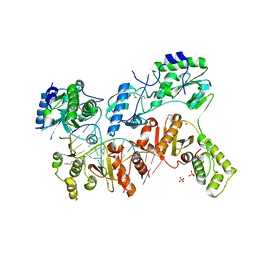 | | Ty3 reverse transcriptase bound to DNA/RNA | | 分子名称: | 5'-D(*CP*AP*TP*CP*TP*TP*CP*CP*TP*CP*TP*CP*TP*CP*TP*C)-3', 5'-R(*CP*UP*GP*AP*GP*AP*GP*AP*GP*AP*GP*GP*AP*AP*GP*AP*UP*G)-3', Reverse transcriptase/ribonuclease H, ... | | 著者 | Nowak, E, Miller, J.T, Bona, M.K, Studnicka, J, Szczepanowski, R.H, Jurkowski, J, Le Grice, S.F.J, Nowotny, M. | | 登録日 | 2014-01-23 | | 公開日 | 2014-03-05 | | 最終更新日 | 2014-04-16 | | 実験手法 | X-RAY DIFFRACTION (3.1 Å) | | 主引用文献 | Ty3 reverse transcriptase complexed with an RNA-DNA hybrid shows structural and functional asymmetry.
Nat.Struct.Mol.Biol., 21, 2014
|
|


