1L56
 
 | |
5CDW
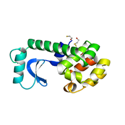
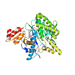
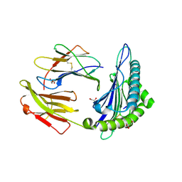
 | | Crystal Structure Analysis of a mutant Grb2 SH2 domain (W121G) with a pYVNV peptide | | 分子名称: | Growth factor receptor-bound protein 2, SER-PTR-VAL-ASN-VAL-GLN | | 著者 | Papaioannou, D, Geibel, S, Kunze, M, Kay, C, Waksman, G. | | 登録日 | 2015-07-05 | | 公開日 | 2016-05-25 | | 最終更新日 | 2024-01-10 | | 実験手法 | X-RAY DIFFRACTION (2.602 Å) | | 主引用文献 | Structural and biophysical investigation of the interaction of a mutant Grb2 SH2 domain (W121G) with its cognate phosphopeptide.
Protein Sci., 25, 2016
|
|
6UGB
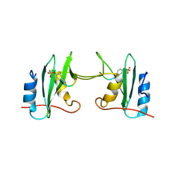
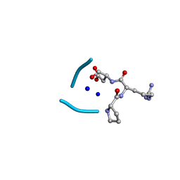 | | C3 symmetric peptide design number 2, Baby Basil | | 分子名称: | C3 symmetric peptide design number 2, Baby Basil, CHLORIDE ION, ... | | 著者 | Mulligan, V.K, Kang, C.S, Antselovich, I, Sawaya, M.R, Yeates, T.O, Baker, D. | | 登録日 | 2019-09-26 | | 公開日 | 2020-12-02 | | 実験手法 | X-RAY DIFFRACTION (0.95 Å) | | 主引用文献 | Computational design of mixed chirality peptide macrocycles with internal symmetry.
Protein Sci., 29, 2020
|
|
3TGJ
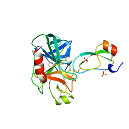 | | S195A TRYPSINOGEN COMPLEXED WITH BOVINE PANCREATIC TRYPSIN INHIBITOR (BPTI) | | 分子名称: | BOVINE PANCREATIC TRYPSIN INHIBITOR, CALCIUM ION, SULFATE ION, ... | | 著者 | Pasternak, A, Ringe, D, Hedstrom, L. | | 登録日 | 1998-07-16 | | 公開日 | 1998-12-23 | | 最終更新日 | 2021-11-03 | | 実験手法 | X-RAY DIFFRACTION (2.2 Å) | | 主引用文献 | Comparison of Anionic and Cationic Trypsinogens: The Anionic Activation Domain is More Flexible in Solution and Differs in its Mode of Bpti Binding in the Crystal Structure
Protein Sci., 8, 1999
|
|
5GDS
 | | HIRUNORMS ARE TRUE HIRUDIN MIMETICS. THE CRYSTAL STRUCTURE OF HUMAN ALPHA-THROMBIN:HIRUNORM V COMPLEX | | 分子名称: | 2-acetamido-2-deoxy-beta-D-glucopyranose, ALPHA-THROMBIN, HIRUNORM V | | 著者 | De Simone, G, Lombardi, A, Galdiero, S, Nastri, F, Della Morte, R, Staiano, N, Pedone, C, Bolognesi, M, Pavone, V. | | 登録日 | 1997-07-17 | | 公開日 | 1998-01-21 | | 最終更新日 | 2023-08-09 | | 実験手法 | X-RAY DIFFRACTION (2.1 Å) | | 主引用文献 | Hirunorms are true hirudin mimetics. The crystal structure of human alpha-thrombin-hirunorm V complex.
Protein Sci., 7, 1998
|
|
5LRT
 | | Structure of the Deamidase-Depupylase Dop of the Prokaryotic Ubiquitin-like Modification Pathway in Complex with ADP and Phosphate | | 分子名称: | ADENOSINE-5'-DIPHOSPHATE, DI(HYDROXYETHYL)ETHER, Depupylase, ... | | 著者 | Bolten, M, Vahlensieck, C, Lipp, C, Leibundgut, M, Ban, N, Weber-Ban, E. | | 登録日 | 2016-08-19 | | 公開日 | 2017-02-01 | | 最終更新日 | 2024-01-17 | | 実験手法 | X-RAY DIFFRACTION (1.85 Å) | | 主引用文献 | Depupylase Dop Requires Inorganic Phosphate in the Active Site for Catalysis.
J. Biol. Chem., 292, 2017
|
|
5F16
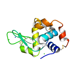 | | CTA-modified hen egg-white lysozyme | | 分子名称: | CHLORIDE ION, Lysozyme C, SODIUM ION | | 著者 | McGlone, C, Nix, J.C, Page, R.C. | | 登録日 | 2015-11-30 | | 公開日 | 2016-02-24 | | 最終更新日 | 2023-09-27 | | 実験手法 | X-RAY DIFFRACTION (1.2 Å) | | 主引用文献 | Investigating the Impact of Polymer Functional Groups on the Stability and Activity of Lysozyme-Polymer Conjugates.
Biomacromolecules, 17, 2016
|
|
6SYE
 | |
5FIR
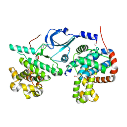
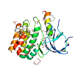
 | | Crystal structure of C. elegans XRN2 in complex with the XRN2-binding domain of PAXT-1 | | 分子名称: | 5'-3' EXORIBONUCLEASE 2 HOMOLOG, PAXT-1, SULFATE ION | | 著者 | Richter, H, Katic, I, Gut, H, Grosshans, H. | | 登録日 | 2015-10-02 | | 公開日 | 2016-01-20 | | 最終更新日 | 2024-05-01 | | 実験手法 | X-RAY DIFFRACTION (2.836 Å) | | 主引用文献 | Structural Basis and Function of Xrn2-Binding by Xtb Domains
Nat.Struct.Mol.Biol., 23, 2016
|
|
5F9J
 | | Structure of HLA-A2:01 with peptide Y9L | | 分子名称: | Beta-2-microglobulin, GLYCEROL, HLA class I histocompatibility antigen, ... | | 著者 | Remesh, S.G, Zajonc, D.M. | | 登録日 | 2015-12-09 | | 公開日 | 2016-12-21 | | 最終更新日 | 2023-09-27 | | 実験手法 | X-RAY DIFFRACTION (2.51 Å) | | 主引用文献 | Unconventional Peptide Presentation by Major Histocompatibility Complex (MHC) Class I Allele HLA-A*02:01: BREAKING CONFINEMENT.
J. Biol. Chem., 292, 2017
|
|
5FA3
 | | Structure of HLA-A2:01 with peptide G9V | | 分子名称: | Beta-2-microglobulin, G9V, GLYCEROL, ... | | 著者 | Zajonc, D.M, Remesh, S.G. | | 登録日 | 2015-12-10 | | 公開日 | 2016-12-21 | | 最終更新日 | 2023-09-27 | | 実験手法 | X-RAY DIFFRACTION (1.86 Å) | | 主引用文献 | Unconventional Peptide Presentation by Major Histocompatibility Complex (MHC) Class I Allele HLA-A*02:01: BREAKING CONFINEMENT.
J. Biol. Chem., 292, 2017
|
|
6SYC
 | | Crystal structure of the lysozyme in presence of bromophenol blue at pH 6.5 | | 分子名称: | CHLORIDE ION, IMIDAZOLE, Lysozyme, ... | | 著者 | Camara-Artigas, A, Plaza-Garrido, M, Salinas-Garcia, M.C. | | 登録日 | 2019-09-27 | | 公開日 | 2020-09-09 | | 最終更新日 | 2024-01-24 | | 実験手法 | X-RAY DIFFRACTION (1.38 Å) | | 主引用文献 | Lysozyme crystals dyed with bromophenol blue: where has the dye gone?
Acta Crystallogr D Struct Biol, 76, 2020
|
|
6T41
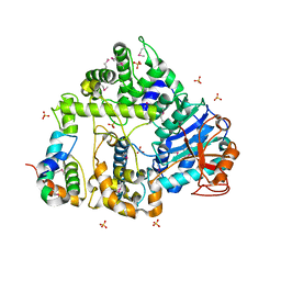
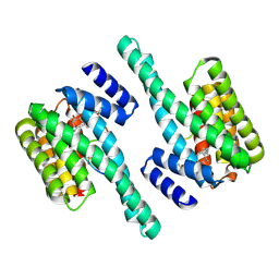
 | | CDK8/Cyclin C in complex with N-(4-chlorobenzyl)isoquinolin-4-amine | | 分子名称: | 1,2-ETHANEDIOL, Cyclin-C, Cyclin-dependent kinase 8, ... | | 著者 | Schneider, E.V, Maskos, K, Huber, R, Kuhn, C.-D. | | 登録日 | 2019-10-11 | | 公開日 | 2020-01-01 | | 最終更新日 | 2024-01-24 | | 実験手法 | X-RAY DIFFRACTION (2.45 Å) | | 主引用文献 | A precisely positioned MED12 activation helix stimulates CDK8 kinase activity.
Proc.Natl.Acad.Sci.USA, 117, 2020
|
|
5EXA
 | |
6TWG
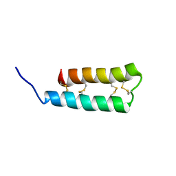
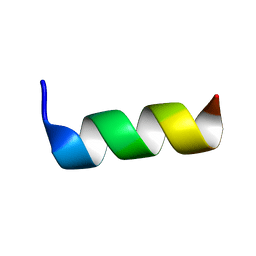 | | Solution structure of antimicrobial peptide, crabrolin Plus in the presence of Lipopolysaccharide | | 分子名称: | Crabrolin Plus, mutant of Crabrolin peptide | | 著者 | Cantini, F, Bouchemal, N, Savarin, P, Sette, M. | | 登録日 | 2020-01-13 | | 公開日 | 2020-07-22 | | 最終更新日 | 2024-06-19 | | 実験手法 | SOLUTION NMR | | 主引用文献 | Effect of positive charges in the structural interaction of crabrolin isoforms with lipopolysaccharide.
J.Pept.Sci., 26, 2020
|
|
5GCH
 | |
5AY8
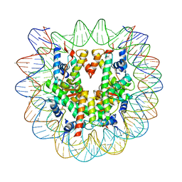 | | Crystal structure of human nucleosome containing H3.Y | | 分子名称: | CHLORIDE ION, DNA (146-MER), H3.Y, ... | | 著者 | Kujirai, T, Horikoshi, N, Sato, K, Maehara, K, Machida, S, Osakabe, A, Kimura, H, Ohkawa, Y, Kurumizaka, H. | | 登録日 | 2015-08-10 | | 公開日 | 2016-04-06 | | 最終更新日 | 2023-11-08 | | 実験手法 | X-RAY DIFFRACTION (2.8 Å) | | 主引用文献 | Structure and function of human histone H3.Y nucleosome
Nucleic Acids Res., 44, 2016
|
|
5LXM
 | |
6UX5
 | |
3SGB
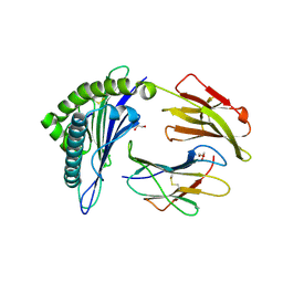
 | | STRUCTURE OF THE COMPLEX OF STREPTOMYCES GRISEUS PROTEASE B AND THE THIRD DOMAIN OF THE TURKEY OVOMUCOID INHIBITOR AT 1.8 ANGSTROMS RESOLUTION | | 分子名称: | PROTEINASE B (SGPB), TURKEY OVOMUCOID INHIBITOR (OMTKY3) | | 著者 | Read, R.J, Fujinaga, M, Sielecki, A.R, James, M.N.G. | | 登録日 | 1983-01-21 | | 公開日 | 1983-07-12 | | 最終更新日 | 2017-11-29 | | 実験手法 | X-RAY DIFFRACTION (1.8 Å) | | 主引用文献 | Structure of the complex of Streptomyces griseus protease B and the third domain of the turkey ovomucoid inhibitor at 1.8-A resolution.
Biochemistry, 22, 1983
|
|
3U92
 | |
6SJG
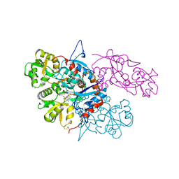
 | | Cryo-EM structure of the RecBCD no Chi negative control complex | | 分子名称: | Forked DNA substrate, RecBCD enzyme subunit RecB, RecBCD enzyme subunit RecC, ... | | 著者 | Cheng, K, Wilkinson, M, Wigley, D.B. | | 登録日 | 2019-08-13 | | 公開日 | 2020-01-01 | | 最終更新日 | 2024-05-22 | | 実験手法 | ELECTRON MICROSCOPY (3.8 Å) | | 主引用文献 | A conformational switch in response to Chi converts RecBCD from phage destruction to DNA repair.
Nat.Struct.Mol.Biol., 27, 2020
|
|
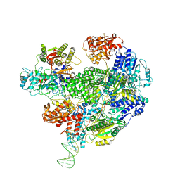
5BTA
 | | Crystal structure of a topoisomerase II complex | | 分子名称: | 1-cyclopropyl-6-fluoro-8-methoxy-7-[(4aS,7aS)-octahydro-6H-pyrrolo[3,4-b]pyridin-6-yl]-4-oxo-1,4-dihydroquinoline-3-carboxylic acid, DNA gyrase subunit A, DNA gyrase subunit B, ... | | 著者 | Blower, T.R, Williamson, B.H, Kerns, R.J, Berger, J.M. | | 登録日 | 2015-06-02 | | 公開日 | 2016-03-02 | | 最終更新日 | 2023-11-15 | | 実験手法 | X-RAY DIFFRACTION (2.55 Å) | | 主引用文献 | Crystal structure and stability of gyrase-fluoroquinolone cleaved complexes from Mycobacterium tuberculosis.
Proc.Natl.Acad.Sci.USA, 113, 2016
|
|
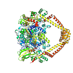
5BTN
 | | Crystal structure of a topoisomerase II complex | | 分子名称: | 1-cyclopropyl-6-fluoro-8-methyl-7-[(4aS,7aS)-octahydro-6H-pyrrolo[3,4-b]pyridin-6-yl]-4-oxo-1,4-dihydroquinoline-3-carboxylic acid, DNA gyrase subunit A, DNA gyrase subunit B, ... | | 著者 | Blower, T.R, Williamson, B.H, Kerns, R.J, Berger, J.M. | | 登録日 | 2015-06-03 | | 公開日 | 2016-03-02 | | 最終更新日 | 2023-11-15 | | 実験手法 | X-RAY DIFFRACTION (2.5 Å) | | 主引用文献 | Crystal structure and stability of gyrase-fluoroquinolone cleaved complexes from Mycobacterium tuberculosis.
Proc.Natl.Acad.Sci.USA, 113, 2016
|
|
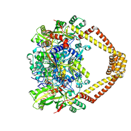
5BS8
 | | Crystal structure of a topoisomerase II complex | | 分子名称: | 1-cyclopropyl-6-fluoro-8-methoxy-7-[(4aS,7aS)-octahydro-6H-pyrrolo[3,4-b]pyridin-6-yl]-4-oxo-1,4-dihydroquinoline-3-carboxylic acid, DNA gyrase subunit A, DNA gyrase subunit B, ... | | 著者 | Blower, T.R, Williamson, B.H, Kerns, R.J, Berger, J.M. | | 登録日 | 2015-06-01 | | 公開日 | 2016-03-02 | | 最終更新日 | 2023-11-15 | | 実験手法 | X-RAY DIFFRACTION (2.399 Å) | | 主引用文献 | Crystal structure and stability of gyrase-fluoroquinolone cleaved complexes from Mycobacterium tuberculosis.
Proc.Natl.Acad.Sci.USA, 113, 2016
|
|


