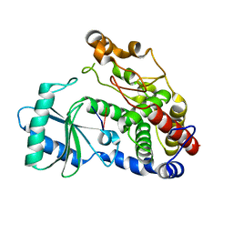
2OUJ
 
 | |
5E4K
 | | Structure of ligand binding region of uPARAP at pH 7.4 | | 分子名称: | 2-acetamido-2-deoxy-beta-D-glucopyranose, 3,6,9,12,15,18,21,24-OCTAOXAHEXACOSAN-1-OL, C-type mannose receptor 2, ... | | 著者 | Yuan, C, Huang, M. | | 登録日 | 2015-10-06 | | 公開日 | 2016-08-10 | | 最終更新日 | 2023-11-08 | | 実験手法 | X-RAY DIFFRACTION (2.58 Å) | | 主引用文献 | Crystal structures of the ligand-binding region of uPARAP: effect of calcium ion binding
Biochem.J., 473, 2016
|
|
3OQG
 | | Restriction endonuclease HPY188I in complex with substrate DNA | | 分子名称: | CHLORIDE ION, DNA 5'-D(*GP*AP*TP*CP*TP*GP*AP*AP*C)-3', DNA 5'-D(*GP*TP*TP*CP*AP*GP*AP*TP*C)-3', ... | | 著者 | Sokolowska, M, Czapinska, H, Bochtler, M. | | 登録日 | 2010-09-03 | | 公開日 | 2010-10-20 | | 最終更新日 | 2017-11-08 | | 実験手法 | X-RAY DIFFRACTION (1.75 Å) | | 主引用文献 | Hpy188I-DNA pre- and post-cleavage complexes--snapshots of the GIY-YIG nuclease mediated catalysis.
Nucleic Acids Res., 39, 2011
|
|
5ELY
 | | X-ray structure of human glutamate carboxypeptidase II (GCPII) in complex with a hydroxamate inhibitor JHU242 | | 分子名称: | 2-acetamido-2-deoxy-beta-D-glucopyranose, 2-acetamido-2-deoxy-beta-D-glucopyranose-(1-4)-2-acetamido-2-deoxy-beta-D-glucopyranose, 4-[(2~{R})-2-carboxy-5-(oxidanylamino)-5-oxidanylidene-pentyl]benzoic acid, ... | | 著者 | Barinka, C, Novakova, Z, Pavlicek, J. | | 登録日 | 2015-11-05 | | 公開日 | 2016-04-27 | | 最終更新日 | 2020-07-29 | | 実験手法 | X-RAY DIFFRACTION (1.81 Å) | | 主引用文献 | Unprecedented Binding Mode of Hydroxamate-Based Inhibitors of Glutamate Carboxypeptidase II: Structural Characterization and Biological Activity.
J.Med.Chem., 59, 2016
|
|
4X9F
 | | Crystal structure of Dscam1 isoform 6.9, N-terminal four Ig domains | | 分子名称: | 4-(2-HYDROXYETHYL)-1-PIPERAZINE ETHANESULFONIC ACID, Down Syndrome Cell Adhesion Molecule isoform 6.9, GLYCEROL, ... | | 著者 | Chen, Q, Yu, Y, Li, S.A, Cheng, L. | | 登録日 | 2014-12-11 | | 公開日 | 2015-12-16 | | 最終更新日 | 2023-11-08 | | 実験手法 | X-RAY DIFFRACTION (2.35 Å) | | 主引用文献 | Structural basis of Dscam1 homodimerization: Insights into context constraint for protein recognition
Sci Adv, 2, 2016
|
|
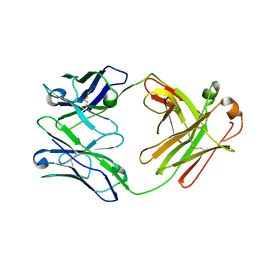
3PMH
 | |
7B01
 | | ADAMTS13-CUB12 | | 分子名称: | 2-acetamido-2-deoxy-beta-D-glucopyranose-(1-4)-2-acetamido-2-deoxy-beta-D-glucopyranose, Maltodextrin-binding protein,Maltodextrin-binding protein,Maltodextrin-binding protein,ADAMTS13 CUB12,A disintegrin and metalloproteinase with thrombospondin motifs 13,A disintegrin and metalloproteinase with thrombospondin motifs 13,A disintegrin and metalloproteinase with thrombospondin motifs 13, alpha-D-glucopyranose-(1-4)-alpha-D-glucopyranose | | 著者 | Kim, H.J, Emsley, J. | | 登録日 | 2020-11-18 | | 公開日 | 2021-04-28 | | 最終更新日 | 2024-01-31 | | 実験手法 | X-RAY DIFFRACTION (2.8 Å) | | 主引用文献 | Crystal structure of ADAMTS13 CUB domains reveals their role in global latency.
Sci Adv, 7, 2021
|
|
7AAF
 | |
7AAO
 | |
6KCU
 | | Shuguo PWWP domain | | 分子名称: | 2-[BIS-(2-HYDROXY-ETHYL)-AMINO]-2-HYDROXYMETHYL-PROPANE-1,3-DIOL, LD23804p | | 著者 | Liu, Y.C, Huang, Y. | | 登録日 | 2019-06-29 | | 公開日 | 2020-07-01 | | 最終更新日 | 2023-11-22 | | 実験手法 | X-RAY DIFFRACTION (1.65 Å) | | 主引用文献 | Shuguo PWWP domain
To Be Published
|
|
7AZB
 | | Structure of DDR2 DS domain in complex with VHH | | 分子名称: | Discoidin domain-containing receptor 2, VHH | | 著者 | Talagas, A, Nawrotek, A, Arrial, A, Vuillard, L.M, Miallau, L. | | 登録日 | 2020-11-16 | | 公開日 | 2020-11-25 | | 最終更新日 | 2024-01-31 | | 実験手法 | X-RAY DIFFRACTION (2.62 Å) | | 主引用文献 | Structure of DDR2 DS domain in complex with VHH
To Be Published
|
|
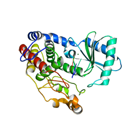
6KD6
 | |
6KCP
 | |
2PVV
 | |
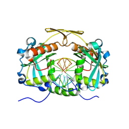
2OUH
 | | Crystal structure of the Thrombospondin-1 N-terminal domain in complex with fractionated Heparin DP10 | | 分子名称: | SULFATE ION, Thrombospondin-1 | | 著者 | Tan, K, Joachimiak, A, Wang, J, Lawler, J. | | 登録日 | 2007-02-11 | | 公開日 | 2008-01-08 | | 最終更新日 | 2023-08-30 | | 実験手法 | X-RAY DIFFRACTION (2.4 Å) | | 主引用文献 | Heparin-induced cis- and trans-Dimerization Modes of the Thrombospondin-1 N-terminal Domain.
J.Biol.Chem., 283, 2008
|
|
2P24
 | | I-Au/MBP125-135 | | 分子名称: | H-2 class II histocompatibility antigen, A-U alpha chain, A-U beta chain | | 著者 | McBeth, C, Strong, R.K. | | 登録日 | 2007-03-06 | | 公開日 | 2008-01-15 | | 最終更新日 | 2023-08-30 | | 実験手法 | X-RAY DIFFRACTION (2.15 Å) | | 主引用文献 | A new twist in TCR diversity revealed by a forbidden alphabeta TCR.
J.Mol.Biol., 375, 2008
|
|
4XGC
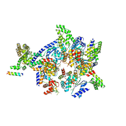 | | Crystal structure of the eukaryotic origin recognition complex | | 分子名称: | CHLORIDE ION, Origin recognition complex subunit 1, Origin recognition complex subunit 2, ... | | 著者 | Bleichert, F, Botchan, M.R, Berger, J.M. | | 登録日 | 2014-12-30 | | 公開日 | 2015-04-01 | | 最終更新日 | 2019-12-04 | | 実験手法 | X-RAY DIFFRACTION (3.5 Å) | | 主引用文献 | Crystal structure of the eukaryotic origin recognition complex.
Nature, 519, 2015
|
|
4XB7
 | | Crystal structure of Dscam1 isoform 4.4, N-terminal four Ig domains | | 分子名称: | 2-acetamido-2-deoxy-beta-D-glucopyranose-(1-4)-2-acetamido-2-deoxy-beta-D-glucopyranose, Down syndrome cell adhesion molecule, isoform 4.4, ... | | 著者 | Chen, Q, Yu, Y, Li, S.A, Cheng, L. | | 登録日 | 2014-12-16 | | 公開日 | 2015-12-16 | | 最終更新日 | 2023-11-08 | | 実験手法 | X-RAY DIFFRACTION (4.004 Å) | | 主引用文献 | Structural basis of Dscam1 homodimerization: Insights into context constraint for protein recognition
Sci Adv, 2, 2016
|
|
2A2E
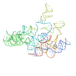 | | Crystal structure of the RNA subunit of Ribonuclease P. Bacterial A-type. | | 分子名称: | OSMIUM ION, RNA subunit of RNase P | | 著者 | Torres-Larios, A, Swinger, K.K, Krasilnikov, A.S, Pan, T, Mondragon, A. | | 登録日 | 2005-06-22 | | 公開日 | 2005-09-06 | | 最終更新日 | 2023-08-23 | | 実験手法 | X-RAY DIFFRACTION (3.85 Å) | | 主引用文献 | Crystal structure of the RNA component of bacterial ribonuclease P.
Nature, 437, 2005
|
|
6ZSZ
 | |
6ZSY
 | |
6I0T
 | |
3EDL
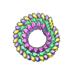 | | Kinesin13-Microtubule Ring complex | | 分子名称: | 2-MERCAPTO-N-[1,2,3,10-TETRAMETHOXY-9-OXO-5,6,7,9-TETRAHYDRO-BENZO[A]HEPTALEN-7-YL]ACETAMIDE, Beta tubulin, GUANOSINE-5'-DIPHOSPHATE, ... | | 著者 | Tan, D, Rice, W.J, Sosa, H. | | 登録日 | 2008-09-03 | | 公開日 | 2009-01-20 | | 最終更新日 | 2024-02-21 | | 実験手法 | ELECTRON MICROSCOPY (28 Å) | | 主引用文献 | Structure of the kinesin13-microtubule ring complex.
Structure, 16, 2008
|
|
6ZTD
 | |
6I0U
 | |


