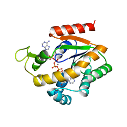
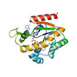
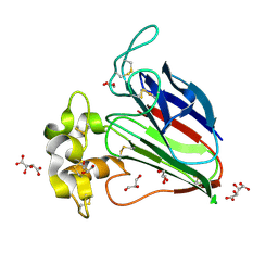
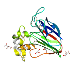
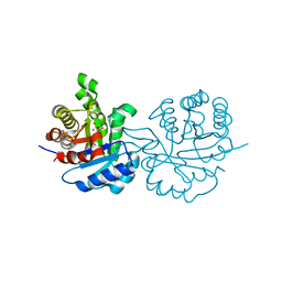
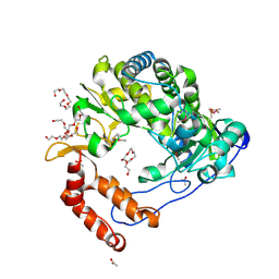
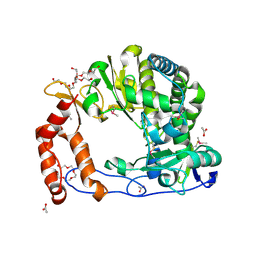
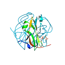
1HDG
 
 | | THE CRYSTAL STRUCTURE OF HOLO-GLYCERALDEHYDE-3-PHOSPHATE DEHYDROGENASE FROM THE HYPERTHERMOPHILIC BACTERIUM THERMOTOGA MARITIMA AT 2.5 ANGSTROMS RESOLUTION | | 分子名称: | HOLO-D-GLYCERALDEHYDE-3-PHOSPHATE DEHYDROGENASE, NICOTINAMIDE-ADENINE-DINUCLEOTIDE, SULFATE ION | | 著者 | Korndoerfer, I, Steipe, B, Huber, R, Tomschy, A, Jaenicke, R. | | 登録日 | 1995-01-17 | | 公開日 | 1995-03-31 | | 最終更新日 | 2024-02-07 | | 実験手法 | X-RAY DIFFRACTION (2.5 Å) | | 主引用文献 | The crystal structure of holo-glyceraldehyde-3-phosphate dehydrogenase from the hyperthermophilic bacterium Thermotoga maritima at 2.5 A resolution.
J.Mol.Biol., 246, 1995
|
|
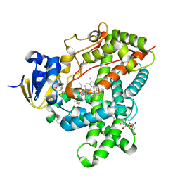
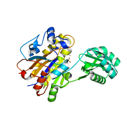
1ZIP
 | | BACILLUS STEAROTHERMOPHILUS ADENYLATE KINASE | | 分子名称: | ADENYLATE KINASE, BIS(ADENOSINE)-5'-PENTAPHOSPHATE, MANGANESE (II) ION, ... | | 著者 | Berry, M.B, Phillips Jr, G.N. | | 登録日 | 1997-05-07 | | 公開日 | 1997-08-20 | | 最終更新日 | 2024-05-22 | | 実験手法 | X-RAY DIFFRACTION (1.85 Å) | | 主引用文献 | Crystal structures of Bacillus stearothermophilus adenylate kinase with bound Ap5A, Mg2+ Ap5A, and Mn2+ Ap5A reveal an intermediate lid position and six coordinate octahedral geometry for bound Mg2+ and Mn2+.
Proteins, 32, 1998
|
|
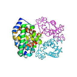
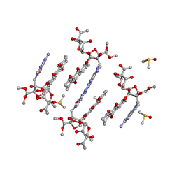
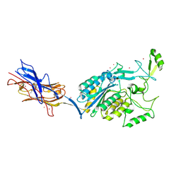
1QS2
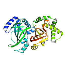 | | CRYSTAL STRUCTURE OF VIP2 WITH NAD | | 分子名称: | ADP-RIBOSYLTRANSFERASE, NICOTINAMIDE-ADENINE-DINUCLEOTIDE | | 著者 | Han, S, Craig, J.A, Putnam, C.D, Carozzi, N.B, Tainer, J.A. | | 登録日 | 1999-06-25 | | 公開日 | 1999-12-29 | | 最終更新日 | 2024-02-14 | | 実験手法 | X-RAY DIFFRACTION (2.7 Å) | | 主引用文献 | Evolution and mechanism from structures of an ADP-ribosylating toxin and NAD complex.
Nat.Struct.Biol., 6, 1999
|
|
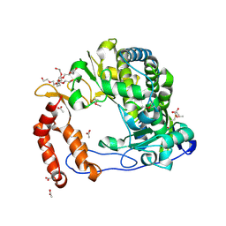
1ZIN
 | | ADENYLATE KINASE WITH BOUND AP5A | | 分子名称: | ADENYLATE KINASE, BIS(ADENOSINE)-5'-PENTAPHOSPHATE, ZINC ION | | 著者 | Berry, M.B, Phillips Jr, G.N. | | 登録日 | 1996-06-07 | | 公開日 | 1997-06-16 | | 最終更新日 | 2024-04-03 | | 実験手法 | X-RAY DIFFRACTION (1.6 Å) | | 主引用文献 | Crystal structures of Bacillus stearothermophilus adenylate kinase with bound Ap5A, Mg2+ Ap5A, and Mn2+ Ap5A reveal an intermediate lid position and six coordinate octahedral geometry for bound Mg2+ and Mn2+.
Proteins, 32, 1998
|
|
1QCP
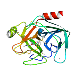 | | CRYSTAL STRUCTURE OF THE RWJ-51084 BOVINE PANCREATIC BETA-TRYPSIN AT 1.8 A | | 分子名称: | CALCIUM ION, CYCLOPENTANECARBOXYLIC ACID [1-(BENZOTHIAZOLE-2-CARBONYL)-4-GUANIDINO-BUTYL]-AMIDE, PROTEIN (BETA-TRYPSIN PROTEIN) | | 著者 | Recacha, R, Carson, M, Costanzo, M.J, Maryanoff, B, Chattopadhyay, D. | | 登録日 | 1999-05-10 | | 公開日 | 1999-05-21 | | 最終更新日 | 2024-10-16 | | 実験手法 | X-RAY DIFFRACTION (1.8 Å) | | 主引用文献 | Structure of the RWJ-51084-bovine pancreatic beta-trypsin complex at 1.8 A.
Acta Crystallogr.,Sect.D, 55, 1999
|
|
3AL7
 | | Recombinant thaumatin I at 1.1 A | | 分子名称: | GLYCEROL, L(+)-TARTARIC ACID, Thaumatin I | | 著者 | Masuda, T, Mikami, B, Kitabatake, N. | | 登録日 | 2010-07-27 | | 公開日 | 2011-06-08 | | 最終更新日 | 2023-11-01 | | 実験手法 | X-RAY DIFFRACTION (1.1 Å) | | 主引用文献 | High-resolution structure of the recombinant sweet-tasting protein thaumatin I
Acta Crystallogr.,Sect.F, 67, 2011
|
|
3P5Q
 | | Ferric R-state human aquomethemoglobin | | 分子名称: | Hemoglobin subunit alpha, Hemoglobin subunit beta, PROTOPORPHYRIN IX CONTAINING FE, ... | | 著者 | Yi, J, Thomas, L.M, Richter-Addo, G.B. | | 登録日 | 2010-10-10 | | 公開日 | 2011-06-08 | | 最終更新日 | 2023-09-06 | | 実験手法 | X-RAY DIFFRACTION (2 Å) | | 主引用文献 | Structure of human R-state aquomethemoglobin at 2.0 A resolution
Acta Crystallogr.,Sect.F, 67, 2011
|
|
3ALD
 | | Crystal structure of sweet-tasting protein Thaumatin I at 1.10 A | | 分子名称: | GLYCEROL, L(+)-TARTARIC ACID, Thaumatin I | | 著者 | Masuda, T, Mikami, B, Kitabatake, N. | | 登録日 | 2010-07-29 | | 公開日 | 2011-06-08 | | 最終更新日 | 2011-11-02 | | 実験手法 | X-RAY DIFFRACTION (1.1 Å) | | 主引用文献 | High-resolution structure of the recombinant sweet-tasting protein thaumatin I
Acta Crystallogr.,Sect.F, 67, 2011
|
|
2OV6
 | |
1QDS
 | | SUPERSTABLE E65Q MUTANT OF LEISHMANIA MEXICANA TRIOSEPHOSPHATE ISOMERASE (TIM) | | 分子名称: | 2-PHOSPHOGLYCOLIC ACID, TRIOSEPHOSPHATE ISOMERASE | | 著者 | Lambeir, A.M, Backmann, J, Ruiz-Sanz, J, Filimonov, V, Nielsen, J.E, Vriend, G, Kursula, I, Norledge, B.V, Wierenga, R.K. | | 登録日 | 1999-07-10 | | 公開日 | 2000-12-13 | | 最終更新日 | 2024-02-14 | | 実験手法 | X-RAY DIFFRACTION (2 Å) | | 主引用文献 | The ionization of a buried glutamic acid is thermodynamically linked to the stability of Leishmania mexicana triose phosphate isomerase.
Eur.J.Biochem., 267, 2000
|
|
4HP7
 | | Trioxacarcin D517 as a product of guanine robbery from d(AACCGGTT) | | 分子名称: | DIMETHYL SULFOXIDE, GUANINE, Trioxacarcin A analogue, ... | | 著者 | Smaltz, D.J, Magauer, T, Proepper, K, Dittrich, B, Myers, A.G. | | 登録日 | 2012-10-23 | | 公開日 | 2013-10-23 | | 最終更新日 | 2024-03-13 | | 実験手法 | X-RAY DIFFRACTION (1.09 Å) | | 主引用文献 | Trioxacarcin D517
TO BE PUBLISHED
|
|
2CTP
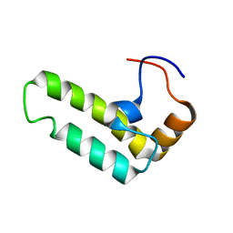 | | Solution structure of J-domain from human DnaJ subfamily B menber 12 | | 分子名称: | DnaJ homolog subfamily B member 12 | | 著者 | Kobayashi, N, Tochio, N, Koshiba, S, Inoue, M, Kigawa, T, Yokoyama, S, RIKEN Structural Genomics/Proteomics Initiative (RSGI) | | 登録日 | 2005-05-24 | | 公開日 | 2005-11-24 | | 最終更新日 | 2024-05-29 | | 実験手法 | SOLUTION NMR | | 主引用文献 | Solution structure of J-domain from human DnaJ subfamily B menber 12
To be Published
|
|
3U97
 | |
3OHR
 | | Crystal structure of fructokinase from bacillus subtilis complexed with ADP | | 分子名称: | ADENOSINE-5'-DIPHOSPHATE, Putative fructokinase, SULFATE ION, ... | | 著者 | Nocek, B, Volkart, L, Cuff, M, Joachimiak, A, Midwest Center for Structural Genomics (MCSG) | | 登録日 | 2010-08-17 | | 公開日 | 2010-09-15 | | 最終更新日 | 2012-10-10 | | 実験手法 | X-RAY DIFFRACTION (1.66 Å) | | 主引用文献 | Structural studies of ROK fructokinase YdhR from Bacillus subtilis: insights into substrate binding and fructose specificity.
J.Mol.Biol., 406, 2011
|
|
3TMZ
 | | Crystal Structure of P450 2B4(H226Y) in complex with Amlodipine | | 分子名称: | 5-CYCLOHEXYL-1-PENTYL-BETA-D-MALTOSIDE, Amlodipine, Cytochrome P450 2B4, ... | | 著者 | Shah, M.B, Pascual, J, Stout, C.D, Halpert, J.R. | | 登録日 | 2011-09-01 | | 公開日 | 2012-09-12 | | 最終更新日 | 2023-09-13 | | 実験手法 | X-RAY DIFFRACTION (2.248 Å) | | 主引用文献 | Conformational Adaptation of Human Cytochrome P450 2B6 and Rabbit Cytochrome P450 2B4 Revealed upon Binding Multiple Amlodipine Molecules.
Biochemistry, 51, 2012
|
|
4NLT
 | |
2DBF
 | | Solution structure of the Death domain in human Nuclear factor NF-kappa-B p105 subunit | | 分子名称: | Nuclear factor NF-kappa-B p105 subunit | | 著者 | Saito, K, Inoue, M, Koshiba, S, Kigawa, T, Yokoyama, S, RIKEN Structural Genomics/Proteomics Initiative (RSGI) | | 登録日 | 2005-12-15 | | 公開日 | 2006-12-19 | | 最終更新日 | 2024-05-29 | | 実験手法 | SOLUTION NMR | | 主引用文献 | Solution structure of the Death domain in human Nuclear factor NF-kappa-B p105 subunit
To be Published
|
|
2DKR
 | |
3TQ3
 | |
4NLS
 | |
3TWZ
 | | Phosphorylated Bacillus cereus phosphopentomutase in space group P212121 | | 分子名称: | MANGANESE (II) ION, Phosphopentomutase | | 著者 | Panosian, T.P, Nanneman, D.P, Bachmann, B.O, Iverson, T.M. | | 登録日 | 2011-09-22 | | 公開日 | 2012-02-29 | | 最終更新日 | 2012-03-21 | | 実験手法 | X-RAY DIFFRACTION (1.75 Å) | | 主引用文献 | Molecular Differences between a Mutase and a Phosphatase: Investigations of the Activation Step in Bacillus cereus Phosphopentomutase.
Biochemistry, 51, 2012
|
|
3TXA
 | | Structural Analysis of Adhesive Tip pilin, GBS104 from Group B Streptococcus agalactiae | | 分子名称: | CADMIUM ION, Cell wall surface anchor family protein, LITHIUM ION, ... | | 著者 | Krishnan, V, Narayana, S.V.L. | | 登録日 | 2011-09-23 | | 公開日 | 2013-03-27 | | 最終更新日 | 2023-09-13 | | 実験手法 | X-RAY DIFFRACTION (2.619 Å) | | 主引用文献 | Structure of Streptococcus agalactiae tip pilin GBS104: a model for GBS pili assembly and host interactions.
Acta Crystallogr.,Sect.D, 69, 2013
|
|
4NLQ
 | |
3TP1
 | |
2DJS
 | | Solution structures of the fn3 domain of human ephrin type-B receptor 1 | | 分子名称: | Ephrin type-B receptor 1 | | 著者 | Sato, M, Tochio, N, Koshiba, S, Inoue, M, Kigawa, T, Yokoyama, S, RIKEN Structural Genomics/Proteomics Initiative (RSGI) | | 登録日 | 2006-04-05 | | 公開日 | 2006-10-05 | | 最終更新日 | 2024-05-29 | | 実験手法 | SOLUTION NMR | | 主引用文献 | Solution structures of the fn3 domain of human ephrin type-B receptor 1
To be Published
|
|


