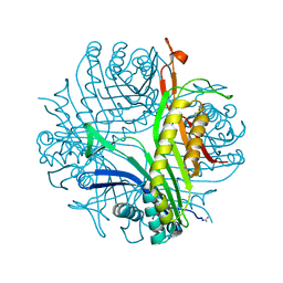
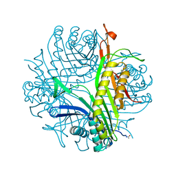
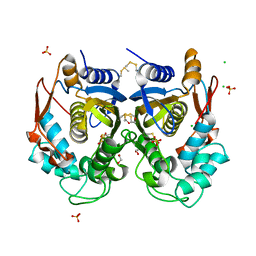
3S2E
 
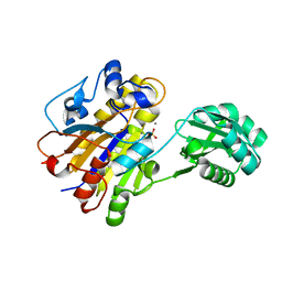
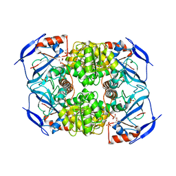 | | Crystal Structure of FurX NADH Complex 1 | | 分子名称: | GLYCEROL, NICOTINAMIDE-ADENINE-DINUCLEOTIDE, SULFATE ION, ... | | 著者 | Hayes, R, Sanchez, E.J, Webb, B.N, Hooper, T, Nissen, M.S, Li, Q, Xun, L. | | 登録日 | 2011-05-16 | | 公開日 | 2012-06-13 | | 最終更新日 | 2024-02-28 | | 実験手法 | X-RAY DIFFRACTION (1.763 Å) | | 主引用文献 | Crystal Structures and furfural reduction mechanism of a bacterial zinc-dependent alcohol dehydrogenase
To be Published
|
|
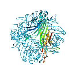
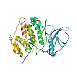
3S1L
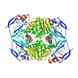 | | Crystal Structure of Apo-form FurX | | 分子名称: | HEXAETHYLENE GLYCOL, ZINC ION, Zinc-containing alcohol dehydrogenase superfamily | | 著者 | Hayes, R, Sanchez, E.J, Webb, B.N, Hooper, T, Nissen, M.S, Li, Q, Xun, L. | | 登録日 | 2011-05-15 | | 公開日 | 2012-04-25 | | 最終更新日 | 2024-02-28 | | 実験手法 | X-RAY DIFFRACTION (1.9 Å) | | 主引用文献 | Furfural reduction mechanism of a zinc-dependent alcohol dehydrogenase from Cupriavidus necator JMP134.
Mol.Microbiol., 83, 2012
|
|
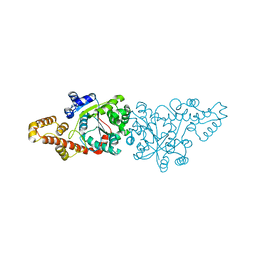
5XD2
 | | Crystal structure of Mycobacterium smegmatis MutT1 in complex with Ap5A, ATP and manganese | | 分子名称: | ADENOSINE-5'-PENTAPHOSPHATE, ADENOSINE-5'-TRIPHOSPHATE, GLYCEROL, ... | | 著者 | Arif, S.M, Varshney, U, Vijayan, M. | | 登録日 | 2017-03-24 | | 公開日 | 2017-08-09 | | 最終更新日 | 2023-11-22 | | 実験手法 | X-RAY DIFFRACTION (1.75 Å) | | 主引用文献 | Hydrolysis of diadenosine polyphosphates. Exploration of an additional role of Mycobacterium smegmatis MutT1
J. Struct. Biol., 199, 2017
|
|
2NUE
 | | Crystal structure of RNase III from Aquifex aeolicus complexed with ds-RNA at 2.9-Angstrom Resolution | | 分子名称: | 46-MER, Ribonuclease III | | 著者 | Gan, J.H, Shaw, G, Tropea, J.E, Waugh, D.S, Court, D.L, Ji, X. | | 登録日 | 2006-11-09 | | 公開日 | 2007-11-20 | | 最終更新日 | 2023-08-30 | | 実験手法 | X-RAY DIFFRACTION (2.9 Å) | | 主引用文献 | A stepwise model for double-stranded RNA processing by ribonuclease III.
Mol.Microbiol., 67, 2007
|
|
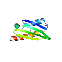
4N3E
 | | Crystal structure of Hyp-1, a St John's wort PR-10 protein, in complex with 8-anilino-1-naphthalene sulfonate (ANS) | | 分子名称: | 4-(2-HYDROXYETHYL)-1-PIPERAZINE ETHANESULFONIC ACID, 8-ANILINO-1-NAPHTHALENE SULFONATE, Phenolic oxidative coupling protein, ... | | 著者 | Sliwiak, J, Dauter, Z, Mccoy, A.J, Read, R.J, Jaskolski, M. | | 登録日 | 2013-10-07 | | 公開日 | 2014-02-26 | | 最終更新日 | 2023-09-20 | | 実験手法 | X-RAY DIFFRACTION (2.43 Å) | | 主引用文献 | Likelihood-based molecular-replacement solution for a highly pathological crystal with tetartohedral twinning and sevenfold translational noncrystallographic symmetry.
Acta Crystallogr.,Sect.D, 70, 2014
|
|
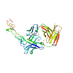
3THM
 | | Crystal structure of Fas receptor extracellular domain in complex with Fab EP6b_B01 | | 分子名称: | 1,2-ETHANEDIOL, 2-acetamido-2-deoxy-beta-D-glucopyranose-(1-4)-[alpha-L-fucopyranose-(1-6)]2-acetamido-2-deoxy-beta-D-glucopyranose, Fab EP6b_B01, ... | | 著者 | Zuger, S, Stirnimann, C, Briand, C, Grutter, M.G. | | 登録日 | 2011-08-19 | | 公開日 | 2012-05-09 | | 最終更新日 | 2023-09-13 | | 実験手法 | X-RAY DIFFRACTION (2.1 Å) | | 主引用文献 | A series of Fas receptor agonist antibodies that demonstrate an inverse correlation between affinity and potency.
Cell Death Differ., 19, 2012
|
|
4N9S
 | | High resolution X-RAY STRUCTURE OF URATE OXIDASE IN COMPLEX WITH 8-HYDROXYXANTHINE | | 分子名称: | 8-hydroxy-3,9-dihydro-1H-purine-2,6-dione, CHLORIDE ION, GLYCEROL, ... | | 著者 | Oksanen, E, Blakeley, M.P, Budayova-Spano, M. | | 登録日 | 2013-10-21 | | 公開日 | 2014-02-05 | | 最終更新日 | 2023-09-20 | | 実験手法 | X-RAY DIFFRACTION (1.06 Å) | | 主引用文献 | The neutron structure of urate oxidase resolves a long-standing mechanistic conundrum and reveals unexpected changes in protonation.
Plos One, 9, 2014
|
|
4N9V
 | | High resolution x-ray structure of urate oxidase in complex with 8-azaxanthine | | 分子名称: | 8-AZAXANTHINE, CHLORIDE ION, GLYCEROL, ... | | 著者 | Oksanen, E, Blakeley, M.P, Budayova-Spano, M. | | 登録日 | 2013-10-21 | | 公開日 | 2014-02-05 | | 最終更新日 | 2023-09-20 | | 実験手法 | X-RAY DIFFRACTION (1.1 Å) | | 主引用文献 | The neutron structure of urate oxidase resolves a long-standing mechanistic conundrum and reveals unexpected changes in protonation.
Plos One, 9, 2014
|
|
3QBW
 | | Crystal structure of pseudomonas aeruginosa 1,6-anhydro-n-actetylmuramic acid kinase (ANMK) bound to adenosine diphosphate | | 分子名称: | ADENOSINE-5'-DIPHOSPHATE, Anhydro-N-acetylmuramic acid kinase, SULFATE ION | | 著者 | Bacik, J.P, Martin, D.R, Mark, B.L. | | 登録日 | 2011-01-14 | | 公開日 | 2011-02-02 | | 最終更新日 | 2023-09-13 | | 実験手法 | X-RAY DIFFRACTION (2.23 Å) | | 主引用文献 | Molecular Basis of 1,6-Anhydro Bond Cleavage and Phosphoryl Transfer by Pseudomonas aeruginosa 1,6-Anhydro-N-acetylmuramic Acid Kinase.
J.Biol.Chem., 286, 2011
|
|
4M0S
 | |
4N9M
 | | Joint neutron/x-ray structure of urate oxidase in complex with 8-hydroxyxanthine | | 分子名称: | 8-hydroxy-3,9-dihydro-1H-purine-2,6-dione, CHLORIDE ION, SODIUM ION, ... | | 著者 | Oksanen, E, Blakeley, M.P, Budayova-Spano, M. | | 登録日 | 2013-10-21 | | 公開日 | 2014-02-05 | | 最終更新日 | 2018-06-13 | | 実験手法 | NEUTRON DIFFRACTION (2.023 Å), X-RAY DIFFRACTION | | 主引用文献 | The neutron structure of urate oxidase resolves a long-standing mechanistic conundrum and reveals unexpected changes in protonation.
Plos One, 9, 2014
|
|
3PPE
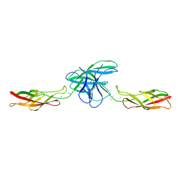 | | Crystal structure of chicken VE-cadherin EC1-2 | | 分子名称: | CALCIUM ION, Vascular endothelial cadherin | | 著者 | Brasch, J, Harrison, O.J, Ahlsen, G, Carnally, S.M, Henderson, R.M, Honig, B, Shapiro, L.S. | | 登録日 | 2010-11-24 | | 公開日 | 2011-02-02 | | 最終更新日 | 2023-09-06 | | 実験手法 | X-RAY DIFFRACTION (2.1 Å) | | 主引用文献 | Structure and binding mechanism of vascular endothelial cadherin: a divergent classical cadherin.
J.Mol.Biol., 408, 2011
|
|
3ROV
 | | Insulin's biosynthesis and activity have opposing structural requirements: a new factor in neonatal diabetes mellitus | | 分子名称: | CHLORIDE ION, Insulin, PHENOL, ... | | 著者 | Weiss, M.A, Wan, Z.L, Dodson, E.J, Liu, M, Xu, B, Hua, Q.X, Turkenburg, M, Whittingham, J, Nakagawa, S.H, Huang, K, Hu, S.Q, Jia, W.H, Wang, S.H, Brange, J, Whittaker, J, Arvan, P, Katsoyannis, P.G, Dodson, G.G. | | 登録日 | 2011-04-26 | | 公開日 | 2012-05-02 | | 最終更新日 | 2023-09-13 | | 実験手法 | X-RAY DIFFRACTION (2.3 Å) | | 主引用文献 | Insulin's biosynthesis and activity have opposing structural requirements: a new factor in neonatal diabetes mellitus
To be Published
|
|
3DAD
 | | Crystal structure of the N-terminal regulatory domains of the formin FHOD1 | | 分子名称: | FH1/FH2 domain-containing protein 1 | | 著者 | Schulte, A, Stolp, B, Schonichen, A, Pylypenko, O, Rak, A, Fackler, O.T, Geyer, M. | | 登録日 | 2008-05-29 | | 公開日 | 2008-09-16 | | 最終更新日 | 2024-02-21 | | 実験手法 | X-RAY DIFFRACTION (2.3 Å) | | 主引用文献 | The Human Formin FHOD1 Contains a Bipartite Structure of FH3 and GTPase-Binding Domains Required for Activation.
Structure, 16, 2008
|
|
2Q6P
 | | The Chemical Control of Protein Folding: Engineering a Superfolder Green Fluorescent Protein | | 分子名称: | Green fluorescent protein mutant 3 | | 著者 | Steiner, T, Hess, P, Bae, J.H, Wiltschi, B, Moroder, L, Budisa, N. | | 登録日 | 2007-06-05 | | 公開日 | 2007-06-19 | | 最終更新日 | 2023-11-15 | | 実験手法 | X-RAY DIFFRACTION (2.1 Å) | | 主引用文献 | Synthetic biology of proteins: tuning GFPs folding and stability with fluoroproline.
Plos One, 3, 2008
|
|
1LT4
 | | HEAT-LABILE ENTEROTOXIN MUTANT S63K | | 分子名称: | HEAT-LABILE ENTEROTOXIN, beta-D-galactopyranose-(1-4)-beta-D-glucopyranose | | 著者 | Van Den Akker, F, Hol, W.G.J. | | 登録日 | 1997-04-14 | | 公開日 | 1997-06-16 | | 最終更新日 | 2023-08-09 | | 実験手法 | X-RAY DIFFRACTION (2 Å) | | 主引用文献 | Crystal structure of a non-toxic mutant of heat-labile enterotoxin, which is a potent mucosal adjuvant.
Protein Sci., 6, 1997
|
|
3U87
 | | Structure of a chimeric construct of human CK2alpha and human CK2alpha' in complex with a non-hydrolysable ATP-analogue | | 分子名称: | CHLORIDE ION, Casein kinase II subunit alpha, GLYCEROL, ... | | 著者 | Niefind, K, Raaf, J, Issinger, O.-G, Olsen, B. | | 登録日 | 2011-10-16 | | 公開日 | 2012-05-30 | | 最終更新日 | 2023-09-13 | | 実験手法 | X-RAY DIFFRACTION (2.9 Å) | | 主引用文献 | Low-density crystal packing of human protein kinase CK2 catalytic subunit in complex with resorufin or other ligands: a tool to study the unique hinge-region plasticity of the enzyme without packing bias.
Acta Crystallogr.,Sect.D, 68, 2012
|
|
1EQT
 | | MET-RANTES | | 分子名称: | SULFATE ION, T-CELL SPECIFIC RANTES PROTEIN | | 著者 | Hoover, D.M, Shaw, J, Gryczynski, Z, Proudfoot, A.E.I, Wells, T. | | 登録日 | 2000-04-06 | | 公開日 | 2000-04-19 | | 最終更新日 | 2023-08-09 | | 実験手法 | X-RAY DIFFRACTION (1.6 Å) | | 主引用文献 | The Crystal Structure of MET-RANTES: Comparison with Native RANTES and AOP-RANTES
PROTEIN PEPT.LETT., 7, 2000
|
|
3TYY
 | | Crystal Structure of Human Lamin-B1 Coil 2 Segment | | 分子名称: | Lamin-B1 | | 著者 | Lam, R, Xu, C, Bian, C.B, Mackenzie, F, Walker, J.R, Bountra, C, Weigelt, J, Arrowsmith, C.H, Edwards, A.M, Bochkarev, A, Min, J, Structural Genomics Consortium (SGC) | | 登録日 | 2011-09-26 | | 公開日 | 2011-10-05 | | 最終更新日 | 2023-09-13 | | 実験手法 | X-RAY DIFFRACTION (2.399 Å) | | 主引用文献 | Crystal structures of the coil 2B fragment and the globular tail domain of human lamin B1.
Febs Lett., 586, 2012
|
|
4O1X
 | |
3U9C
 | | Structure of a C-terminal deletion mutant of human protein kinase CK2 catalytic subunit with the ATP-competitive inhibitor resorufin | | 分子名称: | 7-hydroxy-3H-phenoxazin-3-one, CHLORIDE ION, Casein kinase II subunit alpha, ... | | 著者 | Klopffleisch, K, Issinger, O.-G, Niefind, K. | | 登録日 | 2011-10-18 | | 公開日 | 2012-05-30 | | 最終更新日 | 2023-09-13 | | 実験手法 | X-RAY DIFFRACTION (3.2 Å) | | 主引用文献 | Low-density crystal packing of human protein kinase CK2 catalytic subunit in complex with resorufin or other ligands: a tool to study the unique hinge-region plasticity of the enzyme without packing bias.
Acta Crystallogr.,Sect.D, 68, 2012
|
|
3TQM
 | | Structure of an ribosomal subunit interface protein from Coxiella burnetii | | 分子名称: | Ribosome-associated factor Y, SULFATE ION | | 著者 | Rudolph, M, Cheung, J, Franklin, M.C, Cassidy, M, Gary, E, Burshteyn, F, Love, J. | | 登録日 | 2011-09-09 | | 公開日 | 2011-09-28 | | 最終更新日 | 2017-11-08 | | 実験手法 | X-RAY DIFFRACTION (2.45 Å) | | 主引用文献 | Structural genomics for drug design against the pathogen Coxiella burnetii.
Proteins, 83, 2015
|
|
3TJE
 | | Crystal structure of Fas receptor extracellular domain in complex with Fab E09 | | 分子名称: | 1,2-ETHANEDIOL, CADMIUM ION, CHLORIDE ION, ... | | 著者 | Zuger, S, Stirnimann, C, Briand, C, Grutter, M.G. | | 登録日 | 2011-08-24 | | 公開日 | 2012-05-09 | | 最終更新日 | 2023-09-13 | | 実験手法 | X-RAY DIFFRACTION (1.93 Å) | | 主引用文献 | A series of Fas receptor agonist antibodies that demonstrate an inverse correlation between affinity and potency.
Cell Death Differ., 19, 2012
|
|
3TWZ
 | | Phosphorylated Bacillus cereus phosphopentomutase in space group P212121 | | 分子名称: | MANGANESE (II) ION, Phosphopentomutase | | 著者 | Panosian, T.P, Nanneman, D.P, Bachmann, B.O, Iverson, T.M. | | 登録日 | 2011-09-22 | | 公開日 | 2012-02-29 | | 最終更新日 | 2012-03-21 | | 実験手法 | X-RAY DIFFRACTION (1.75 Å) | | 主引用文献 | Molecular Differences between a Mutase and a Phosphatase: Investigations of the Activation Step in Bacillus cereus Phosphopentomutase.
Biochemistry, 51, 2012
|
|
2QUK
 | |


