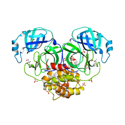
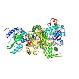
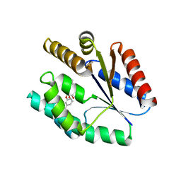
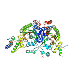
3ZOU
 
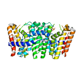 | | Native structure of Farnesyl Pyrophosphate Synthase from Pseudomonas aeruginosa PA01, with bound fragment SPB02696, and substrate geranyl pyrophosphate. | | 分子名称: | 3-(2-oxo-1,3-benzoxazol-3(2H)-yl)propanoic acid, DIMETHYL SULFOXIDE, FARNESYL PYROPHOSPHATE SYNTHASE, ... | | 著者 | Schmidberger, J.W, Schnell, R, Schneider, G. | | 登録日 | 2013-02-25 | | 公開日 | 2014-03-12 | | 最終更新日 | 2023-12-20 | | 実験手法 | X-RAY DIFFRACTION (1.55 Å) | | 主引用文献 | Structural Characterization of Substrate and Inhibitor Binding to Farnesyl Pyrophosphate Synthase from Pseudomonas Aeruginosa
Acta Crystallogr.,Sect.D, 71, 2015
|
|
1C26
 | |
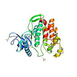
6ITV
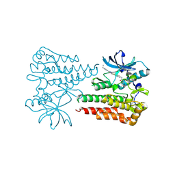 | | Crystal structure of activated c-KIT in complex with compound | | 分子名称: | Mast/stem cell growth factor receptor Kit, N-(5-ethyl-1,2-oxazol-3-yl)-N'-[4-(2-{[6-(4-ethylpyrazin-1(4H)-yl)pyrimidin-4-yl]amino}-1,3-thiazol-5-yl)phenyl]urea | | 著者 | Wu, T.S, Wu, S.Y. | | 登録日 | 2018-11-26 | | 公開日 | 2019-11-27 | | 最終更新日 | 2024-11-13 | | 実験手法 | X-RAY DIFFRACTION (1.881 Å) | | 主引用文献 | Crystal structure of activated c-KIT in complex with compound
To Be Published
|
|
2B83
 | |
1LVO
 | | Structure of coronavirus main proteinase reveals combination of a chymotrypsin fold with an extra alpha-helical domain | | 分子名称: | (4R)-2-METHYLPENTANE-2,4-DIOL, 1,4-DIETHYLENE DIOXIDE, Replicase, ... | | 著者 | Anand, K, Palm, G.J, Mesters, J.R, Siddell, S.G, Ziebuhr, J, Hilgenfeld, R. | | 登録日 | 2002-05-29 | | 公開日 | 2002-07-17 | | 最終更新日 | 2024-03-13 | | 実験手法 | X-RAY DIFFRACTION (1.96 Å) | | 主引用文献 | Structure of coronavirus main proteinase reveals combination of a chymotrypsin fold with an extra alpha-helical domain.
EMBO J., 21, 2002
|
|
4ABQ
 | | CRYSTAL STRUCTURE OF TRANSTHYRETIN IN COMPLEX WITH LIGAND C-1 | | 分子名称: | 3-(5-MERCAPTO-1,3,4-OXADIAZOL-2-YL)-PHENOL, TRANSTHYRETIN | | 著者 | Tomar, D, Khan, T, Singh, R.R, Mishra, S, Gupta, S, Surolia, A, Salunke, D.M. | | 登録日 | 2011-12-10 | | 公開日 | 2012-09-26 | | 最終更新日 | 2023-12-20 | | 実験手法 | X-RAY DIFFRACTION (1.7 Å) | | 主引用文献 | Crystallographic Study of Novel Transthyretin Ligands Exhibiting Negative-Cooperativity between Two Thyroxine Binding Sites.
Plos One, 7, 2012
|
|
6IWI
 | | Crystal structure of PDE5A in complex with a novel inhibitor | | 分子名称: | MAGNESIUM ION, N-[3-(4,5-diethyl-6-oxo-1,6-dihydropyrimidin-2-yl)-4-propoxyphenyl]-2-(4-methylpiperazin-1-yl)acetamide, ZINC ION, ... | | 著者 | Zhang, X.L, Xu, Y.C. | | 登録日 | 2018-12-05 | | 公開日 | 2019-12-11 | | 最終更新日 | 2024-03-27 | | 実験手法 | X-RAY DIFFRACTION (2.155 Å) | | 主引用文献 | Pharmacokinetics-Driven Optimization of 4(3 H)-Pyrimidinones as Phosphodiesterase Type 5 Inhibitors Leading to TPN171, a Clinical Candidate for the Treatment of Pulmonary Arterial Hypertension.
J.Med.Chem., 62, 2019
|
|
4GY5
 | | Crystal structure of the tandem tudor domain and plant homeodomain of UHRF1 with Histone H3K9me3 | | 分子名称: | E3 ubiquitin-protein ligase UHRF1, Peptide from Histone H3.3, ZINC ION | | 著者 | Cheng, J, Yang, Y, Fang, J, Xiao, J, Zhu, T, Chen, F, Wang, P, Xu, Y. | | 登録日 | 2012-09-05 | | 公開日 | 2012-11-14 | | 最終更新日 | 2023-11-08 | | 実験手法 | X-RAY DIFFRACTION (2.956 Å) | | 主引用文献 | Structural insight into coordinated recognition of trimethylated histone H3 lysine 9 (H3K9me3) by the plant homeodomain (PHD) and tandem tudor domain (TTD) of UHRF1 (ubiquitin-like, containing PHD and RING finger domains, 1) protein
J.Biol.Chem., 288, 2013
|
|
4H0X
 | | Crystal structure of NAD+-Ia(E380A)-actin complex | | 分子名称: | 1,2-ETHANEDIOL, ADENOSINE-5'-TRIPHOSPHATE, Actin, ... | | 著者 | Tsurumura, T, Oda, M, Nagahama, M, Tsuge, H. | | 登録日 | 2012-09-10 | | 公開日 | 2013-02-20 | | 最終更新日 | 2023-11-08 | | 実験手法 | X-RAY DIFFRACTION (2.33 Å) | | 主引用文献 | Arginine ADP-ribosylation mechanism based on structural snapshots of iota-toxin and actin complex
Proc.Natl.Acad.Sci.USA, 110, 2013
|
|
2YKP
 | |
6I5L
 | | Crystal structure of CLK1 in complexed with furo[3,2-b]pyridine compound VN316 (derivative of compound 12h) | | 分子名称: | 3-(3-cyclobutylphenyl)-5-(1-methylpyrazol-4-yl)furo[3,2-b]pyridine, Dual specificity protein kinase CLK1, GLYCEROL, ... | | 著者 | Chaikuad, A, Arrowsmith, C.H, Edwards, A.M, Bountra, C, Paruch, K, Knapp, S, Structural Genomics Consortium (SGC) | | 登録日 | 2018-11-13 | | 公開日 | 2019-01-09 | | 最終更新日 | 2024-01-24 | | 実験手法 | X-RAY DIFFRACTION (2.55 Å) | | 主引用文献 | Furo[3,2-b]pyridine: A Privileged Scaffold for Highly Selective Kinase Inhibitors and Effective Modulators of the Hedgehog Pathway.
Angew. Chem. Int. Ed. Engl., 58, 2019
|
|
2OXG
 | | The SoxYZ Complex of Paracoccus pantotrophus | | 分子名称: | 1,2-ETHANEDIOL, SULFATE ION, SoxY protein, ... | | 著者 | Sauve, V, Berks, B.C, Hemmings, A.M. | | 登録日 | 2007-02-20 | | 公開日 | 2007-05-22 | | 最終更新日 | 2023-08-30 | | 実験手法 | X-RAY DIFFRACTION (1.4 Å) | | 主引用文献 | The SoxYZ Complex Carries Sulfur Cycle Intermediates on a Peptide Swinging Arm.
J.Biol.Chem., 282, 2007
|
|
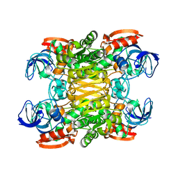
6GIQ
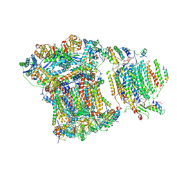 | | Saccharomyces cerevisiae respiratory supercomplex III2IV | | 分子名称: | (1R)-2-(dodecanoyloxy)-1-[(phosphonooxy)methyl]ethyl tetradecanoate, (1R)-2-(phosphonooxy)-1-[(tridecanoyloxy)methyl]ethyl pentadecanoate, (1R)-2-{[(S)-(2-aminoethoxy)(hydroxy)phosphoryl]oxy}-1-[(heptanoyloxy)methyl]ethyl octadecanoate, ... | | 著者 | Rathore, S, Berndtsson, J, Conrad, J, Ott, M. | | 登録日 | 2018-05-15 | | 公開日 | 2019-01-02 | | 最終更新日 | 2024-10-09 | | 実験手法 | ELECTRON MICROSCOPY (3.23 Å) | | 主引用文献 | Cryo-EM structure of the yeast respiratory supercomplex.
Nat. Struct. Mol. Biol., 26, 2019
|
|
7HTZ
 | | PanDDA analysis group deposition -- Crystal Structure of FatA in complex with Z69092635 | | 分子名称: | N-[2-(cyclohex-1-en-1-yl)ethyl]-3,5-dimethyl-1,2-oxazole-4-carboxamide, Oleoyl-acyl carrier protein thioesterase 1, chloroplastic, ... | | 著者 | Kot, E, Ni, X, Tomlinson, C.W.E, Fearon, D, Aschenbrenner, J.C, Fairhead, M, Koekemoer, L, Marx, M.L, Wright, N.D, Mulholland, N.P, Montgomery, M.G, von Delft, F. | | 登録日 | 2024-12-23 | | 公開日 | 2025-08-13 | | 実験手法 | X-RAY DIFFRACTION (2.07 Å) | | 主引用文献 | PanDDA analysis group deposition
To Be Published
|
|
7WRH
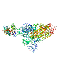 | | Cryo-EM structure of SARS-CoV-2 Omicron BA.1 spike protein in complex with mouse ACE2 | | 分子名称: | 2-acetamido-2-deoxy-beta-D-glucopyranose, 2-acetamido-2-deoxy-beta-D-glucopyranose-(1-4)-2-acetamido-2-deoxy-beta-D-glucopyranose, Processed angiotensin-converting enzyme 2, ... | | 著者 | Han, P, Xie, Y, Qi, J. | | 登録日 | 2022-01-26 | | 公開日 | 2023-02-01 | | 最終更新日 | 2024-10-30 | | 実験手法 | ELECTRON MICROSCOPY (2.66 Å) | | 主引用文献 | Broader-species receptor binding and structural bases of Omicron SARS-CoV-2 to both mouse and palm-civet ACE2s.
Cell Discov, 8, 2022
|
|
1BX7
 | | HIRUSTASIN FROM HIRUDO MEDICINALIS AT 1.2 ANGSTROMS | | 分子名称: | HIRUSTASIN, SULFATE ION | | 著者 | Uson, I, Sheldrick, G.M, De La Fortelle, E, Bricogne, G, Di Marco, S, Priestle, J.P, Gruetter, M.G, Mittl, P.R.E. | | 登録日 | 1998-10-14 | | 公開日 | 1999-04-27 | | 最終更新日 | 2024-10-23 | | 実験手法 | X-RAY DIFFRACTION (1.2 Å) | | 主引用文献 | The 1.2 A crystal structure of hirustasin reveals the intrinsic flexibility of a family of highly disulphide-bridged inhibitors.
Structure Fold.Des., 7, 1999
|
|
1RG1
 | | Crystal structure of human Tyrosyl-DNA Phosphodiesterase complexed with vanadate, octopamine, and tetranucleotide AGTT | | 分子名称: | 4-(2S-AMINO-1-HYDROXYETHYL)PHENOL, 5'-D(*AP*GP*TP*T)-3', SPERMINE, ... | | 著者 | Davies, D.R, Interthal, H, Champoux, J.J, Hol, W.G. | | 登録日 | 2003-11-10 | | 公開日 | 2004-03-02 | | 最終更新日 | 2023-08-23 | | 実験手法 | X-RAY DIFFRACTION (2.1 Å) | | 主引用文献 | Explorations of peptide and oligonucleotide binding sites of tyrosyl-DNA phosphodiesterase using vanadate complexes.
J.Med.Chem., 47, 2004
|
|
3ARY
 | | Crystal Structure Analysis of Chitinase A from Vibrio harveyi with novel inhibitors - complex structure with 2-(imidazolin-2-yl)-5-isothiocyanatobenzofuran | | 分子名称: | 2-(5-isothiocyanato-1-benzofuran-2-yl)-4,5-dihydro-1H-imidazole, Chitinase A, GLYCEROL | | 著者 | Pantoom, S, Vetter, I.R, Prinz, H, Suginta, W. | | 登録日 | 2010-12-09 | | 公開日 | 2011-04-20 | | 最終更新日 | 2024-10-23 | | 実験手法 | X-RAY DIFFRACTION (1.35 Å) | | 主引用文献 | Potent family-18 chitinase inhibitors: x-ray structures, affinities, and binding mechanisms
J.Biol.Chem., 286, 2011
|
|
4GSY
 | |
1CL7
 | | ANTI HIV1 PROTEASE FAB | | 分子名称: | PROTEIN (IGG1 ANTIBODY 1696 (constant heavy chain)), PROTEIN (IGG1 ANTIBODY 1696 (light chain)), PROTEIN (IGG1 ANTIBODY 1696 (variable heavy chain)) | | 著者 | Lescar, J, Bentley, G.A. | | 登録日 | 1999-05-06 | | 公開日 | 2000-01-12 | | 最終更新日 | 2024-11-13 | | 実験手法 | X-RAY DIFFRACTION (3 Å) | | 主引用文献 | Inhibition of the HIV-1 and HIV-2 proteases by a monoclonal antibody.
Protein Sci., 8, 1999
|
|
1RL0
 | | Crystal structure of a new ribosome-inactivating protein (RIP): dianthin 30 | | 分子名称: | Antiviral protein DAP-30 | | 著者 | Fermani, S, Falini, G, Ripamonti, A, Bolognesi, A, Polito, L, Stirpe, F. | | 登録日 | 2003-11-24 | | 公開日 | 2004-12-07 | | 最終更新日 | 2023-08-23 | | 実験手法 | X-RAY DIFFRACTION (1.4 Å) | | 主引用文献 | The 1.4A structure of dianthin 30 indicates a role of surface potential at the active site of type 1 ribosome inactivating proteins
J.Struct.Biol., 149, 2005
|
|
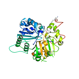
3AQV
 | | Human AMP-activated protein kinase alpha 2 subunit kinase domain (T172D) complexed with compound C | | 分子名称: | 5'-AMP-activated protein kinase catalytic subunit alpha-2, 6-[4-(2-piperidin-1-ylethoxy)phenyl]-3-pyridin-4-ylpyrazolo[1,5-a]pyrimidine | | 著者 | Handa, N, Takagi, T, Saijo, S, Kishishita, S, Toyama, M, Terada, T, Shirouzu, M, Yokoyama, S, RIKEN Structural Genomics/Proteomics Initiative (RSGI) | | 登録日 | 2010-11-19 | | 公開日 | 2011-04-27 | | 最終更新日 | 2023-11-01 | | 実験手法 | X-RAY DIFFRACTION (2.08 Å) | | 主引用文献 | Structural basis for compound C inhibition of the human AMP-activated protein kinase alpha 2 subunit kinase domain
Acta Crystallogr.,Sect.D, 67, 2011
|
|
4H0T
 | | Crystal structure of Ia-ADPR-actin complex | | 分子名称: | 1,2-ETHANEDIOL, ADENOSINE-5'-TRIPHOSPHATE, Actin, ... | | 著者 | Tsurumura, T, Oda, M, Nagahama, M, Tsuge, H. | | 登録日 | 2012-09-10 | | 公開日 | 2013-02-20 | | 最終更新日 | 2024-10-30 | | 実験手法 | X-RAY DIFFRACTION (2.2 Å) | | 主引用文献 | Arginine ADP-ribosylation mechanism based on structural snapshots of iota-toxin and actin complex
Proc.Natl.Acad.Sci.USA, 110, 2013
|
|
4H2V
 | | Crystal structure of Bradyrhizobium japonicum glycine:[carrier protein] ligase complexed with glycylated carrier protein | | 分子名称: | 4'-PHOSPHOPANTETHEINE, ACETATE ION, ADENOSINE MONOPHOSPHATE, ... | | 著者 | Luic, M, Weygand-Durasevic, I, Ivic, N, Mocibob, M. | | 登録日 | 2012-09-13 | | 公開日 | 2013-03-06 | | 最終更新日 | 2025-03-26 | | 実験手法 | X-RAY DIFFRACTION (2 Å) | | 主引用文献 | Adaptation of aminoacyl-tRNA synthetase catalytic core to carrier protein aminoacylation.
Structure, 21, 2013
|
|
4DX0
 | | Structure of the 14-3-3/PMA2 complex stabilized by a pyrazole derivative | | 分子名称: | 14-3-3-like protein E, 4-[(4R)-4-(4-nitrophenyl)-6-oxidanylidene-3-phenyl-1,4-dihydropyrrolo[3,4-c]pyrazol-5-yl]benzoic acid, N.plumbaginifolia H+-translocating ATPase mRNA | | 著者 | Richter, A, Rose, R, Hedberg, C, Waldmann, H, Ottmann, C. | | 登録日 | 2012-02-27 | | 公開日 | 2012-05-30 | | 最終更新日 | 2023-12-13 | | 実験手法 | X-RAY DIFFRACTION (3.4 Å) | | 主引用文献 | An Optimised Small-Molecule Stabiliser of the 14-3-3-PMA2 Protein-Protein Interaction.
Chemistry, 18, 2012
|
|


