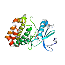
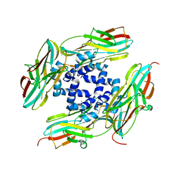
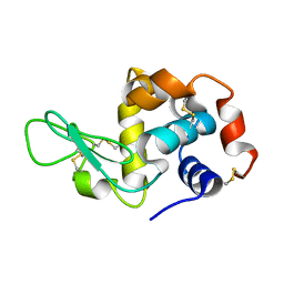
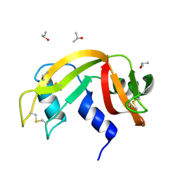
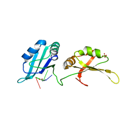
4YFE
 
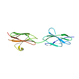 | | Crystal structure of PTP delta Fn1-Fn2 | | 分子名称: | Receptor-type tyrosine-protein phosphatase delta | | 著者 | Yamagata, A, Fukai, S. | | 登録日 | 2015-02-25 | | 公開日 | 2015-05-06 | | 最終更新日 | 2023-11-08 | | 実験手法 | X-RAY DIFFRACTION (1.972 Å) | | 主引用文献 | Mechanisms of splicing-dependent trans-synaptic adhesion by PTP delta-IL1RAPL1/IL-1RAcP for synaptic differentiation.
Nat Commun, 6, 2015
|
|
6RAT
 | |
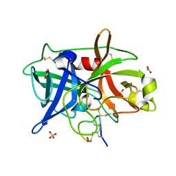
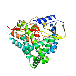
4YDF
 | | Crystal structure of compound 9 in complex with HTLV-1 Protease | | 分子名称: | HTLV-1 Protease, N-benzyl-N-[(3S,4S)-4-{benzyl[(4-nitrophenyl)sulfonyl]amino}pyrrolidin-3-yl]-3-nitrobenzenesulfonamide, SULFATE ION | | 著者 | Kuhnert, M, Blum, A, Steuber, H, Diederich, W.E. | | 登録日 | 2015-02-22 | | 公開日 | 2016-02-03 | | 最終更新日 | 2024-01-10 | | 実験手法 | X-RAY DIFFRACTION (2.804 Å) | | 主引用文献 | Privileged Structures Meet Human T-Cell Leukemia Virus-1 (HTLV-1): C2-Symmetric 3,4-Disubstituted Pyrrolidines as Nonpeptidic HTLV-1 Protease Inhibitors.
J.Med.Chem., 58, 2015
|
|
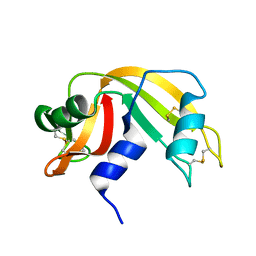
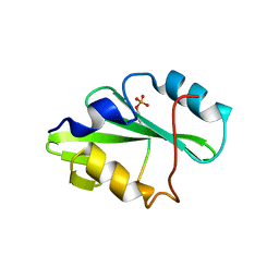
6EW2
 | | Human myelin protein P2 F57A mutant, tetragonal crystal form | | 分子名称: | CHLORIDE ION, Myelin P2 protein, PALMITIC ACID | | 著者 | Laulumaa, S, Lehtimaki, M, Kursula, P. | | 登録日 | 2017-11-03 | | 公開日 | 2018-07-11 | | 最終更新日 | 2024-01-17 | | 実験手法 | X-RAY DIFFRACTION (1.588 Å) | | 主引用文献 | Structure and dynamics of a human myelin protein P2 portal region mutant indicate opening of the beta barrel in fatty acid binding proteins.
BMC Struct. Biol., 18, 2018
|
|
6EW5
 | |
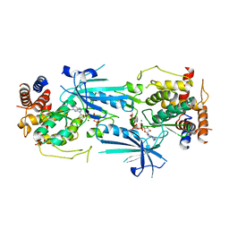
6R49
 | | Aurora-A in complex with shape-diverse fragment 39 | | 分子名称: | (1~{S},10~{S})-12-cyclopropyl-1-oxidanyl-10-propan-2-yl-9,12-diazatricyclo[8.2.1.0^{2,7}]trideca-2(7),3,5-trien-11-one, ADENOSINE-5'-DIPHOSPHATE, Aurora kinase A, ... | | 著者 | Bayliss, R, McIntyre, P.J. | | 登録日 | 2019-03-22 | | 公開日 | 2019-05-08 | | 最終更新日 | 2024-01-24 | | 実験手法 | X-RAY DIFFRACTION (2.209 Å) | | 主引用文献 | Construction of a Shape-Diverse Fragment Set: Design, Synthesis and Screen against Aurora-A Kinase.
Chemistry, 25, 2019
|
|
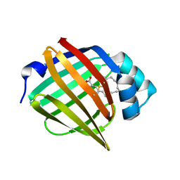
4YGW
 | | RNase S in complex with stabilized S peptide | | 分子名称: | 1-hydroxypropan-2-one, Ribonuclease A C2, S-peptide: ACE-LYS-GLU-THR-ALA-ALA-HCS-LYS-PHE-GLU-HCS-GLN-HIS-MET-ASP-SER, ... | | 著者 | Assem, N, Ferreira, D, Wolan, D.W, Dawson, P.E. | | 登録日 | 2015-02-26 | | 公開日 | 2015-07-01 | | 最終更新日 | 2024-04-03 | | 実験手法 | X-RAY DIFFRACTION (2.18 Å) | | 主引用文献 | Acetone-Linked Peptides: A Convergent Approach for Peptide Macrocyclization and Labeling.
Angew.Chem.Int.Ed.Engl., 54, 2015
|
|
4YEH
 | | Crystal structure of Mg2+ ion containing hemopexin fold from Kabuli chana (chickpea white) at 2.45A resolution reveals a structural basis of metal ion transport | | 分子名称: | Lectin, MAGNESIUM ION | | 著者 | Kumar, S, Singh, A, Yamini, S, Bhushan, A, Dey, S, Sharma, S, Singh, T.P. | | 登録日 | 2015-02-24 | | 公開日 | 2015-03-25 | | 最終更新日 | 2023-11-08 | | 実験手法 | X-RAY DIFFRACTION (2.45 Å) | | 主引用文献 | Crystal Structure of Mg(2+) Containing Hemopexin-Fold Protein from Kabuli Chana (Chickpea-White, CW-25) at 2.45 angstrom Resolution Reveals Its Metal Ion Transport Property
Protein J., 34, 2015
|
|
4YEO
 | | Triclinic HEWL co-crystallised with cisplatin, studied at a data collection temperature of 150K - new refinement | | 分子名称: | 1,2-ETHANEDIOL, ACETATE ION, Cisplatin, ... | | 著者 | Shabalin, I.G, Dauter, Z, Jaskolski, M, Minor, W, Wlodawer, A. | | 登録日 | 2015-02-24 | | 公開日 | 2015-03-04 | | 最終更新日 | 2023-09-27 | | 実験手法 | X-RAY DIFFRACTION (0.98 Å) | | 主引用文献 | Crystallography and chemistry should always go together: a cautionary tale of protein complexes with cisplatin and carboplatin.
Acta Crystallogr.,Sect.D, 71, 2015
|
|
8W8S
 | | Cryo-EM structure of the AA14-bound GPR101 complex | | 分子名称: | 1-(4-methylpyridin-2-yl)-3-[3-(trifluoromethyl)phenyl]thiourea, Probable G-protein coupled receptor 101 | | 著者 | Sun, J.P, Yu, X, Gao, N, Yang, F, Wang, J.Y, Yang, Z, Guan, Y, Wang, G.P. | | 登録日 | 2023-09-04 | | 公開日 | 2024-01-03 | | 最終更新日 | 2024-04-10 | | 実験手法 | ELECTRON MICROSCOPY (3.3 Å) | | 主引用文献 | Structure of GPR101-Gs enables identification of ligands with rejuvenating potential.
Nat.Chem.Biol., 20, 2024
|
|
4YKO
 | |
4YGZ
 | |
4YLC
 | |
7S5A
 | | Crystal structure of human chemokine CCL8 | | 分子名称: | C-C motif chemokine 8 | | 著者 | Bhusal, R.P, Devkota, S.R, Aryal, P, Wilce, M.C.J, Stone, M.J. | | 登録日 | 2021-09-10 | | 公開日 | 2021-09-29 | | 最終更新日 | 2023-10-18 | | 実験手法 | X-RAY DIFFRACTION (1.37 Å) | | 主引用文献 | Structure-guided engineering of tick evasins for targeting chemokines in inflammatory diseases.
Proc.Natl.Acad.Sci.USA, 119, 2022
|
|
4OS2
 | | Crystal structure of urokinase-type plasminogen activator (uPA) complexed with bicyclic peptide UK602 (bicyclic 1) | | 分子名称: | ACETATE ION, SULFATE ION, Urokinase-type plasminogen activator, ... | | 著者 | Chen, S, Pojer, F, Heinis, C. | | 登録日 | 2014-02-12 | | 公開日 | 2014-09-24 | | 最終更新日 | 2021-06-02 | | 実験手法 | X-RAY DIFFRACTION (1.79 Å) | | 主引用文献 | Dithiol amino acids can structurally shape and enhance the ligand-binding properties of polypeptides.
Nat Chem, 6, 2014
|
|
5TPK
 | |
8W6K
 | | in situ room temperature Laue crystallography | | 分子名称: | Lysozyme C | | 著者 | Wang, Z.J, Wang, S.S, Pan, Q.Y, Yu, L, Su, Z.H, Yang, T.Y, Wang, Y.Z, Zhang, W.Z, Hao, Q, Gao, X.Y. | | 登録日 | 2023-08-29 | | 公開日 | 2024-01-17 | | 実験手法 | X-RAY DIFFRACTION (2 Å) | | 主引用文献 | BL03HB: Laue crystallography beamline at SSRF
To Be Published
|
|
4P0O
 | | Cleaved Serpin 42Da | | 分子名称: | Serine protease inhibitor 4, isoform B | | 著者 | Ellisdon, A.M, Whisstock, J.C. | | 登録日 | 2014-02-21 | | 公開日 | 2014-05-07 | | 最終更新日 | 2023-12-27 | | 実験手法 | X-RAY DIFFRACTION (1.8 Å) | | 主引用文献 | High resolution structure of cleaved Serpin 42 Da from Drosophila melanogaster.
Bmc Struct.Biol., 14, 2014
|
|
6F8C
 | |
5TQS
 | |
6ETP
 | |
8UYI
 | | Structure of ADP-bound and phosphorylated Pediculus humanus (Ph) PINK1 dimer | | 分子名称: | ADENOSINE-5'-DIPHOSPHATE, MAGNESIUM ION, Serine/threonine-protein kinase Pink1, ... | | 著者 | Gan, Z.Y, Kirk, N.S, Leis, A, Komander, D. | | 登録日 | 2023-11-13 | | 公開日 | 2024-01-31 | | 実験手法 | ELECTRON MICROSCOPY (3.13 Å) | | 主引用文献 | Interaction of PINK1 with nucleotides and kinetin.
Sci Adv, 10, 2024
|
|
8UZ1
 | | Straight actin filament from Arp2/3 branch junction sample (ADP-BeFx) | | 分子名称: | ADENOSINE-5'-DIPHOSPHATE, Actin, alpha skeletal muscle, ... | | 著者 | Chavali, S.S, Chou, S.Z, Sindelar, C.V. | | 登録日 | 2023-11-14 | | 公開日 | 2024-01-31 | | 最終更新日 | 2024-03-20 | | 実験手法 | ELECTRON MICROSCOPY (3.6 Å) | | 主引用文献 | Cryo-EM structures reveal how phosphate release from Arp3 weakens actin filament branches formed by Arp2/3 complex.
Nat Commun, 15, 2024
|
|
6FAD
 | | SR protein kinase 1 (SRPK1) in complex with the RGG-box of HSV1 ICP27 | | 分子名称: | PHOSPHATE ION, SRSF protein kinase 1, mRNA export factor | | 著者 | Tunnicliffe, R.B, Levy, C, Mould, A.P, Mckenzie, E.A, Sandri-Goldin, R.M, Golovanov, A.P. | | 登録日 | 2017-12-15 | | 公開日 | 2019-09-25 | | 最終更新日 | 2024-01-17 | | 実験手法 | X-RAY DIFFRACTION (2.801 Å) | | 主引用文献 | Molecular Mechanism of SR Protein Kinase 1 Inhibition by the Herpes Virus Protein ICP27.
Mbio, 10, 2019
|
|
4YOE
 | | Structure of UP1 bound to RNA 5'-AGU-3' | | 分子名称: | ACETATE ION, Heterogeneous nuclear ribonucleoprotein A1, RNA AGU, ... | | 著者 | Meagher, J.L, Stuckey, J.A. | | 登録日 | 2015-03-11 | | 公開日 | 2015-06-10 | | 最終更新日 | 2023-09-27 | | 実験手法 | X-RAY DIFFRACTION (1.92 Å) | | 主引用文献 | The First Crystal Structure of the UP1 Domain of hnRNP A1 Bound to RNA Reveals a New Look for an Old RNA Binding Protein.
J.Mol.Biol., 427, 2015
|
|


