2OSB
 
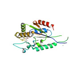
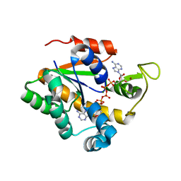 | | Crystal structure of a thermostable mutant of Bacillus subtilis Adenylate Kinase (Q16L/Q199R/) | | 分子名称: | Adenylate kinase, BIS(ADENOSINE)-5'-PENTAPHOSPHATE, MAGNESIUM ION, ... | | 著者 | Myers, J, Counago, R, Wilson, C.J, Wu, G, Shamoo, Y. | | 登録日 | 2007-02-05 | | 公開日 | 2008-01-15 | | 最終更新日 | 2023-08-30 | | 実験手法 | X-RAY DIFFRACTION (1.8 Å) | | 主引用文献 | Crystal structure of a thermostable mutant of Bacillus subtilis Adenylate Kinase (Q16L/Q199R/)
To be Published
|
|
8INJ
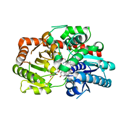 | | Crystal structure of UGT74AN3-UDP-DIG | | 分子名称: | 2-AMINO-2-HYDROXYMETHYL-PROPANE-1,3-DIOL, DIGITOXIGENIN, Glycosyltransferase, ... | | 著者 | Feng, L, Wei, H. | | 登録日 | 2023-03-10 | | 公開日 | 2024-02-14 | | 実験手法 | X-RAY DIFFRACTION (1.76 Å) | | 主引用文献 | Crystal structure of UGT74AN3-UDP-DIG
To Be Published
|
|
2OYT
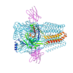
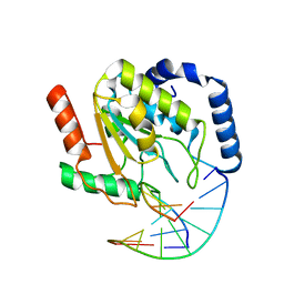 | | Crystal Structure of UNG2/DNA(TM) | | 分子名称: | DNA strand1, DNA strand2, Uracil-DNA glycosylase | | 著者 | Bianchet, M.A, Krosky, D.J, Stivers, J.T, Amzel, L.M. | | 登録日 | 2007-02-22 | | 公開日 | 2007-10-30 | | 最終更新日 | 2023-08-30 | | 実験手法 | X-RAY DIFFRACTION (2 Å) | | 主引用文献 | Enzymatic capture of an extrahelical thymine in the search for uracil in DNA.
Nature, 449, 2007
|
|
8IV3
 | |
2OZO
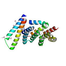
 | | Autoinhibited intact human ZAP-70 | | 分子名称: | MAGNESIUM ION, PHOSPHOAMINOPHOSPHONIC ACID-ADENYLATE ESTER, Tyrosine-protein kinase ZAP-70 | | 著者 | Deindl, S, Kadlecek, T.A, Brdicka, T, Cao, X, Weiss, A, Kuriyan, J. | | 登録日 | 2007-02-26 | | 公開日 | 2007-05-22 | | 最終更新日 | 2024-02-21 | | 実験手法 | X-RAY DIFFRACTION (2.6 Å) | | 主引用文献 | Structural Basis for the Inhibition of Tyrosine Kinase Activity of ZAP-70.
Cell(Cambridge,Mass.), 129, 2007
|
|
8I78
 | | Meso-Diaminopimelate dehydrogenase | | 分子名称: | Meso-diaminopimelate D-dehydrogenase | | 著者 | Wei, S, Wu, T.F. | | 登録日 | 2023-01-31 | | 公開日 | 2024-02-07 | | 実験手法 | X-RAY DIFFRACTION (2.64 Å) | | 主引用文献 | Analysis of the catalytic mechanism of meso-DAPDH and extension of D-aromatic amino acid substrate scope
To Be Published
|
|
2P09
 | | Structural Insights into the Evolution of a Non-Biological Protein | | 分子名称: | ADENOSINE-5'-TRIPHOSPHATE, CHLORIDE ION, PENTAETHYLENE GLYCOL, ... | | 著者 | Smith, M, Rosenow, M, Wang, M, Allen, J.P, Szostak, J.W, Chaput, J.C. | | 登録日 | 2007-02-28 | | 公開日 | 2007-06-05 | | 最終更新日 | 2024-02-21 | | 実験手法 | X-RAY DIFFRACTION (1.65 Å) | | 主引用文献 | Structural insights into the evolution of a non-biological protein: importance of surface residues in protein fold optimization.
PLoS ONE, 2, 2007
|
|
2P0G
 | | Crystal structure of Selenoprotein W-related protein from Vibrio cholerae. Northeast Structural Genomics target VcR75 | | 分子名称: | Selenoprotein W-related protein | | 著者 | Benach, J, Neely, H, Seetharaman, J, Ho, C.K, Janjua, H, Cunningham, K, Ma, L, Xiao, R, Liu, J, Baran, M.C, Acton, T.B, Rost, B, Montelione, G.T, Hunt, J.F, Tong, L, Northeast Structural Genomics Consortium (NESG) | | 登録日 | 2007-02-28 | | 公開日 | 2007-03-20 | | 最終更新日 | 2024-10-09 | | 実験手法 | X-RAY DIFFRACTION (2.3 Å) | | 主引用文献 | Crystal structure of Selenoprotein W-related protein from Vibrio cholerae.
To be Published
|
|
2P0Y
 | | Crystal structure of Q88YI3_LACPL from Lactobacillus plantarum. Northeast Structural Genomics Consortium target LpR6 | | 分子名称: | Hypothetical protein lp_0780 | | 著者 | Benach, J, Chen, Y, Seetharaman, J, Chi, K.H, Janjua, H, Cunningham, K, Ma, L.C, Xiao, R, Liu, J, Baran, M.C, Acton, T.B, Rost, B, Montelione, G.T, Tong, L, Hunt, J.F, Northeast Structural Genomics Consortium (NESG) | | 登録日 | 2007-03-01 | | 公開日 | 2007-03-27 | | 最終更新日 | 2024-11-20 | | 実験手法 | X-RAY DIFFRACTION (3 Å) | | 主引用文献 | Crystal structure of Q88YI3_LACPL from Lactobacillus plantarum.
To be Published
|
|
8IQJ
 | | Crystal structure of SARS-CoV2 N-NTD | | 分子名称: | Nucleoprotein | | 著者 | Hong, J.Y, Hou, M.H. | | 登録日 | 2023-03-16 | | 公開日 | 2024-02-07 | | 最終更新日 | 2024-03-06 | | 実験手法 | X-RAY DIFFRACTION (2.3 Å) | | 主引用文献 | Targeting protein-protein interaction interfaces with antiviral N protein inhibitor in SARS-CoV-2.
Biophys.J., 123, 2024
|
|
2P15
 | | Crystal structure of the ER alpha ligand binding domain with the agonist ortho-trifluoromethylphenylvinyl estradiol | | 分子名称: | (17BETA)-17-{(E)-2-[2-(TRIFLUOROMETHYL)PHENYL]VINYL}ESTRA-1(10),2,4-TRIENE-3,17-DIOL, Estrogen receptor, GRIP peptide | | 著者 | Bruning, J.B, Nettles, K.W, Greene, G.L, Kim, Y. | | 登録日 | 2007-03-02 | | 公開日 | 2007-05-01 | | 最終更新日 | 2024-02-21 | | 実験手法 | X-RAY DIFFRACTION (1.94 Å) | | 主引用文献 | Structural plasticity in the oestrogen receptor ligand-binding domain.
Embo Rep., 8, 2007
|
|
2P1H
 | | Rapid Folding and Unfolding of Apaf-1 CARD | | 分子名称: | Apoptotic protease-activating factor 1, ZINC ION | | 著者 | Milam, S.L, Nicely, N.I, Feeney, B, Mattos, C, Clark, A.C. | | 登録日 | 2007-03-05 | | 公開日 | 2007-05-15 | | 最終更新日 | 2023-08-30 | | 実験手法 | X-RAY DIFFRACTION (1.59 Å) | | 主引用文献 | Rapid Folding and Unfolding of Apaf-1 CARD.
J.Mol.Biol., 369, 2007
|
|
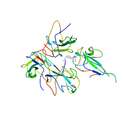
2P2F
 | | Acetyl-CoA Synthetase, wild-type with acetate, AMP, and CoA bound | | 分子名称: | ACETATE ION, ADENOSINE MONOPHOSPHATE, Acetyl-coenzyme A synthetase, ... | | 著者 | Reger, A.S, Gulick, A.M. | | 登録日 | 2007-03-07 | | 公開日 | 2007-05-29 | | 最終更新日 | 2024-03-13 | | 実験手法 | X-RAY DIFFRACTION (2.58 Å) | | 主引用文献 | Biochemical and Crystallographic Analysis of Substrate Binding and Conformational Changes in Acetyl-CoA Synthetase.
Biochemistry, 46, 2007
|
|
2OYP
 | | T Cell Immunoglobulin Mucin-3 Crystal Structure Revealed a Galectin-9-independent Binding Surface | | 分子名称: | Hepatitis A virus cellular receptor 2, SULFATE ION | | 著者 | Cao, E, Ramagopal, U.A, Fedorov, A.A, Fedorov, E.V, Nathenson, S.G, Almo, S.C. | | 登録日 | 2007-02-22 | | 公開日 | 2007-04-10 | | 最終更新日 | 2024-10-30 | | 実験手法 | X-RAY DIFFRACTION (1.952 Å) | | 主引用文献 | T cell immunoglobulin mucin-3 crystal structure reveals a galectin-9-independent ligand-binding surface
Immunity, 26, 2007
|
|
2P32
 | |
2P4S
 | | Structure of Purine Nucleoside Phosphorylase from Anopheles gambiae in complex with DADMe-ImmH | | 分子名称: | 7-[[(3R,4R)-3-(hydroxymethyl)-4-oxidanyl-pyrrolidin-1-ium-1-yl]methyl]-3,5-dihydropyrrolo[3,2-d]pyrimidin-4-one, PHOSPHATE ION, Purine nucleoside phosphorylase | | 著者 | Rinaldo-Matthis, A, Almo, S.C, Schramm, V.L. | | 登録日 | 2007-03-13 | | 公開日 | 2008-01-15 | | 最終更新日 | 2023-08-30 | | 実験手法 | X-RAY DIFFRACTION (2.2 Å) | | 主引用文献 | Anopheles gambiae purine nucleoside phosphorylase: catalysis, structure, and inhibition.
Biochemistry, 46, 2007
|
|
2OZJ
 | |
8IR0
 | |
8IQX
 | |
2P58
 | | Structure of the Yersinia pestis Type III secretion system needle protein YscF in complex with its chaperones YscE/YscG | | 分子名称: | Putative type III secretion protein YscE, Putative type III secretion protein YscF, Putative type III secretion protein YscG | | 著者 | Sun, P, Austin, B.P, Tropea, J.E, Waugh, D.S. | | 登録日 | 2007-03-14 | | 公開日 | 2008-03-04 | | 最終更新日 | 2024-11-13 | | 実験手法 | X-RAY DIFFRACTION (1.8 Å) | | 主引用文献 | Structural characterization of the Yersinia pestis type III secretion system needle protein YscF in complex with its heterodimeric chaperone YscE/YscG.
J.Mol.Biol., 377, 2008
|
|
8PVB
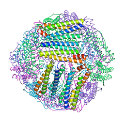
 | | Structure of GABAAR determined by cryoEM at 100 keV | | 分子名称: | 2-acetamido-2-deoxy-beta-D-glucopyranose-(1-4)-2-acetamido-2-deoxy-beta-D-glucopyranose, CHLORIDE ION, DECANE, ... | | 著者 | McMullan, G, Naydenova, K, Mihaylov, D, Peet, M.J, Wilson, H, Yamashita, K, Dickerson, J.L, Chen, S, Cannone, G, Lee, Y, Hutchings, K.A, Gittins, O, Sobhy, M, Wells, T, El-Gomati, M.M, Dalby, J, Meffert, M, Schulze-Briese, C, Henderson, R, Russo, C.J. | | 登録日 | 2023-07-17 | | 公開日 | 2023-11-29 | | 最終更新日 | 2024-10-09 | | 実験手法 | ELECTRON MICROSCOPY (3.6 Å) | | 主引用文献 | Structure determination by cryoEM at 100 keV.
Proc.Natl.Acad.Sci.USA, 120, 2023
|
|
2P0E
 | | Human nicotinamide riboside kinase 1 in complex with tiazofurin | | 分子名称: | (1R)-1-[4-(AMINOCARBONYL)-1,3-THIAZOL-2-YL]-1,4-ANHYDRO-D-RIBITOL, CHLORIDE ION, Nicotinamide riboside kinase 1, ... | | 著者 | Rabeh, W.M, Tempel, W, Nedyalkova, L, Landry, R, Arrowsmith, C.H, Edwards, A.M, Sundstrom, M, Weigelt, J, Bochkarev, A, Brenner, C, Park, H, Structural Genomics Consortium (SGC) | | 登録日 | 2007-02-28 | | 公開日 | 2007-05-08 | | 最終更新日 | 2024-11-20 | | 実験手法 | X-RAY DIFFRACTION (1.8 Å) | | 主引用文献 | Nicotinamide Riboside Kinase Structures Reveal New Pathways to NAD(+).
Plos Biol., 5, 2007
|
|
2P16
 | | Factor Xa in Complex with the Inhibitor APIXABAN (BMS-562247) AKA 1-(4-METHOXYPHENYL)-7-OXO-6-(4-(2-OXO-1-PIPERIDINYL)PHENYL)-4,5,6,7-TETRAHYDRO-1H-PYRAZOLO[3, 4-C]PYRIDINE-3-CARBOXAMIDE | | 分子名称: | 1-(4-METHOXYPHENYL)-7-OXO-6-[4-(2-OXOPIPERIDIN-1-YL)PHENYL]-4,5,6,7-TETRAHYDRO-1H-PYRAZOLO[3,4-C]PYRIDINE-3-CARBOXAMIDE, CALCIUM ION, Coagulation factor X (EC 3.4.21.6) (Stuart factor) (Stuart-Prower factor) | | 著者 | Alexander, R. | | 登録日 | 2007-03-02 | | 公開日 | 2007-10-16 | | 最終更新日 | 2024-10-09 | | 実験手法 | X-RAY DIFFRACTION (2.3 Å) | | 主引用文献 | Discovery of 1-(4-Methoxyphenyl)-7-oxo-6-(4-(2-oxopiperidin-1-yl)phenyl)-4,5,6,7-tetrahydro- 1H-pyrazolo[3,4-c]pyridine-3-carboxamide (Apixaban, BMS-562247), a Highly Potent, Selective, Efficacious, and Orally Bioavailable Inhibitor of Blood Coagulation Factor Xa.
J.Med.Chem., 50, 2007
|
|
2P1E
 | | Crystal structure of the Leishmania infantum glyoxalase II with D-Lactate at the active site | | 分子名称: | Glyoxalase II, LACTIC ACID, SPERMIDINE, ... | | 著者 | Trincao, J, Barata, L, Najmudin, S, Bonifacio, C, Romao, M.J. | | 登録日 | 2007-03-05 | | 公開日 | 2008-01-15 | | 最終更新日 | 2023-11-15 | | 実験手法 | X-RAY DIFFRACTION (1.9 Å) | | 主引用文献 | Catalysis and Structural Properties of Leishmania infantum Glyoxalase II: Trypanothione Specificity and Phylogeny.
Biochemistry, 47, 2008
|
|
2OSL
 | | Crystal structure of Rituximab Fab in complex with an epitope peptide | | 分子名称: | B-lymphocyte antigen CD20, heavy chain of the Rituximab Fab fragment,heavy chain of the Rituximab Fab fragment, light chain of the Rituximab Fab fragment,light chain of the Rituximab Fab fragment | | 著者 | Du, J, Zhong, C, Ding, J. | | 登録日 | 2007-02-06 | | 公開日 | 2007-04-10 | | 最終更新日 | 2024-10-16 | | 実験手法 | X-RAY DIFFRACTION (2.6 Å) | | 主引用文献 | Structural basis for recognition of CD20 by therapeutic antibody Rituximab
J.Biol.Chem., 282, 2007
|
|


