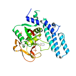
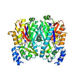
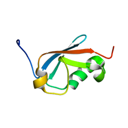
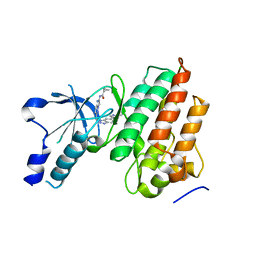
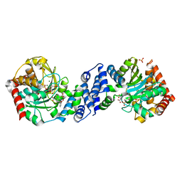
7H6G
 
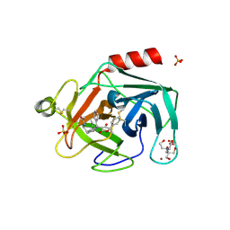 | | THE 1.21 A CRYSTAL STRUCTURE OF HUMAN CATHEPSIN G IN COMPLEX WITH N-[2-[6-fluoro-2-[(4-hydroxy-5-methyl-2-oxo-5-phenylfuran-3-yl)-phenylmethyl]-1H-indol-3-yl]ethyl]acetamide | | 分子名称: | 2-[BIS-(2-HYDROXY-ETHYL)-AMINO]-2-HYDROXYMETHYL-PROPANE-1,3-DIOL, Cathepsin G, N-(2-{6-fluoro-2-[(R)-[(5R)-4-hydroxy-5-methyl-2-oxo-5-phenyl-2,5-dihydrofuran-3-yl](phenyl)methyl]-1H-indol-3-yl}ethyl)acetamide, ... | | 著者 | Banner, D.W, Benz, J.M, Schlatter, D, Hilpert, H. | | 登録日 | 2024-04-19 | | 公開日 | 2025-03-05 | | 実験手法 | X-RAY DIFFRACTION (1.21 Å) | | 主引用文献 | Crystal structures of human Chymase and Cathepsin G
To be published
|
|
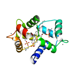
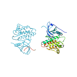
6HTR
 | | Yeast 20S proteasome with human beta2c (S171G) in complex with 13 | | 分子名称: | (2~{S})-~{N}-[(2~{S})-1-[[(2~{S})-1-[4-(aminomethyl)phenyl]-4-methylsulfonyl-butan-2-yl]amino]-3-oxidanyl-1-oxidanylidene-propan-2-yl]-2-[[(2~{S})-2-azido-3-phenyl-propanoyl]amino]-4-methyl-pentanamide, MAGNESIUM ION, PROTEASOME SUBUNIT ALPHA TYPE-1, ... | | 著者 | Huber, E.M, Groll, M. | | 登録日 | 2018-10-04 | | 公開日 | 2019-01-30 | | 最終更新日 | 2024-10-23 | | 実験手法 | X-RAY DIFFRACTION (2.6 Å) | | 主引用文献 | Structure-Based Design of Inhibitors Selective for Human Proteasome beta 2c or beta 2i Subunits.
J.Med.Chem., 62, 2019
|
|
4JH2
 | | Crystal Structure of FosB from Bacillus cereus with Zinc and Sulfate at 1.27 A Resolution | | 分子名称: | FORMIC ACID, MAGNESIUM ION, Metallothiol transferase FosB, ... | | 著者 | Thompson, M.K, Harp, J, Keithly, M.E, Jagessar, K, Cook, P.D, Armstrong, R.N. | | 登録日 | 2013-03-04 | | 公開日 | 2013-10-02 | | 最終更新日 | 2024-02-28 | | 実験手法 | X-RAY DIFFRACTION (1.27 Å) | | 主引用文献 | Structural and Chemical Aspects of Resistance to the Antibiotic Fosfomycin Conferred by FosB from Bacillus cereus.
Biochemistry, 52, 2013
|
|
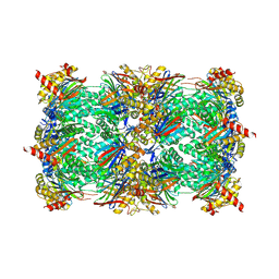
6HW3
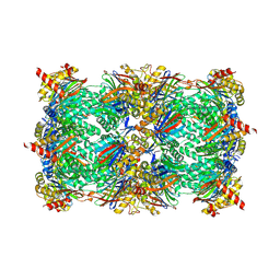 | | Yeast 20S proteasome in complex with 13 | | 分子名称: | (2~{S})-~{N}-[(2~{S})-1-[[(2~{S})-1-[4-(aminomethyl)phenyl]-4-methylsulfonyl-butan-2-yl]amino]-3-oxidanyl-1-oxidanylidene-propan-2-yl]-2-[[(2~{S})-2-azido-3-phenyl-propanoyl]amino]-4-methyl-pentanamide, CHLORIDE ION, MAGNESIUM ION, ... | | 著者 | Huber, E.M, Groll, M. | | 登録日 | 2018-10-11 | | 公開日 | 2019-01-30 | | 最終更新日 | 2024-10-09 | | 実験手法 | X-RAY DIFFRACTION (2.6 Å) | | 主引用文献 | Structure-Based Design of Inhibitors Selective for Human Proteasome beta 2c or beta 2i Subunits.
J.Med.Chem., 62, 2019
|
|
5XSU
 | | novel orally efficacious inhibitors complexed with PARP1 | | 分子名称: | 6-fluoranyl-2-(4,5,6,7-tetrahydrofuro[2,3-c]pyridin-2-yl)-1~{H}-benzimidazole-4-carboxamide, Poly [ADP-ribose] polymerase 1, SULFATE ION | | 著者 | Liu, Q, Xu, Y. | | 登録日 | 2017-06-15 | | 公開日 | 2018-04-25 | | 最終更新日 | 2025-09-17 | | 実験手法 | X-RAY DIFFRACTION (2.4 Å) | | 主引用文献 | Design and synthesis of 2-(4,5,6,7-tetrahydrothienopyridin-2-yl)-benzoimidazole carboxamides as novel orally efficacious Poly(ADP-ribose)polymerase (PARP) inhibitors
Eur J Med Chem, 145, 2018
|
|
3I5N
 | | Crystal structure of c-Met with triazolopyridazine inhibitor 13 | | 分子名称: | 7-methoxy-N-[(6-phenyl[1,2,4]triazolo[4,3-b]pyridazin-3-yl)methyl]-1,5-naphthyridin-4-amine, Hepatocyte growth factor receptor | | 著者 | Bellon, S.F, Whittington, D.A, Long, A.M, Boezio, A.A. | | 登録日 | 2009-07-06 | | 公開日 | 2010-01-12 | | 最終更新日 | 2023-09-06 | | 実験手法 | X-RAY DIFFRACTION (2 Å) | | 主引用文献 | Discovery and optimization of potent and selective triazolopyridazine series of c-Met inhibitors
Bioorg.Med.Chem.Lett., 19, 2009
|
|
7EJV
 | | The co-crystal structure of DYRK2 with YK-2-69 | | 分子名称: | Dual specificity tyrosine-phosphorylation-regulated kinase 2, [6-[[4-[2-(dimethylamino)-1,3-benzothiazol-6-yl]-5-fluoranyl-pyrimidin-2-yl]amino]pyridin-3-yl]-(4-ethylpiperazin-1-yl)methanone | | 著者 | Li, Z, Xiao, Y, Yuan, K, Kuang, W, Xiuquan, Y, Yang, P. | | 登録日 | 2021-04-02 | | 公開日 | 2022-04-06 | | 最終更新日 | 2024-11-13 | | 実験手法 | X-RAY DIFFRACTION (2.5 Å) | | 主引用文献 | Targeting dual-specificity tyrosine phosphorylation-regulated kinase 2 with a highly selective inhibitor for the treatment of prostate cancer.
Nat Commun, 13, 2022
|
|
3HLA
 | |
4JAO
 | |
3V7R
 | | Crystal structure of Staphylococcus aureus biotin protein ligase in complex with inhibitor | | 分子名称: | (3aS,4S,6aR)-4-(5-{1-[4-(6-amino-9H-purin-9-yl)butyl]-1H-1,2,3-triazol-4-yl}pentyl)tetrahydro-1H-thieno[3,4-d]imidazol-2(3H)-one, Biotin ligase | | 著者 | Yap, M.Y, Pendini, N.R. | | 登録日 | 2011-12-21 | | 公開日 | 2012-12-26 | | 最終更新日 | 2023-11-08 | | 実験手法 | X-RAY DIFFRACTION (2.61 Å) | | 主引用文献 | Selective inhibition of biotin protein ligase from Staphylococcus aureus.
J.Biol.Chem., 287, 2012
|
|
9IZO
 | | Apg mutant enzyme D336A of acarbose hydrolase from human gut flora K. grimontii TD1, complex with acarviosine | | 分子名称: | (2~{S},3~{R},4~{S},5~{R},6~{R})-5-[[(1~{S},4~{R},5~{S},6~{S})-3-(hydroxymethyl)-4,5,6-tris(oxidanyl)cyclohex-2-en-1-yl]amino]-6-methyl-oxane-2,3,4-triol, Maltodextrin glucosidase | | 著者 | Zhou, J.H, Huang, J.Y. | | 登録日 | 2024-08-01 | | 公開日 | 2025-08-06 | | 最終更新日 | 2025-09-03 | | 実験手法 | X-RAY DIFFRACTION (2.13 Å) | | 主引用文献 | Molecular insights of acarbose metabolization catalyzed by acarbose-preferred glucosidase.
Nat Commun, 16, 2025
|
|
4OWZ
 | |
6KTV
 | | The structure of EanB complex with hercynine and persulfided Cys412 | | 分子名称: | 1,2-ETHANEDIOL, 1,3-PROPANDIOL, CHLORIDE ION, ... | | 著者 | Wu, L, Liu, P.H, Zhou, J.H. | | 登録日 | 2019-08-29 | | 公開日 | 2020-08-26 | | 最終更新日 | 2024-11-13 | | 実験手法 | X-RAY DIFFRACTION (2.2 Å) | | 主引用文献 | Single-Step Replacement of an Unreactive C-H Bond by a C-S Bond Using Polysulfide as the Direct Sulfur Source in the Anaerobic Ergothioneine Biosynthesis
Acs Catalysis, 10, 2020
|
|
6KUZ
 | | E.coli beta-galactosidase (E537Q) in complex with fluorescent probe KSL01 | | 分子名称: | 3-(1,3-benzothiazol-2-yl)-2-[[4-[(2~{S},3~{R},4~{S},5~{R},6~{R})-6-(hydroxymethyl)-3,4,5-tris(oxidanyl)oxan-2-yl]oxyphenyl]methoxy]-5-methyl-benzaldehyde, Beta-galactosidase, DIMETHYL SULFOXIDE, ... | | 著者 | Chen, X, Hu, Y.L, Li, X.K, Guo, Y, Li, J. | | 登録日 | 2019-09-03 | | 公開日 | 2020-07-08 | | 最終更新日 | 2023-11-22 | | 実験手法 | X-RAY DIFFRACTION (2.83 Å) | | 主引用文献 | First-generation species-selective chemical probes for fluorescence imaging of human senescence-associated beta-galactosidase.
Chem Sci, 11, 2020
|
|
5QIL
 | |
5XQM
 | |
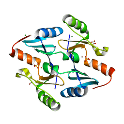
3IF7
 | | Structure of Calmodulin complexed with its first endogenous inhibitor, sphingosylphosphorylcholine | | 分子名称: | 2-{[(R)-{[(2S,3R,4E)-2-amino-3-hydroxyoctadec-4-en-1-yl]oxy}(hydroxy)phosphoryl]oxy}-N,N,N-trimethylethanaminium, CALCIUM ION, Calmodulin | | 著者 | Kovacs, E, Harmat, V, Toth, J, Vertessy, B.G, Modos, K, Kardos, J, Liliom, K. | | 登録日 | 2009-07-24 | | 公開日 | 2010-06-30 | | 最終更新日 | 2023-11-01 | | 実験手法 | X-RAY DIFFRACTION (1.6 Å) | | 主引用文献 | Structure and mechanism of calmodulin binding to a signaling sphingolipid reveal new aspects of lipid-protein interactions
Faseb J., 24, 2010
|
|
1MMD
 | | TRUNCATED HEAD OF MYOSIN FROM DICTYOSTELIUM DISCOIDEUM COMPLEXED WITH MGADP-BEF3 | | 分子名称: | ADENOSINE-5'-DIPHOSPHATE, BERYLLIUM TRIFLUORIDE ION, MAGNESIUM ION, ... | | 著者 | Fisher, A.J, Holden, H.M, Rayment, I. | | 登録日 | 1995-03-21 | | 公開日 | 1996-08-17 | | 最終更新日 | 2024-02-14 | | 実験手法 | X-RAY DIFFRACTION (2 Å) | | 主引用文献 | X-ray structures of the myosin motor domain of Dictyostelium discoideum complexed with MgADP.BeFx and MgADP.AlF4-.
Biochemistry, 34, 1995
|
|
2EHG
 | |
5UAD
 | |
4JQ8
 | | Crystal structure of EGFR kinase domain in complex with compound 4b | | 分子名称: | Epidermal growth factor receptor, N-[3-(4-{[(1S)-2-hydroxy-1-phenylethyl]amino}-6-phenylfuro[2,3-d]pyrimidin-5-yl)phenyl]-N~3~,N~3~-dimethyl-beta-alaninamide | | 著者 | Peng, Y.H, Wu, J.S. | | 登録日 | 2013-03-20 | | 公開日 | 2013-06-19 | | 最終更新日 | 2023-11-08 | | 実験手法 | X-RAY DIFFRACTION (2.83 Å) | | 主引用文献 | Protein Kinase Inhibitor Design by Targeting the Asp-Phe-Gly (DFG) Motif: The Role of the DFG Motif in the Design of Epidermal Growth Factor Receptor Inhibitors
J.Med.Chem., 56, 2013
|
|
3ILA
 | |
4JR3
 | | Crystal structure of EGFR kinase domain in complex with compound 3g | | 分子名称: | Epidermal growth factor receptor, N-[3-(4-{[(1S)-2-hydroxy-1-phenylethyl]amino}-6-phenylfuro[2,3-d]pyrimidin-5-yl)phenyl]acetamide | | 著者 | Peng, Y.H, Wu, J.S. | | 登録日 | 2013-03-21 | | 公開日 | 2013-06-19 | | 最終更新日 | 2023-11-08 | | 実験手法 | X-RAY DIFFRACTION (2.7 Å) | | 主引用文献 | Protein Kinase Inhibitor Design by Targeting the Asp-Phe-Gly (DFG) Motif: The Role of the DFG Motif in the Design of Epidermal Growth Factor Receptor Inhibitors
J.Med.Chem., 56, 2013
|
|
4ZOF
 | |
1M7E
 | | Crystal structure of the phosphotyrosine binding domain(PTB) of mouse Disabled 2(Dab2):implications for Reeling signaling | | 分子名称: | Disabled homolog 2, NGYENPTYK peptide | | 著者 | Yun, M, Keshvara, L, Park, C.-G, Zhang, Y.-M, Dickerson, J.B, Zheng, J, Rock, C.O, Curran, T, Park, H.-W. | | 登録日 | 2002-07-19 | | 公開日 | 2003-08-05 | | 最終更新日 | 2024-05-22 | | 実験手法 | X-RAY DIFFRACTION (2.45 Å) | | 主引用文献 | Crystal structures of the Dab homology domains of mouse disabled 1 and 2
J.Biol.Chem., 278, 2003
|
|


