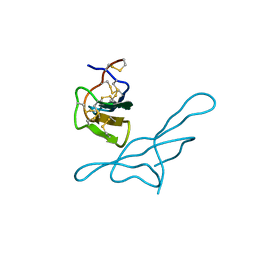
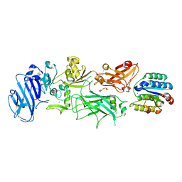
1SGE
 
 | | GLU 18 VARIANT OF TURKEY OVOMUCOID INHIBITOR THIRD DOMAIN COMPLEXED WITH STREPTOMYCES GRISEUS PROTEINASE B AT PH 6.5 | | 分子名称: | Ovomucoid, PHOSPHATE ION, Streptogrisin B | | 著者 | Huang, K, Lu, W, Anderson, S, Laskowski Jr, M, James, M.N.G. | | 登録日 | 1999-03-25 | | 公開日 | 2003-08-26 | | 最終更新日 | 2023-08-23 | | 実験手法 | X-RAY DIFFRACTION (1.8 Å) | | 主引用文献 | Recruitment of a Buried K+ Ion to Stabilize the Negative Charge of Ionized P1 in the Hydrophobic Pocket: Crystal Structures of Glu18, Gln18, Asp18 and Asn18 Variants of Turkey Ovomucoid Inhibitor Third Domain Complexed with Streptomyces griseus Protease B at Various pHs
To be Published
|
|
1SGQ
 | |
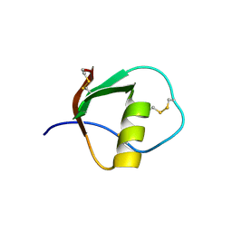
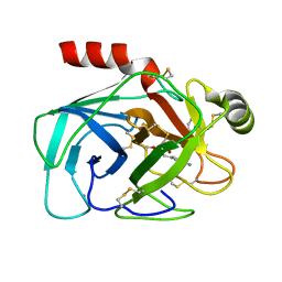
1SGD
 | | ASP 18 VARIANT OF TURKEY OVOMUCOID INHIBITOR THIRD DOMAIN COMPLEXED WITH STREPTOMYCES GRISEUS PROTEINASE B AT PH 6.5 | | 分子名称: | Ovomucoid, PHOSPHATE ION, Streptogrisin B | | 著者 | Huang, K, Lu, W, Anderson, S, Laskowski Jr, M, James, M.N.G. | | 登録日 | 1999-03-25 | | 公開日 | 2003-08-26 | | 最終更新日 | 2023-08-23 | | 実験手法 | X-RAY DIFFRACTION (1.8 Å) | | 主引用文献 | Recruitment of a Buried K+ Ion to Stabilize the Negative Charge of Ionized P1 in the Hydrophobic Pocket: Crystal Structures of Glu18, Gln18, Asp18 and Asn18 Variants of Turkey Ovomucoid Inhibitor Third Domain Complexed with Streptomyces griseus Protease B at Various pH's
To be Published
|
|
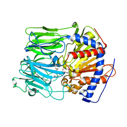
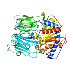
1FXY
 | | COAGULATION FACTOR XA-TRYPSIN CHIMERA INHIBITED WITH D-PHE-PRO-ARG-CHLOROMETHYLKETONE | | 分子名称: | COAGULATION FACTOR XA-TRYPSIN CHIMERA, D-phenylalanyl-N-[(2S,3S)-6-{[amino(iminio)methyl]amino}-1-chloro-2-hydroxyhexan-3-yl]-L-prolinamide | | 著者 | Hopfner, K.P, Kopetzki, E, Kresse, G.-B, Huber, R, Bode, W, Engh, R.A. | | 登録日 | 1998-04-22 | | 公開日 | 1998-06-17 | | 最終更新日 | 2013-02-27 | | 実験手法 | X-RAY DIFFRACTION (2.15 Å) | | 主引用文献 | New enzyme lineages by subdomain shuffling.
Proc.Natl.Acad.Sci.USA, 95, 1998
|
|
3G2G
 | | S437Y Mutant of human muscle pyruvate kinase, isoform M2 | | 分子名称: | Pyruvate kinase isozymes M1/M2, SULFATE ION, UNKNOWN ATOM OR ION | | 著者 | Hong, B, Dimov, S, Allali-Hassani, A, Tempel, W, MacKenzie, F, Arrowsmith, C.H, Edwards, A.M, Bountra, c, Weigelt, J, Bochkarev, A, Vedadi, M, Park, H, Structural Genomics Consortium (SGC) | | 登録日 | 2009-01-31 | | 公開日 | 2009-03-03 | | 最終更新日 | 2023-09-06 | | 実験手法 | X-RAY DIFFRACTION (2 Å) | | 主引用文献 | S437Y Mutant of human muscle pyruvate kinase, isoform M2
To be Published
|
|
1R8N
 | |
1GNV
 | |
1UOP
 | |
1GNS
 | | SUBTILISIN BPN' | | 分子名称: | ACETONE, SUBTILISIN BPN' | | 著者 | Almog, O, Gallagher, D.T, Ladner, J.E, Strausberg, S, Alexander, P. | | 登録日 | 2001-10-06 | | 公開日 | 2002-06-27 | | 最終更新日 | 2023-12-13 | | 実験手法 | X-RAY DIFFRACTION (1.8 Å) | | 主引用文献 | Structural Basis of Thermostability. Analysis of Stabilizing Mutations in Subtilisin Bpn'.
J.Biol.Chem., 277, 2002
|
|
1V6D
 | |
1TUS
 | |
1TZN
 | | Crystal Structure of the Anthrax Toxin Protective Antigen Heptameric Prepore bound to the VWA domain of CMG2, an anthrax toxin receptor | | 分子名称: | Anthrax toxin receptor 2, CALCIUM ION, MAGNESIUM ION, ... | | 著者 | Lacy, D.B, Wigelsworth, D.J, Melnyk, R.A, Collier, R.J. | | 登録日 | 2004-07-10 | | 公開日 | 2004-08-17 | | 最終更新日 | 2024-04-03 | | 実験手法 | X-RAY DIFFRACTION (4.3 Å) | | 主引用文献 | Structure of heptameric protective antigen bound to an anthrax toxin receptor: A role for receptor in pH-dependent pore formation
Proc.Natl.Acad.Sci.USA, 101, 2004
|
|
1TZO
 | | Crystal Structure of the Anthrax Toxin Protective Antigen Heptameric Prepore | | 分子名称: | CALCIUM ION, Protective antigen | | 著者 | Lacy, D.B, Wigelsworth, D.J, Melnyk, R.A, Collier, R.J. | | 登録日 | 2004-07-10 | | 公開日 | 2004-08-17 | | 最終更新日 | 2024-04-03 | | 実験手法 | X-RAY DIFFRACTION (3.6 Å) | | 主引用文献 | Structure of heptameric protective antigen bound to an anthrax toxin receptor: A role for receptor in pH-dependent pore formation
Proc.Natl.Acad.Sci.USA, 101, 2004
|
|
1H34
 | | Crystal structure of lima bean trypsin inhibitor | | 分子名称: | BOWMAN-BIRK TYPE PROTEINASE INHIBITOR | | 著者 | Debreczeni, J.E, Bunkoczi, G, Girmann, B, Sheldrick, G.M. | | 登録日 | 2002-08-21 | | 公開日 | 2003-02-06 | | 最終更新日 | 2019-07-24 | | 実験手法 | X-RAY DIFFRACTION (2.04 Å) | | 主引用文献 | In-House Phase Determination of the Lima Bean Trypsin Inhibitor: A Low-Resolution Sulfur-Sad Case
Acta Crystallogr.,Sect.D, 59, 2003
|
|
1RR9
 | | Catalytic domain of E.coli Lon protease | | 分子名称: | ATP-dependent protease La, SULFATE ION | | 著者 | Botos, I, Melnikov, E.E, Cherry, S, Tropea, J.E, Khalatova, A.G, Dauter, Z, Maurizi, M.R, Rotanova, T.V, Wlodawer, A, Gustchina, A. | | 登録日 | 2003-12-08 | | 公開日 | 2003-12-23 | | 最終更新日 | 2021-10-27 | | 実験手法 | X-RAY DIFFRACTION (2.1 Å) | | 主引用文献 | The catalytic domain of Escherichia coli Lon protease has a unique fold and a Ser-Lys dyad in the active site
J.Biol.Chem., 279, 2004
|
|
1UOQ
 | |
1UOO
 | |
1TAB
 | | STRUCTURE OF THE TRYPSIN-BINDING DOMAIN OF BOWMAN-BIRK TYPE PROTEASE INHIBITOR AND ITS INTERACTION WITH TRYPSIN | | 分子名称: | BOWMAN-BIRK TYPE PROTEINASE INHIBITOR, TRYPSIN | | 著者 | Tsunogae, Y, Tanaka, I, Yamane, T, Kikkawa, J.-I, Ashida, T, Ishikawa, C, Watanabe, K, Nakamura, S, Takahashi, K. | | 登録日 | 1990-10-15 | | 公開日 | 1992-01-15 | | 最終更新日 | 2024-06-05 | | 実験手法 | X-RAY DIFFRACTION (2.3 Å) | | 主引用文献 | Structure of the trypsin-binding domain of Bowman-Birk type protease inhibitor and its interaction with trypsin.
J.Biochem.(Tokyo), 100, 1986
|
|
1TUR
 | | SOLUTION STRUCTURE OF TURKEY OVOMUCOID THIRD DOMAIN AS DETERMINED FROM NUCLEAR MAGNETIC RESONANCE DATA | | 分子名称: | OVOMUCOID | | 著者 | Krezel, A.M, Darba, P, Robertson, A.D, Fejzo, J, Macura, S, Markley, J.L. | | 登録日 | 1994-07-06 | | 公開日 | 1994-10-15 | | 最終更新日 | 2017-11-29 | | 実験手法 | SOLUTION NMR | | 主引用文献 | Solution structure of turkey ovomucoid third domain as determined from nuclear magnetic resonance data.
J.Mol.Biol., 242, 1994
|
|
1T6B
 | | Crystal structure of B. anthracis Protective Antigen complexed with human Anthrax toxin receptor | | 分子名称: | Anthrax toxin receptor 2, CALCIUM ION, MANGANESE (II) ION, ... | | 著者 | Santelli, E, Bankston, L.A, Leppla, S.H, Liddington, R.C. | | 登録日 | 2004-05-05 | | 公開日 | 2004-07-20 | | 最終更新日 | 2024-02-14 | | 実験手法 | X-RAY DIFFRACTION (2.5 Å) | | 主引用文献 | Crystal structure of a complex between anthrax toxin and its host cell receptor
Nature, 430, 2004
|
|
1AUJ
 | | BOVINE TRYPSIN COMPLEXED TO META-CYANO-BENZYLIC INHIBITOR | | 分子名称: | 1-{[1-(2-AMINO-3-PHENYL-PROPIONYL)-PYRROLIDINE-2-CARBONYL]-AMINO}-2-(3-CYANO-PHENYL)-ETHANEBORONIC ACID, CALCIUM ION, TRYPSIN | | 著者 | Alexander, R, Smallwood, A, Kettner, C. | | 登録日 | 1997-08-28 | | 公開日 | 1998-10-14 | | 最終更新日 | 2024-06-05 | | 実験手法 | X-RAY DIFFRACTION (2.1 Å) | | 主引用文献 | New inhibitors of thrombin and other trypsin-like proteases: hydrogen bonding of an aromatic cyano group with a backbone amide of the P1 binding site replaces binding of a basic side chain.
Biochemistry, 36, 1997
|
|
1ATA
 | |
1BIK
 | | X-RAY STRUCTURE OF BIKUNIN FROM THE HUMAN INTER-ALPHA-INHIBITOR COMPLEX | | 分子名称: | 2-acetamido-2-deoxy-beta-D-glucopyranose, BIKUNIN, SULFATE ION | | 著者 | Xu, Y, Carr, P.D, Guss, J.M, Ollis, D.L. | | 登録日 | 1997-11-26 | | 公開日 | 1999-03-16 | | 最終更新日 | 2023-08-02 | | 実験手法 | X-RAY DIFFRACTION (2.5 Å) | | 主引用文献 | The crystal structure of bikunin from the inter-alpha-inhibitor complex: a serine protease inhibitor with two Kunitz domains.
J.Mol.Biol., 276, 1998
|
|
1ATB
 | |
1ATD
 | |


