5AAN
 
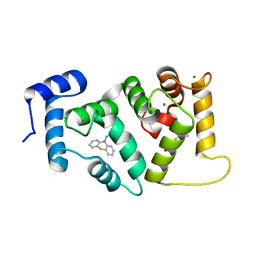 | | Crystal structure of Drosophila NCS-1 bound to penothiazine FD44 | | 分子名称: | 2-(2-METHOXYETHOXY)ETHANOL, CALCIUM ION, CG5907-PA, ... | | 著者 | Chaves-Sanjuan, A, Infantes, L, Sanchez-Barrena, M.J. | | 登録日 | 2015-07-27 | | 公開日 | 2017-01-25 | | 最終更新日 | 2024-01-10 | | 実験手法 | X-RAY DIFFRACTION (1.6 Å) | | 主引用文献 | Interference of the complex between NCS-1 and Ric8a with phenothiazines regulates synaptic function and is an approach for fragile X syndrome.
Proc. Natl. Acad. Sci. U.S.A., 114, 2017
|
|
2BD1
 
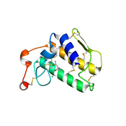 | | A possible role of the second calcium ion in interfacial binding: Atomic and medium resolution crystal structures of the quadruple mutant of phospholipase A2 | | 分子名称: | (4S)-2-METHYL-2,4-PENTANEDIOL, CALCIUM ION, Phospholipase A2 | | 著者 | Sekar, K, Velmurugan, D, Tsai, M.D. | | 登録日 | 2005-10-19 | | 公開日 | 2006-07-04 | | 最終更新日 | 2023-08-23 | | 実験手法 | X-RAY DIFFRACTION (1.9 Å) | | 主引用文献 | Suggestive evidence for the involvement of the second calcium and surface loop in interfacial binding: monoclinic and trigonal crystal structures of a quadruple mutant of phospholipase A(2).
Acta Crystallogr.,Sect.D, 62, 2006
|
|
2BCH
 
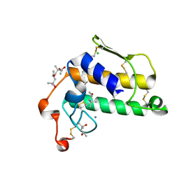 | | A possible of Second calcium ion in interfacial binding: Atomic and Medium resolution crystal structures of the quadruple mutant of phospholipase A2 | | 分子名称: | (4S)-2-METHYL-2,4-PENTANEDIOL, CALCIUM ION, CHLORIDE ION, ... | | 著者 | Sekar, K, Yogavel, M, Velmurugan, D, Poi, M.J, Dauter, Z, Tsai, M.D. | | 登録日 | 2005-10-19 | | 公開日 | 2006-07-04 | | 最終更新日 | 2023-08-23 | | 実験手法 | X-RAY DIFFRACTION (1.1 Å) | | 主引用文献 | Suggestive evidence for the involvement of the second calcium and surface loop in interfacial binding: monoclinic and trigonal crystal structures of a quadruple mutant of phospholipase A(2).
Acta Crystallogr.,Sect.D, 62, 2006
|
|
2BCN
 
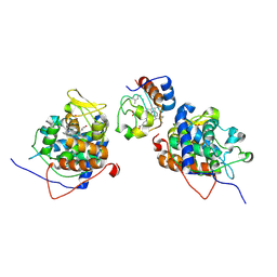 | |
6RK5
 
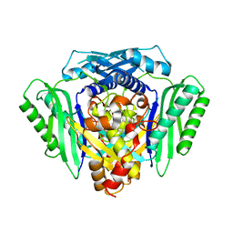 | | Inter-dimeric interface controls function and stability of S-methionine adenosyltransferase from U. urealiticum | | 分子名称: | Methionine adenosyltransferase | | 著者 | Shahar, A, Zarivach, R, Bershtein, S, Kleiner, D, Shmulevich, F. | | 登録日 | 2019-04-30 | | 公開日 | 2019-09-25 | | 最終更新日 | 2024-01-24 | | 実験手法 | X-RAY DIFFRACTION (2.6 Å) | | 主引用文献 | The interdimeric interface controls function and stability of Ureaplasma urealiticum methionine S-adenosyltransferase.
J.Mol.Biol., 431, 2019
|
|
6RJS
 
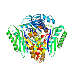 | | Inter-dimeric interface controls function and stability of S-methionine adenosyltransferase from U. urealiticum | | 分子名称: | Methionine adenosyltransferase | | 著者 | Shahar, A, Zarivach, R, Bershtein, S, Kleiner, D, Shmulevich, F. | | 登録日 | 2019-04-29 | | 公開日 | 2019-09-25 | | 最終更新日 | 2024-01-24 | | 実験手法 | X-RAY DIFFRACTION (2.6 Å) | | 主引用文献 | The interdimeric interface controls function and stability of Ureaplasma urealiticum methionine S-adenosyltransferase.
J.Mol.Biol., 431, 2019
|
|
6RK7
 
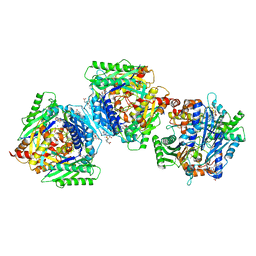 | | Inter-dimeric interface controls function and stability of S-methionine adenosyltransferase from U. urealiticum | | 分子名称: | CHLORIDE ION, Methionine adenosyltransferase, S-ADENOSYLMETHIONINE | | 著者 | Shahar, A, Zarivach, R, Bershtein, S, Kleiner, D, Shmulevich, F. | | 登録日 | 2019-04-30 | | 公開日 | 2019-09-25 | | 最終更新日 | 2024-01-24 | | 実験手法 | X-RAY DIFFRACTION (1.8 Å) | | 主引用文献 | The interdimeric interface controls function and stability of Ureaplasma urealiticum methionine S-adenosyltransferase.
J.Mol.Biol., 431, 2019
|
|
6RKC
 
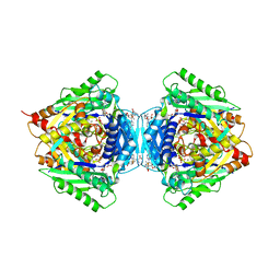 | | Inter-dimeric interface controls function and stability of S-methionine adenosyltransferase from U. urealiticum | | 分子名称: | (DIPHOSPHONO)AMINOPHOSPHONIC ACID, MAGNESIUM ION, Methionine adenosyltransferase, ... | | 著者 | Shahar, A, Zarivach, R, Bershtein, S, Kleiner, D, Shmulevich, F. | | 登録日 | 2019-04-30 | | 公開日 | 2019-09-25 | | 最終更新日 | 2024-01-24 | | 実験手法 | X-RAY DIFFRACTION (2.56 Å) | | 主引用文献 | The interdimeric interface controls function and stability of Ureaplasma urealiticum methionine S-adenosyltransferase.
J.Mol.Biol., 431, 2019
|
|
3P56
 
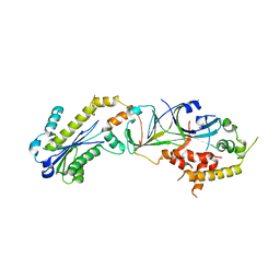 | |
6RKA
 
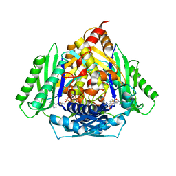 | | Inter-dimeric interface controls function and stability of S-methionine adenosyltransferase from U. urealiticum | | 分子名称: | ADENOSINE-5'-TRIPHOSPHATE, Methionine adenosyltransferase, PHOSPHATE ION, ... | | 著者 | Shahar, A, Zarivach, R, Bershtein, S, Kleiner, D, Shmulevich, F. | | 登録日 | 2019-04-30 | | 公開日 | 2019-09-25 | | 最終更新日 | 2024-01-24 | | 実験手法 | X-RAY DIFFRACTION (2.5 Å) | | 主引用文献 | The interdimeric interface controls function and stability of Ureaplasma urealiticum methionine S-adenosyltransferase.
J.Mol.Biol., 431, 2019
|
|
3P5J
 
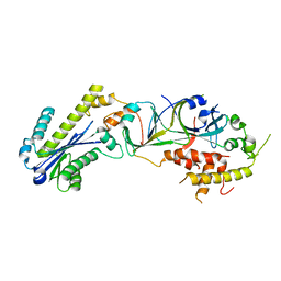 | |
4JMF
 
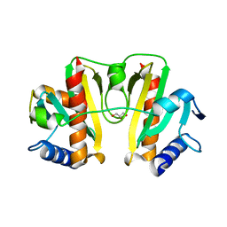 | |
2M2D
 
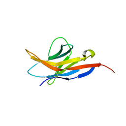 | | Human programmed cell death 1 receptor | | 分子名称: | Programmed cell death protein 1 | | 著者 | Veverka, V, Cheng, X, Waters, L.C, Muskett, F.W, Morgan, S, Lesley, A, Griffiths, M, Stubberfield, C, Griffin, R, Henry, A.J, Robinson, M.K, Jansson, A, Ladbury, J.E, Ikemizu, S, Davis, S.J, Carr, M.D. | | 登録日 | 2012-12-18 | | 公開日 | 2013-02-27 | | 最終更新日 | 2013-05-15 | | 実験手法 | SOLUTION NMR | | 主引用文献 | Structure and interactions of the human programmed cell death 1 receptor.
J.Biol.Chem., 288, 2013
|
|
5TGL
 
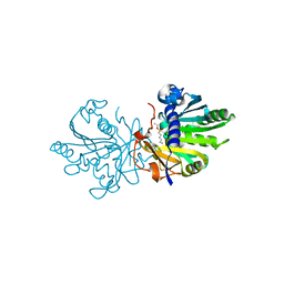 | | A MODEL FOR INTERFACIAL ACTIVATION IN LIPASES FROM THE STRUCTURE OF A FUNGAL LIPASE-INHIBITOR COMPLEX | | 分子名称: | LIPASE, N-HEXYLPHOSPHONATE ETHYL ESTER | | 著者 | Brzozowski, A.M, Derewenda, U, Derewenda, Z.S, Dodson, G.G, Lawson, D, Turkenburg, J.P, Bjorkling, F, Huge-Jensen, B, Patkar, S.R, Thim, L. | | 登録日 | 1991-10-30 | | 公開日 | 1994-01-31 | | 最終更新日 | 2024-03-06 | | 実験手法 | X-RAY DIFFRACTION (3 Å) | | 主引用文献 | A model for interfacial activation in lipases from the structure of a fungal lipase-inhibitor complex.
Nature, 351, 1991
|
|
2EYN
 
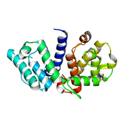 | | Crystal structure of the actin-binding domain of human alpha-actinin 1 at 1.8 Angstrom resolution | | 分子名称: | Alpha-actinin 1 | | 著者 | Borrego-Diaz, E, Kerff, F, Lee, S.H, Ferron, F, Li, Y, Dominguez, R. | | 登録日 | 2005-11-09 | | 公開日 | 2006-08-29 | | 最終更新日 | 2023-08-23 | | 実験手法 | X-RAY DIFFRACTION (1.8 Å) | | 主引用文献 | Crystal structure of the actin-binding domain of alpha-actinin 1: Evaluating two competing actin-binding models.
J.Struct.Biol., 155, 2006
|
|
2EYI
 
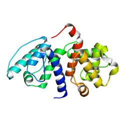 | | Crystal structure of the actin-binding domain of human alpha-actinin 1 at 1.7 Angstrom resolution | | 分子名称: | Alpha-actinin 1 | | 著者 | Borrego-Diaz, E, Kerff, F, Lee, S.H, Ferron, F, Li, Y, Dominguez, R. | | 登録日 | 2005-11-09 | | 公開日 | 2006-08-29 | | 最終更新日 | 2023-08-23 | | 実験手法 | X-RAY DIFFRACTION (1.7 Å) | | 主引用文献 | Crystal structure of the actin-binding domain of alpha-actinin 1: Evaluating two competing actin-binding models.
J.Struct.Biol., 155, 2006
|
|
3TVL
 
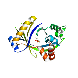 | | Complex between the human thiamine triphosphatase and triphosphate | | 分子名称: | 1,2-ETHANEDIOL, TRIPHOSPHATE, Thiamine-triphosphatase | | 著者 | Delvaux, D, Herman, R, Sauvage, E, Wins, P, Bettendorff, L, Charlier, P, Kerff, F. | | 登録日 | 2011-09-20 | | 公開日 | 2012-10-10 | | 最終更新日 | 2023-09-13 | | 実験手法 | X-RAY DIFFRACTION (2.3 Å) | | 主引用文献 | Structural determinants of specificity and catalytic mechanism in mammalian 25-kDa thiamine triphosphatase.
Biochim.Biophys.Acta, 1830, 2013
|
|
5MP6
 
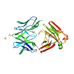 | | Structure of the Unliganded Fab from HIV-1 Neutralizing Antibody CAP248-2B that Binds to the gp120 C-terminus - gp41 Interface, at two Angstrom resolution. | | 分子名称: | CAP248-2B Heavy Chain, CAP248-2B Light Chain, SULFATE ION | | 著者 | Wibmer, C.K, Gorman, J, Kwong, P.D. | | 登録日 | 2016-12-15 | | 公開日 | 2016-12-28 | | 最終更新日 | 2024-01-17 | | 実験手法 | X-RAY DIFFRACTION (1.959 Å) | | 主引用文献 | Structure and Recognition of a Novel HIV-1 gp120-gp41 Interface Antibody that Caused MPER Exposure through Viral Escape.
PLoS Pathog., 13, 2017
|
|
1ZM0
 
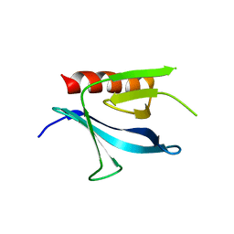 | | Crystal Structure of the Carboxyl Terminal PH Domain of Pleckstrin To 2.1 Angstroms | | 分子名称: | Pleckstrin | | 著者 | Jackson, S.G, Zhang, Y, Zhang, K, Summerfield, R, Haslam, R.J, Junop, M.S. | | 登録日 | 2005-05-09 | | 公開日 | 2006-02-28 | | 最終更新日 | 2024-02-14 | | 実験手法 | X-RAY DIFFRACTION (2.1 Å) | | 主引用文献 | Structure of the carboxy-terminal PH domain of pleckstrin at 2.1 Angstroms.
Acta Crystallogr.,Sect.D, 62, 2006
|
|
5OUP
 
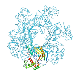 | | Structure of TgPLP1 MACPF domain | | 分子名称: | 2-acetamido-2-deoxy-beta-D-glucopyranose, Perforin-like protein 1 | | 著者 | Ni, T, Gilbert, R.J.C. | | 登録日 | 2017-08-24 | | 公開日 | 2018-04-11 | | 最終更新日 | 2020-07-29 | | 実験手法 | X-RAY DIFFRACTION (2.03 Å) | | 主引用文献 | Structures of monomeric and oligomeric forms of theToxoplasma gondiiperforin-like protein 1.
Sci Adv, 4, 2018
|
|
5OUO
 
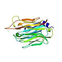 | | Structure of TgPLP1 APCbeta domain | | 分子名称: | CHLORIDE ION, MAGNESIUM ION, Perforin-like protein 1 | | 著者 | Ni, T, Gilbert, R.J.C. | | 登録日 | 2017-08-24 | | 公開日 | 2018-04-11 | | 最終更新日 | 2019-10-16 | | 実験手法 | X-RAY DIFFRACTION (1.11 Å) | | 主引用文献 | Structures of monomeric and oligomeric forms of theToxoplasma gondiiperforin-like protein 1.
Sci Adv, 4, 2018
|
|
5OUQ
 
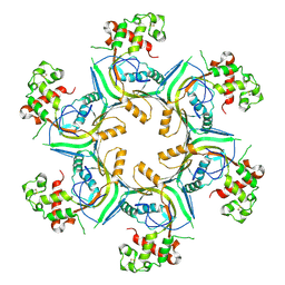 | | Structure of TgPLP1 MACPF domain | | 分子名称: | 2-acetamido-2-deoxy-beta-D-glucopyranose, Perforin-like protein 1 | | 著者 | Ni, T, Gilbert, R.J.C. | | 登録日 | 2017-08-24 | | 公開日 | 2018-04-11 | | 最終更新日 | 2024-01-17 | | 実験手法 | X-RAY DIFFRACTION (5.11 Å) | | 主引用文献 | Structures of monomeric and oligomeric forms of theToxoplasma gondiiperforin-like protein 1.
Sci Adv, 4, 2018
|
|
5OWN
 
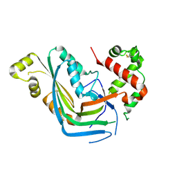 | | Structure of TgPLP1 MACPF domain | | 分子名称: | 2-acetamido-2-deoxy-beta-D-glucopyranose, Perforin-like protein 1 | | 著者 | Ni, T, Gilbert, R.J.C. | | 登録日 | 2017-09-01 | | 公開日 | 2018-04-11 | | 最終更新日 | 2024-01-17 | | 実験手法 | X-RAY DIFFRACTION (3.11 Å) | | 主引用文献 | Structures of monomeric and oligomeric forms of theToxoplasma gondiiperforin-like protein 1.
Sci Adv, 4, 2018
|
|
4NI2
 
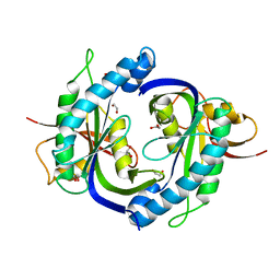 | | Crystal structure of the heterodimeric catalytic domain of wild-type human soluble guanylate cyclase | | 分子名称: | 1,2-ETHANEDIOL, Guanylate cyclase soluble subunit alpha-3, Guanylate cyclase soluble subunit beta-1 | | 著者 | Seeger, F, Williams, G.J, Tainer, J.A, Garcin, E.D. | | 登録日 | 2013-11-05 | | 公開日 | 2014-04-16 | | 最終更新日 | 2023-09-20 | | 実験手法 | X-RAY DIFFRACTION (1.9 Å) | | 主引用文献 | Interfacial residues promote an optimal alignment of the catalytic center in human soluble guanylate cyclase: heterodimerization is required but not sufficient for activity.
Biochemistry, 53, 2014
|
|
7RYD
 
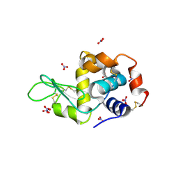 | | Hen egg-white lysozyme with ionic liquid butylammonium nitrate 1 mol% | | 分子名称: | Lysozyme C, NITRATE ION | | 著者 | Han, Q, Darmanin, C, Smith, K, Drummond, C, Greaves, T. | | 登録日 | 2021-08-25 | | 公開日 | 2023-03-01 | | 最終更新日 | 2023-10-25 | | 実験手法 | X-RAY DIFFRACTION (1.18 Å) | | 主引用文献 | Probing ion-binding at a protein interface: Modulation of protein properties by ionic liquids.
J Colloid Interface Sci, 650, 2023
|
|
