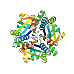
2QRX
 
 | |
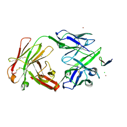
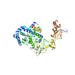
2QRY
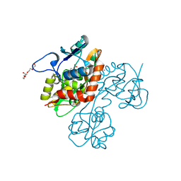
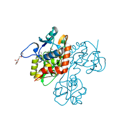
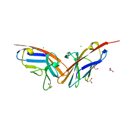
 | | Periplasmic thiamin binding protein | | 分子名称: | THIAMIN PHOSPHATE, Thiamine-binding periplasmic protein | | 著者 | Ealick, S.E, Soriano, E.V. | | 登録日 | 2007-07-30 | | 公開日 | 2008-02-05 | | 最終更新日 | 2011-07-13 | | 実験手法 | X-RAY DIFFRACTION (2.25 Å) | | 主引用文献 | Structural Similarities between Thiamin-Binding Protein and Thiaminase-I Suggest a Common Ancestor
Biochemistry, 47, 2008
|
|
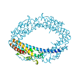
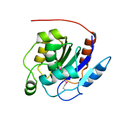
2QRZ
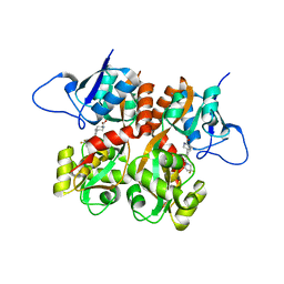
 | | Cdc42 bound to GMP-PCP: Induced Fit by Effector is Required | | 分子名称: | Cell division control protein 42 homolog precursor, MAGNESIUM ION, PHOSPHOMETHYLPHOSPHONIC ACID GUANYLATE ESTER, ... | | 著者 | Phillips, M.J, Calero, G, Chan, B, Cerione, R.A. | | 登録日 | 2007-07-30 | | 公開日 | 2008-03-18 | | 最終更新日 | 2011-07-13 | | 実験手法 | X-RAY DIFFRACTION (2.4 Å) | | 主引用文献 | Effector Proteins Exert an Important Influence on the Signaling-active State of the Small GTPase Cdc42.
J.Biol.Chem., 283, 2008
|
|
2QS1
 | | Crystal structure of the GluR5 ligand binding core dimer in complex with UBP315 at 1.80 Angstroms resolution | | 分子名称: | 3-({3-[(2S)-2-amino-2-carboxyethyl]-5-methyl-2,6-dioxo-3,6-dihydropyrimidin-1(2H)-yl}methyl)-4,5-dibromothiophene-2-carboxylic acid, CHLORIDE ION, Glutamate receptor, ... | | 著者 | Alushin, G.M, Jane, D.E, Mayer, M.L. | | 登録日 | 2007-07-30 | | 公開日 | 2008-08-05 | | 最終更新日 | 2023-08-30 | | 実験手法 | X-RAY DIFFRACTION (1.8 Å) | | 主引用文献 | Binding site and ligand flexibility revealed by high resolution crystal structures of GluK1 competitive antagonists.
Neuropharmacology, 60, 2011
|
|
2QS2
 | | Crystal structure of the GluR5 ligand binding core dimer in complex with UBP318 at 1.80 Angstroms resolution | | 分子名称: | 3-({3-[(2S)-2-amino-2-carboxyethyl]-5-bromo-2,6-dioxo-3,6-dihydropyrimidin-1(2H)-yl}methyl)thiophene-2-carboxylic acid, CHLORIDE ION, Glutamate receptor, ... | | 著者 | Alushin, G.M, Jane, D.E, Mayer, M.L. | | 登録日 | 2007-07-30 | | 公開日 | 2008-08-05 | | 最終更新日 | 2023-08-30 | | 実験手法 | X-RAY DIFFRACTION (1.8 Å) | | 主引用文献 | Binding site and ligand flexibility revealed by high resolution crystal structures of GluK1 competitive antagonists.
Neuropharmacology, 60, 2011
|
|
2QS3
 | | Crystal structure of the GluR5 ligand binding core dimer in complex with UBP316 at 1.76 Angstroms resolution | | 分子名称: | 3-({3-[(2S)-2-amino-2-carboxyethyl]-5-methyl-2,6-dioxo-3,6-dihydropyrimidin-1(2H)-yl}methyl)-5-phenylthiophene-2-carboxylic acid, CHLORIDE ION, Glutamate receptor, ... | | 著者 | Alushin, G.M, Jane, D.E, Mayer, M.L. | | 登録日 | 2007-07-30 | | 公開日 | 2008-08-05 | | 最終更新日 | 2023-08-30 | | 実験手法 | X-RAY DIFFRACTION (1.76 Å) | | 主引用文献 | ACET is a highly potent and specific kainate receptor antagonist: characterisation and effects on hippocampal mossy fibre function.
Neuropharmacology, 56, 2009
|
|
2QS4
 | | Crystal structure of the GluR5 ligand binding core dimer in complex with LY466195 at 1.58 Angstroms resolution | | 分子名称: | (3S,4aR,6S,8aR)-6-{[(2S)-2-carboxy-4,4-difluoropyrrolidin-1-yl]methyl}decahydroisoquinoline-3-carboxylic acid, AMMONIUM ION, GLYCEROL, ... | | 著者 | Alushin, G.M, Jane, D.E, Mayer, M.L. | | 登録日 | 2007-07-30 | | 公開日 | 2008-08-05 | | 最終更新日 | 2023-08-30 | | 実験手法 | X-RAY DIFFRACTION (1.58 Å) | | 主引用文献 | Binding site and ligand flexibility revealed by high resolution crystal structures of GluK1 competitive antagonists.
Neuropharmacology, 60, 2011
|
|
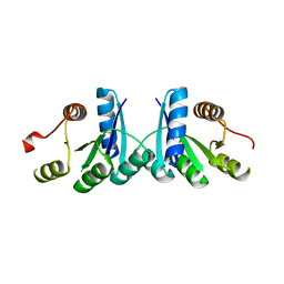
2QS6
 | | Structure of a Hoogsteen antiparallel duplex with extra-helical thymines | | 分子名称: | DNA (5'-D(*DAP*DTP*DAP*DTP*DAP*DTP*DCP*DT)-3') | | 著者 | Pous, J, Urpi, L, Subirana, J.A, Gouyette, C, Navaza, J, Campos, J.L. | | 登録日 | 2007-07-30 | | 公開日 | 2008-03-25 | | 最終更新日 | 2024-04-03 | | 実験手法 | X-RAY DIFFRACTION (3.08 Å) | | 主引用文献 | Stabilization by extra-helical thymines of a DNA duplex with Hoogsteen base pairs.
J.Am.Chem.Soc., 130, 2008
|
|
2QS7
 | |
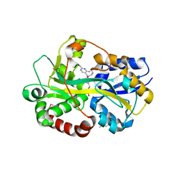
2QS8
 | | Crystal structure of a Xaa-Pro dipeptidase with bound methionine in the active site | | 分子名称: | MAGNESIUM ION, METHIONINE, Xaa-Pro Dipeptidase | | 著者 | Kumaran, D, Burley, S.K, Swaminathan, S, New York SGX Research Center for Structural Genomics (NYSGXRC) | | 登録日 | 2007-07-30 | | 公開日 | 2007-08-21 | | 最終更新日 | 2021-02-03 | | 実験手法 | X-RAY DIFFRACTION (2.33 Å) | | 主引用文献 | Functional annotation of two new carboxypeptidases from the amidohydrolase superfamily of enzymes.
Biochemistry, 48, 2009
|
|
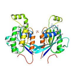
2QS9
 | | Crystal structure of the human retinoblastoma-binding protein 9 (RBBP-9). NESG target HR2978 | | 分子名称: | Retinoblastoma-binding protein 9 | | 著者 | Vorobiev, S.M, Su, M, Seetharaman, J, Kuzin, A, Chen, C.X, Cunningham, K, Owens, L, Maglaqui, M, Xiao, R, Acton, T.B, Montelione, G.T, Hunt, J.F, Tong, L, Northeast Structural Genomics Consortium (NESG) | | 登録日 | 2007-07-30 | | 公開日 | 2007-08-14 | | 最終更新日 | 2011-07-13 | | 実験手法 | X-RAY DIFFRACTION (1.72 Å) | | 主引用文献 | Crystal structure of human retinoblastoma binding protein 9.
Proteins, 74, 2008
|
|
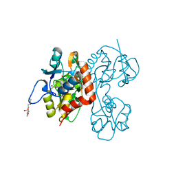
2QSA
 | | Crystal structure of J-domain of DnaJ homolog dnj-2 precursor from C.elegans. | | 分子名称: | CHLORIDE ION, DnaJ homolog dnj-2 | | 著者 | Osipiuk, J, Mulligan, R, Gu, M, Voisine, C, Morimoto, R.I, Joachimiak, A, Midwest Center for Structural Genomics (MCSG) | | 登録日 | 2007-07-30 | | 公開日 | 2007-08-14 | | 最終更新日 | 2023-08-30 | | 実験手法 | X-RAY DIFFRACTION (1.68 Å) | | 主引用文献 | X-ray crystal structure of J-domain of DnaJ homolog dnj-2 precursor from C.elegans.
To be Published
|
|
2QSB
 | |
2QSC
 | | Crystal structure analysis of anti-HIV-1 V3-Fab F425-B4e8 in complex with a V3-peptide | | 分子名称: | CHLORIDE ION, Envelope glycoprotein gp120, Fab F425-B4e8, ... | | 著者 | Bell, C.H, Schiefner, A, Stanfield, R.L, Wilson, I.A. | | 登録日 | 2007-07-30 | | 公開日 | 2008-01-15 | | 最終更新日 | 2020-07-29 | | 実験手法 | X-RAY DIFFRACTION (2.8 Å) | | 主引用文献 | Structure of antibody F425-B4e8 in complex with a V3 peptide reveals a new binding mode for HIV-1 neutralization.
J.Mol.Biol., 375, 2008
|
|
2QSD
 | | Crystal structure of a protein Il1583 from Idiomarina loihiensis | | 分子名称: | GLYCEROL, Uncharacterized conserved protein | | 著者 | Patskovsky, Y, Bonanno, J, Sauder, J.M, Romero, R, Rutter, M, Koss, J, Mckenzie, C, Gheyi, T, Bain, K, Wasserman, S.R, Burley, S.K, Almo, S.C, New York SGX Research Center for Structural Genomics (NYSGXRC) | | 登録日 | 2007-07-30 | | 公開日 | 2007-08-14 | | 最終更新日 | 2021-02-03 | | 実験手法 | X-RAY DIFFRACTION (2.5 Å) | | 主引用文献 | Crystal Structure of a Protein Il1583 from Idiomarina loihiensis.
To be Published
|
|
2QSE
 | |
2QSF
 | |
2QSG
 | |
2QSH
 | |
2QSI
 | | Crystal structure of putative hydrogenase expression/formation protein hupG from Rhodopseudomonas palustris CGA009 | | 分子名称: | 2,3-DIHYDROXY-1,4-DITHIOBUTANE, GLYCEROL, Putative hydrogenase expression/formation protein hupG | | 著者 | Nocek, B, Skarina, T, Kagan, O, Savchenko, A, Edwards, A, Joachimiak, A, Midwest Center for Structural Genomics (MCSG) | | 登録日 | 2007-07-31 | | 公開日 | 2007-08-14 | | 最終更新日 | 2011-07-13 | | 実験手法 | X-RAY DIFFRACTION (1.8 Å) | | 主引用文献 | Structure of putative hydrogenase expression/formation protein hupG from Rhodopseudomonas palustris CGA009.
To be Published
|
|
2QSJ
 | | Crystal structure of a LuxR family DNA-binding response regulator from Silicibacter pomeroyi | | 分子名称: | DNA-binding response regulator, LuxR family | | 著者 | Bonanno, J.B, Freeman, J, Bain, K.T, Mendoza, M, Romero, R, Smith, D, Wasserman, S, Sauder, J.M, Burley, S.K, Almo, S.C, New York SGX Research Center for Structural Genomics (NYSGXRC) | | 登録日 | 2007-07-31 | | 公開日 | 2007-08-14 | | 最終更新日 | 2024-02-21 | | 実験手法 | X-RAY DIFFRACTION (2.1 Å) | | 主引用文献 | Crystal structure of a LuxR family DNA-binding response regulator from Silicibacter pomeroyi.
To be Published
|
|
2QSK
 | | Atomic-resolution crystal structure of the Recombinant form of Scytovirin | | 分子名称: | CHLORIDE ION, GLYCEROL, scytovirin | | 著者 | Moulaei, T, Botos, I, Ziolkowska, N.E, Dauter, Z, Wlodawer, A. | | 登録日 | 2007-07-31 | | 公開日 | 2007-11-27 | | 最終更新日 | 2023-08-30 | | 実験手法 | X-RAY DIFFRACTION (1 Å) | | 主引用文献 | Atomic-resolution crystal structure of the antiviral lectin scytovirin.
Protein Sci., 16, 2007
|
|
2QSP
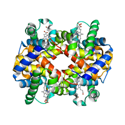 | | Bovine Hemoglobin at pH 5.7 | | 分子名称: | Hemoglobin subunit alpha, Hemoglobin subunit beta, PROTOPORPHYRIN IX CONTAINING FE | | 著者 | Aranda IV, R, Richards, M.P, Phillips Jr, G.N. | | 登録日 | 2007-07-31 | | 公開日 | 2008-09-02 | | 最終更新日 | 2023-08-30 | | 実験手法 | X-RAY DIFFRACTION (1.85 Å) | | 主引用文献 | Structural analysis of fish versus mammalian hemoglobins: Effect of the heme pocket environment on autooxidation and hemin loss.
Proteins, 75, 2008
|
|
2QSQ
 | | Crystal structure of the N-terminal domain of carcinoembryonic antigen (CEA) | | 分子名称: | CHLORIDE ION, Carcinoembryonic antigen-related cell adhesion molecule 5, GLYCEROL | | 著者 | Le Trong, I, Korotkova, N, Moseley, S.L, Stenkamp, R.E. | | 登録日 | 2007-07-31 | | 公開日 | 2008-01-01 | | 最終更新日 | 2024-02-21 | | 実験手法 | X-RAY DIFFRACTION (1.95 Å) | | 主引用文献 | Binding of Dr adhesins of Escherichia coli to carcinoembryonic antigen triggers receptor dissociation.
Mol.Microbiol., 67, 2008
|
|
2QSR
 | | Crystal structure of C-terminal domain of transcription-repair coupling factor | | 分子名称: | Transcription-repair coupling factor | | 著者 | Ramagopal, U.A, Toro, R, Gilmore, M, Bain, K, Iizuka, M, Wasserman, S, Rodgers, L, Sauder, J.M, Burley, S.K, Almo, S.C, New York SGX Research Center for Structural Genomics (NYSGXRC) | | 登録日 | 2007-07-31 | | 公開日 | 2007-09-11 | | 最終更新日 | 2021-02-03 | | 実験手法 | X-RAY DIFFRACTION (3.1 Å) | | 主引用文献 | Structure of C-terminal domain of transcription-repair coupling factor.
To be Published
|
|


