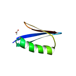
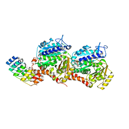
7Z5S
 
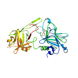 | | Crystal Structure of botulinum neurotoxin A2 cell binding domain in complex with GD1a | | 分子名称: | 1,2-ETHANEDIOL, Botulinum neurotoxin, HEXAETHYLENE GLYCOL, ... | | 著者 | Gregory, K.S, Acharya, K.R, Liu, S.M, Mahadeva, T.B. | | 登録日 | 2022-03-10 | | 公開日 | 2022-06-08 | | 最終更新日 | 2024-01-31 | | 実験手法 | X-RAY DIFFRACTION (2.1 Å) | | 主引用文献 | Structural Features of Clostridium botulinum Neurotoxin Subtype A2 Cell Binding Domain.
Toxins, 14, 2022
|
|
7Z5T
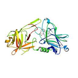 | | Crystal Structure of botulinum neurotoxin A2 cell binding domain | | 分子名称: | ACETATE ION, Botulinum neurotoxin, PENTAETHYLENE GLYCOL | | 著者 | Gregory, K.S, Acharya, K.R, Liu, S.M, Mahadeva, T.B. | | 登録日 | 2022-03-10 | | 公開日 | 2022-06-08 | | 最終更新日 | 2024-01-31 | | 実験手法 | X-RAY DIFFRACTION (1.63 Å) | | 主引用文献 | Structural Features of Clostridium botulinum Neurotoxin Subtype A2 Cell Binding Domain.
Toxins, 14, 2022
|
|
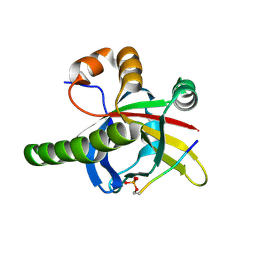
6M8M
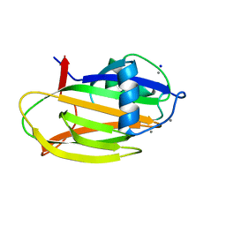 | | PA14 sugar-binding domain from RTX adhesin | | 分子名称: | CALCIUM ION, Putative large adhesion protein (Lap) involved in biofilm formation, SODIUM ION, ... | | 著者 | Vance, T.D.R, Conroy, B, Davies, P.L. | | 登録日 | 2018-08-22 | | 公開日 | 2019-09-04 | | 最終更新日 | 2023-10-11 | | 実験手法 | X-RAY DIFFRACTION (1.2 Å) | | 主引用文献 | Structure and functional analysis of a bacterial adhesin sugar-binding domain.
Plos One, 14, 2019
|
|
7YPX
 | | Cyanophage Pam3 fiber | | 分子名称: | Pam3 tail fiber proreins, tail fiber chaperone | | 著者 | Wei, Z.L, Jiang, Y.L, Zhou, C.Z. | | 登録日 | 2022-08-04 | | 公開日 | 2022-11-09 | | 最終更新日 | 2024-07-03 | | 実験手法 | ELECTRON MICROSCOPY (3.12 Å) | | 主引用文献 | Structural Insights into the Chaperone-Assisted Assembly of a Simplified Tail Fiber of the Myocyanophage Pam3.
Viruses, 14, 2022
|
|
7Z1K
 | | Crystal structure of the SPOC domain of human SHARP (SPEN) in complex with RNA polymerase II CTD heptapeptide phosphorylated on Ser5 | | 分子名称: | Msx2-interacting protein, SER-TYR-SER-PRO-THR-SEP | | 著者 | Appel, L, Grishkovskaya, I, Slade, D, Djinovic-Carugo, K. | | 登録日 | 2022-02-24 | | 公開日 | 2022-12-07 | | 最終更新日 | 2024-02-07 | | 実験手法 | X-RAY DIFFRACTION (1.55 Å) | | 主引用文献 | The SPOC domain is a phosphoserine binding module that bridges transcription machinery with co- and post-transcriptional regulators.
Nat Commun, 14, 2023
|
|
7Z27
 | |
7YOT
 | |
7YOU
 | |
6KMC
 | |
7YOV
 | |
7YYW
 | | Molecular snapshots of drug release from tubulin: 10 nanoseconds after photoactivation. | | 分子名称: | Azo-Combretastatin A4 (trans), CALCIUM ION, Designed Ankyrin Repeat Protein (DARPIN) D1, ... | | 著者 | Wranik, M, Weinert, T, Standfuss, J. | | 登録日 | 2022-02-18 | | 公開日 | 2023-02-22 | | 最終更新日 | 2023-09-27 | | 実験手法 | X-RAY DIFFRACTION (2.2 Å) | | 主引用文献 | Watching the release of a photopharmacological drug from tubulin using time-resolved serial crystallography.
Nat Commun, 14, 2023
|
|
7YZ5
 | | Molecular snapshots of drug release from tubulin: 100 milliseconds (steady state) | | 分子名称: | CALCIUM ION, Designed Ankyrin Repeat Protein (DARPIN) D1, GUANOSINE-5'-DIPHOSPHATE, ... | | 著者 | Wranik, M, Weinert, T, Standfuss, J. | | 登録日 | 2022-02-18 | | 公開日 | 2023-02-22 | | 最終更新日 | 2024-02-07 | | 実験手法 | X-RAY DIFFRACTION (2.11 Å) | | 主引用文献 | Watching the release of a photopharmacological drug from tubulin using time-resolved serial crystallography.
Nat Commun, 14, 2023
|
|
7YYV
 | | Molecular snapshots of drug release from tubulin: 1 nanosecond after photoactivation. | | 分子名称: | Azo-Combretastatin A4 (trans), CALCIUM ION, Designed Ankyrin Repeat Protein (DARPIN) D1, ... | | 著者 | Wranik, M, Weinert, T, Standfuss, J. | | 登録日 | 2022-02-18 | | 公開日 | 2023-02-22 | | 最終更新日 | 2024-02-07 | | 実験手法 | X-RAY DIFFRACTION (2.2 Å) | | 主引用文献 | Watching the release of a photopharmacological drug from tubulin using time-resolved serial crystallography.
Nat Commun, 14, 2023
|
|
7YZ2
 | | Molecular snapshots of drug release from tubulin: 10 milliseconds after photoactivation. | | 分子名称: | Designed Ankyrin Repeat Protein (DARPIN) D1, GUANOSINE-5'-DIPHOSPHATE, GUANOSINE-5'-TRIPHOSPHATE, ... | | 著者 | Wranik, M, Weinert, T, Standfuss, J. | | 登録日 | 2022-02-18 | | 公開日 | 2023-02-22 | | 最終更新日 | 2024-02-07 | | 実験手法 | X-RAY DIFFRACTION (2.2 Å) | | 主引用文献 | Watching the release of a photopharmacological drug from tubulin using time-resolved serial crystallography.
Nat Commun, 14, 2023
|
|
7YYZ
 | | Molecular snapshots of drug release from tubulin: 10 microseconds after photoactivation. | | 分子名称: | Azo-Combretastatin A4 (trans), CALCIUM ION, Designed Ankyrin Repeat Protein (DARPIN) D1, ... | | 著者 | Wranik, M, Weinert, T, Standfuss, J. | | 登録日 | 2022-02-18 | | 公開日 | 2023-02-22 | | 最終更新日 | 2024-02-07 | | 実験手法 | X-RAY DIFFRACTION (2.2 Å) | | 主引用文献 | Watching the release of a photopharmacological drug from tubulin using time-resolved serial crystallography.
Nat Commun, 14, 2023
|
|
7YZ6
 | | Molecular snapshots of drug release from tubulin: Dark (steady state) | | 分子名称: | Azo-Combretastatin A4 (cis), CALCIUM ION, Designed Ankyrin Repeat Protein (DARPIN) D1, ... | | 著者 | Wranik, M, Weinert, T, Standfuss, J. | | 登録日 | 2022-02-18 | | 公開日 | 2023-02-22 | | 最終更新日 | 2023-09-27 | | 実験手法 | X-RAY DIFFRACTION (2.1 Å) | | 主引用文献 | Watching the release of a photopharmacological drug from tubulin using time-resolved serial crystallography.
Nat Commun, 14, 2023
|
|
7YZ1
 | | Molecular snapshots of drug release from tubulin: 1 millisecond after photoactivation. | | 分子名称: | Azo-Combretastatin A4 (trans), CALCIUM ION, Designed Ankyrin Repeat Protein (DARPIN) D1, ... | | 著者 | Wranik, M, Weinert, T, Standfuss, J. | | 登録日 | 2022-02-18 | | 公開日 | 2023-02-22 | | 最終更新日 | 2023-09-27 | | 実験手法 | X-RAY DIFFRACTION (2.2 Å) | | 主引用文献 | Watching the release of a photopharmacological drug from tubulin using time-resolved serial crystallography.
Nat Commun, 14, 2023
|
|
7YYX
 | | Molecular snapshots of drug release from tubulin: 100 nanoseconds after photoactivation. | | 分子名称: | Azo-Combretastatin A4 (trans), CALCIUM ION, Designed Ankyrin Repeat Protein (DARPIN) D1, ... | | 著者 | Wranik, M, Weinert, T, Standfuss, J. | | 登録日 | 2022-02-18 | | 公開日 | 2023-02-22 | | 最終更新日 | 2023-09-27 | | 実験手法 | X-RAY DIFFRACTION (2.2 Å) | | 主引用文献 | Watching the release of a photopharmacological drug from tubulin using time-resolved serial crystallography.
Nat Commun, 14, 2023
|
|
7YZ0
 | | Molecular snapshots of drug release from tubulin: 100 microseconds after photoactivation. | | 分子名称: | Azo-Combretastatin A4 (trans), CALCIUM ION, Designed Ankyrin Repeat Protein (DARPIN) D1, ... | | 著者 | Wranik, M, Weinert, T, Standfuss, J. | | 登録日 | 2022-02-18 | | 公開日 | 2023-02-22 | | 最終更新日 | 2024-02-07 | | 実験手法 | X-RAY DIFFRACTION (2.2 Å) | | 主引用文献 | Watching the release of a photopharmacological drug from tubulin using time-resolved serial crystallography.
Nat Commun, 14, 2023
|
|
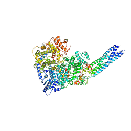
7YTJ
 | | Cryo-EM structure of VTC complex | | 分子名称: | 1,2-DIACYL-SN-GLYCERO-3-PHOSPHOCHOLINE, INOSITOL HEXAKISPHOSPHATE, PHOSPHATE ION, ... | | 著者 | Guan, Z.Y, Chen, J, Liu, R.W, Chen, Y.K, Xing, Q, Du, Z.M, Liu, Z. | | 登録日 | 2022-08-15 | | 公開日 | 2023-02-22 | | 最終更新日 | 2024-07-03 | | 実験手法 | ELECTRON MICROSCOPY (3 Å) | | 主引用文献 | The cytoplasmic synthesis and coupled membrane translocation of eukaryotic polyphosphate by signal-activated VTC complex.
Nat Commun, 14, 2023
|
|
7YZ3
 | | Molecular snapshots of drug release from tubulin: Apo state | | 分子名称: | CALCIUM ION, Designed Ankyrin Repeat Protein (DARPIN) D1, GUANOSINE-5'-DIPHOSPHATE, ... | | 著者 | Wranik, M, Weinert, T, Standfuss, J. | | 登録日 | 2022-02-18 | | 公開日 | 2023-02-22 | | 最終更新日 | 2024-02-07 | | 実験手法 | X-RAY DIFFRACTION (1.8 Å) | | 主引用文献 | Watching the release of a photopharmacological drug from tubulin using time-resolved serial crystallography.
Nat Commun, 14, 2023
|
|
7YYQ
 | | Molecular snapshots of drug release from tubulin: Dark state | | 分子名称: | Azo-Combretastatin A4 (cis), CALCIUM ION, Designed Ankyrin Repeat Protein (DARPIN) D1, ... | | 著者 | Wranik, M, Weinert, T, Standfuss, J. | | 登録日 | 2022-02-18 | | 公開日 | 2023-02-22 | | 最終更新日 | 2023-09-27 | | 実験手法 | X-RAY DIFFRACTION (1.7 Å) | | 主引用文献 | Watching the release of a photopharmacological drug from tubulin using time-resolved serial crystallography.
Nat Commun, 14, 2023
|
|
7YYY
 | | Molecular snapshots of drug release from tubulin: 1 microsecond after photoactivation | | 分子名称: | Azo-Combretastatin A4 (trans), CALCIUM ION, Designed Ankyrin Repeat Protein (DARPIN) D1, ... | | 著者 | Wranik, M, Weinert, T, Standfuss, J. | | 登録日 | 2022-02-18 | | 公開日 | 2023-02-22 | | 最終更新日 | 2023-09-27 | | 実験手法 | X-RAY DIFFRACTION (2.2 Å) | | 主引用文献 | Watching the release of a photopharmacological drug from tubulin using time-resolved serial crystallography.
Nat Commun, 14, 2023
|
|
6K3K
 | | Solution structure of APOBEC3G-CD2 with ssDNA, Product B | | 分子名称: | DNA dC->dU-editing enzyme APOBEC-3G, DNA/RNA (5'-D(*AP*TP*TP*CP*UP*(ICY)P*AP*AP*TP*T)-3'), ZINC ION | | 著者 | Cao, C, Yan, X, Lan, W, Wang, C. | | 登録日 | 2019-05-19 | | 公開日 | 2019-06-12 | | 最終更新日 | 2024-05-15 | | 実験手法 | SOLUTION NMR | | 主引用文献 | Structural Investigations on the Interactions between Cytidine Deaminase Human APOBEC3G and DNA.
Chem Asian J, 14, 2019
|
|
6K3J
 | | Solution structure of APOBEC3G-CD2 with ssDNA, Product A | | 分子名称: | DNA (5'-D(*AP*TP*TP*CP*UP*(IUR)P*AP*AP*TP*T)-3'), DNA dC->dU-editing enzyme APOBEC-3G, ZINC ION | | 著者 | Cao, C, Yan, X, Lan, W, Wang, C. | | 登録日 | 2019-05-19 | | 公開日 | 2019-06-12 | | 最終更新日 | 2024-05-15 | | 実験手法 | SOLUTION NMR | | 主引用文献 | Structural Investigations on the Interactions between Cytidine Deaminase Human APOBEC3G and DNA.
Chem Asian J, 14, 2019
|
|


