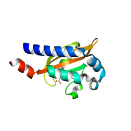
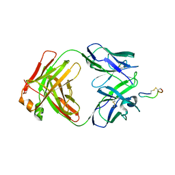
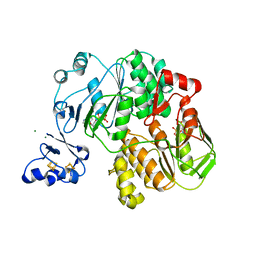
3BUW
 
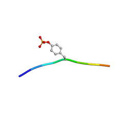 | | Crystal structure of c-Cbl-TKB domain complexed with its binding motif in Syk | | 分子名称: | 13-meric peptide from Tyrosine-protein kinase SYK, E3 ubiquitin-protein ligase CBL | | 著者 | Ng, C, Jackson, R.A, Buschdorf, J.P, Sun, Q, Guy, G.R, Sivaraman, J. | | 登録日 | 2008-01-03 | | 公開日 | 2008-02-26 | | 最終更新日 | 2023-11-15 | | 実験手法 | X-RAY DIFFRACTION (1.45 Å) | | 主引用文献 | Structural basis for a novel intrapeptidyl H-bond and reverse binding of c-Cbl-TKB domain substrates
Embo J., 27, 2008
|
|
4MDW
 | |
3C1E
 | |
4LQZ
 | |
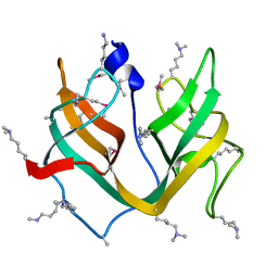
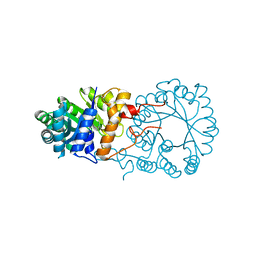
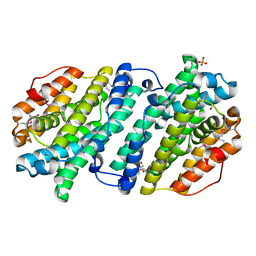
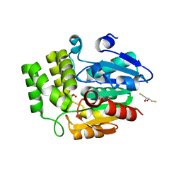
3BWY
 | | Crystal Structure of Human 108M Catechol O-methyltransferase bound with S-adenosylmethionine and inhibitor dinitrocatechol | | 分子名称: | (4S)-2-METHYL-2,4-PENTANEDIOL, 3,5-DINITROCATECHOL, COMT protein, ... | | 著者 | Rutherford, K, Le Trong, I, Stenkamp, R.E, Parson, W.W. | | 登録日 | 2008-01-10 | | 公開日 | 2008-06-03 | | 最終更新日 | 2023-08-30 | | 実験手法 | X-RAY DIFFRACTION (1.3 Å) | | 主引用文献 | Crystal structures of human 108V and 108M catechol O-methyltransferase.
J.Mol.Biol., 380, 2008
|
|
3BY8
 | |
3BZ3
 | |
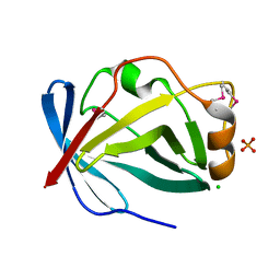
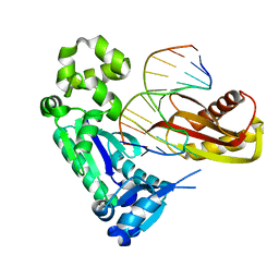
4DOQ
 | | Crystal structure of the complex of Porcine Pancreatic Trypsin with 1/2SLPI | | 分子名称: | 3,6,9,12,15,18,21,24,27-NONAOXANONACOSANE-1,29-DIOL, Antileukoproteinase, CALCIUM ION, ... | | 著者 | Fukushima, K, Takimoto-Kamimura, M. | | 登録日 | 2012-02-10 | | 公開日 | 2013-08-14 | | 最終更新日 | 2023-11-08 | | 実験手法 | X-RAY DIFFRACTION (2 Å) | | 主引用文献 | Structure basis 1/2SLPI and porcine pancreas trypsin interaction
J.SYNCHROTRON RADIAT., 20, 2013
|
|
3C2O
 | |
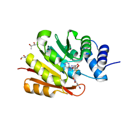
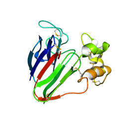
3BKY
 | | Crystal Structure of Chimeric Antibody C2H7 Fab in complex with a CD20 Peptide | | 分子名称: | B-lymphocyte antigen CD20, the Fab fragment of chimeric 2H7, heavy chain, ... | | 著者 | Du, J, Zhong, C, Ding, J. | | 登録日 | 2007-12-07 | | 公開日 | 2008-04-29 | | 最終更新日 | 2024-04-10 | | 実験手法 | X-RAY DIFFRACTION (2.61 Å) | | 主引用文献 | Crystal structure of chimeric antibody C2H7 Fab in complex with a CD20 peptide
Mol.Immunol., 45, 2008
|
|
3BLE
 | |
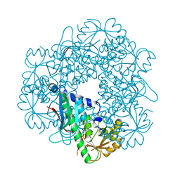
5J4C
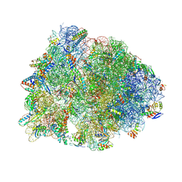 | | Crystal structure of the Thermus thermophilus 70S ribosome in complex with cisplatin (soaked) and bound to mRNA and A-, P- and E-site tRNAs at 2.8A resolution | | 分子名称: | 16S ribosomal RNA, 23S ribosomal RNA, 30S ribosomal protein S10, ... | | 著者 | Melnikov, S.V, Soll, D, Steitz, T.A, Polikanov, Y.S. | | 登録日 | 2016-03-31 | | 公開日 | 2016-04-27 | | 最終更新日 | 2023-11-15 | | 実験手法 | X-RAY DIFFRACTION (2.8 Å) | | 主引用文献 | Insights into RNA binding by the anticancer drug cisplatin from the crystal structure of cisplatin-modified ribosome.
Nucleic Acids Res., 44, 2016
|
|
3BOR
 | | Crystal structure of the DEADc domain of human translation initiation factor 4A-2 | | 分子名称: | Human initiation factor 4A-II | | 著者 | Dimov, S, Hong, B, Tempel, W, MacKenzie, F, Karlberg, T, Arrowsmith, C.H, Edwards, A.M, Weigelt, J, Bochkarev, A, Park, H, Structural Genomics Consortium (SGC) | | 登録日 | 2007-12-17 | | 公開日 | 2008-01-01 | | 最終更新日 | 2023-08-30 | | 実験手法 | X-RAY DIFFRACTION (1.85 Å) | | 主引用文献 | Comparative Structural Analysis of Human DEAD-Box RNA Helicases.
Plos One, 5, 2010
|
|
3BRS
 | | Crystal structure of sugar transporter from Clostridium phytofermentans | | 分子名称: | Periplasmic binding protein/LacI transcriptional regulator | | 著者 | Malashkevich, V.N, Patskovsky, Y, Toro, R, Meyers, A.J, Wasserman, S, Sauder, J.M, Burley, S.K, Almo, S.C, New York SGX Research Center for Structural Genomics (NYSGXRC) | | 登録日 | 2007-12-21 | | 公開日 | 2008-02-05 | | 最終更新日 | 2024-02-21 | | 実験手法 | X-RAY DIFFRACTION (2 Å) | | 主引用文献 | Crystal structure of sugar transporter from Clostridium phytofermentans.
To be Published
|
|
3BUJ
 | | Crystal Structure of CalO2 | | 分子名称: | CalO2, PROTOPORPHYRIN IX CONTAINING FE | | 著者 | McCoy, J.G, Johnson, H.D, Singh, S, Bingman, C.A, Thorson, J.S, Phillips Jr, G.N. | | 登録日 | 2008-01-02 | | 公開日 | 2008-04-29 | | 最終更新日 | 2023-08-30 | | 実験手法 | X-RAY DIFFRACTION (2.47 Å) | | 主引用文献 | Structural characterization of CalO2: a putative orsellinic acid P450 oxidase in the calicheamicin biosynthetic pathway.
Proteins, 74, 2009
|
|
3BYI
 | | Crystal structure of human Rho GTPase activating protein 15 (ARHGAP15) | | 分子名称: | Rho GTPase activating protein 15 | | 著者 | Shrestha, L, Tickle, J, Elkins, J, Burgess-Brown, N, Johansson, C, Papagrigoriou, E, Kavanagh, K, Pike, A.C.W, Ugochukwu, E, Uppenberg, J, von Delft, F, Arrowsmith, C.H, Edwards, A.M, Weigelt, J, Doyle, D, Structural Genomics Consortium (SGC) | | 登録日 | 2008-01-16 | | 公開日 | 2008-02-26 | | 最終更新日 | 2023-08-30 | | 実験手法 | X-RAY DIFFRACTION (2.25 Å) | | 主引用文献 | Crystal Structure of Human Rho GTPase Activating Protein 15 (ARHGAP15).
To be Published
|
|
3BZ8
 | | Crystal Structures of (S)-(-)-Blebbistatin Analogs bound to Dictyostelium discoideum myosin II | | 分子名称: | (3aS)-3a-hydroxy-7-methyl-1-phenyl-1,2,3,3a-tetrahydro-4H-pyrrolo[2,3-b]quinolin-4-one, 1,2-ETHANEDIOL, ADENOSINE-5'-DIPHOSPHATE, ... | | 著者 | Allingham, J.S, Rayment, I. | | 登録日 | 2008-01-17 | | 公開日 | 2008-02-19 | | 最終更新日 | 2023-08-30 | | 実験手法 | X-RAY DIFFRACTION (2.2 Å) | | 主引用文献 | The small molecule tool (S)-(-)-blebbistatin: novel insights of relevance to myosin inhibitor design.
Org.Biomol.Chem., 6, 2008
|
|
3BNO
 | |
4DJ1
 | |
3C2F
 | |
4DJN
 | |
3BK7
 | |
4DNP
 | | Crystal Structure of DAD2 | | 分子名称: | (2S,3S)-1,4-DIMERCAPTOBUTANE-2,3-DIOL, DAD2, GLYCEROL | | 著者 | Hamiaux, C. | | 登録日 | 2012-02-08 | | 公開日 | 2012-11-14 | | 最終更新日 | 2024-02-28 | | 実験手法 | X-RAY DIFFRACTION (2.15 Å) | | 主引用文献 | DAD2 Is an alpha/beta Hydrolase likely to Be Involved in the Perception of the Plant Branching Hormone, Strigolactone
Curr.Biol., 22, 2012
|
|
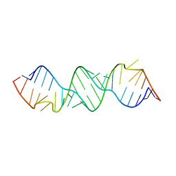
3BQ0
 | | Pre-insertion binary complex of Dbh DNA polymerase | | 分子名称: | CALCIUM ION, DNA (5'-D(*DGP*DAP*DAP*DGP*DCP*DCP*DGP*DGP*DCP*DG)-3'), DNA (5'-D(*DT*DTP*DCP*DCP*DGP*DCP*DCP*DCP*DGP*DGP*DCP*DTP*DTP*DCP*DC)-3'), ... | | 著者 | Pata, J.D, Wilson, R.C. | | 登録日 | 2007-12-19 | | 公開日 | 2008-04-08 | | 最終更新日 | 2023-08-30 | | 実験手法 | X-RAY DIFFRACTION (2.6 Å) | | 主引用文献 | Structural insights into the generation of single-base deletions by the Y family DNA polymerase dbh.
Mol.Cell, 29, 2008
|
|
4DA2
 | |


