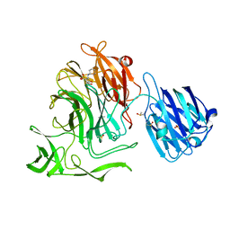
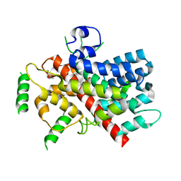
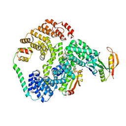
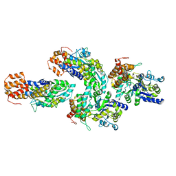
6ZJ5
 
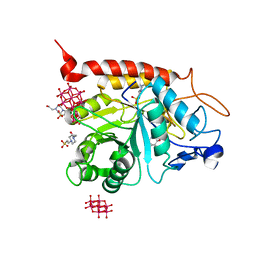 | | Structure of the catalytic domain of human endo-alpha-mannosidase MANEA in complex with GlcDMJ and hexatungstotellurate(VI) TEW | | 分子名称: | 1-DEOXYMANNOJIRIMYCIN, 4-(2-HYDROXYETHYL)-1-PIPERAZINE ETHANESULFONIC ACID, 6-tungstotellurate(VI), ... | | 著者 | Sobala, L.F, Fernandes, P.Z, Hakki, Z, Thompson, A.J, Howe, J.D, Hill, M, Zitzmann, N, Davies, S, Stamataki, Z, Butters, T.D, Alonzi, D.S, Williams, S.J, Davies, G.J. | | 登録日 | 2020-06-27 | | 公開日 | 2020-12-09 | | 最終更新日 | 2024-01-31 | | 実験手法 | X-RAY DIFFRACTION (2.269 Å) | | 主引用文献 | Structure of human endo-alpha-1,2-mannosidase (MANEA), an antiviral host-glycosylation target.
Proc.Natl.Acad.Sci.USA, 117, 2020
|
|
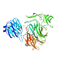
1OT7
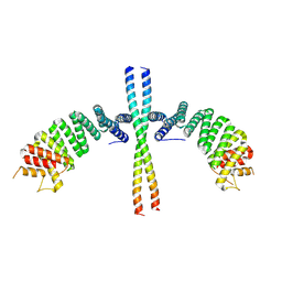
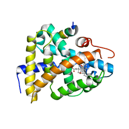 | | Structural Basis for 3-deoxy-CDCA Binding and Activation of FXR | | 分子名称: | 6-ETHYL-CHENODEOXYCHOLIC ACID, Bile Acid Receptor, ISO-URSODEOXYCHOLIC ACID, ... | | 著者 | Mi, L.Z, Devarakonda, S, Harp, J.M, Han, Q, Pellicciari, R, Willson, T.M, Khorasanizadeh, S, Rastinejad, F. | | 登録日 | 2003-03-21 | | 公開日 | 2004-03-23 | | 最終更新日 | 2024-04-03 | | 実験手法 | X-RAY DIFFRACTION (2.9 Å) | | 主引用文献 | Structural Basis for Bile Acid Binding and Activation of the Nuclear Receptor FXR
Mol.Cell, 11, 2003
|
|
5XYF
 | |
4XE9
 | |
4XJW
 | |
4XOG
 | |
4XJ9
 | |
4XHX
 | |
5T0N
 | |
4XIL
 | |
4XMA
 | |
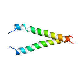
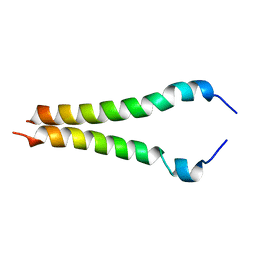
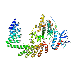
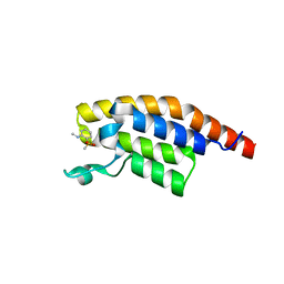
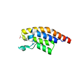
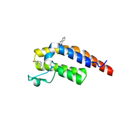
5FMU
 | | MmIFT54 CH-domain | | 分子名称: | 2-(N-MORPHOLINO)-ETHANESULFONIC ACID, TRAF3-INTERACTING PROTEIN 1 | | 著者 | Weber, K, Lorentzen, E. | | 登録日 | 2015-11-09 | | 公開日 | 2016-03-09 | | 最終更新日 | 2024-01-10 | | 実験手法 | X-RAY DIFFRACTION (1.593 Å) | | 主引用文献 | Intraflagellar Transport Proteins 172, 80, 57, 54, 38, and 20 Form a Stable Tubulin-Binding Ift-B2 Complex.
Embo J., 35, 2016
|
|
6PW9
 | | Cryo-EM structure of human NatE/HYPK complex | | 分子名称: | ACETYL COENZYME *A, Huntingtin-interacting protein K, INOSITOL HEXAKISPHOSPHATE, ... | | 著者 | Deng, S, Marmorstein, R. | | 登録日 | 2019-07-22 | | 公開日 | 2020-02-19 | | 最終更新日 | 2020-10-14 | | 実験手法 | ELECTRON MICROSCOPY (4.03 Å) | | 主引用文献 | Molecular basis for N-terminal acetylation by human NatE and its modulation by HYPK.
Nat Commun, 11, 2020
|
|
2KA2
 | |
2KA1
 | |
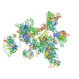
7ABI
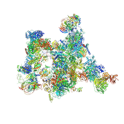 | | Human pre-Bact-2 spliceosome | | 分子名称: | 116 kDa U5 small nuclear ribonucleoprotein component, BUD13 homolog, Beta-catenin-like protein 1, ... | | 著者 | Townsend, C, Kastner, B, Leelaram, M.N, Bertram, K, Stark, H, Luehrmann, R. | | 登録日 | 2020-09-07 | | 公開日 | 2021-02-10 | | 実験手法 | ELECTRON MICROSCOPY (8 Å) | | 主引用文献 | Mechanism of protein-guided folding of the active site U2/U6 RNA during spliceosome activation.
Science, 370, 2020
|
|
6EJN
 | |
6F5E
 | | Crystal structure of DARPin-DARPin rigid fusion, variant DD_D12_10_47 in complex JNK1a1 and JIP1 peptide | | 分子名称: | C-Jun-amino-terminal kinase-interacting protein 1, DD_D12_10_47, Mitogen-activated protein kinase 8 | | 著者 | Wu, Y, Mittl, P.R, Honegger, A, Batyuk, A, Plueckthun, A. | | 登録日 | 2017-12-01 | | 公開日 | 2017-12-13 | | 最終更新日 | 2024-05-08 | | 実験手法 | X-RAY DIFFRACTION (2.7 Å) | | 主引用文献 | Crystal structure of DARPin-DARPin rigid fusion, variant DD_D12_10_47 in complex JNK1a1 and JIP1 peptide
To be published
|
|
7ABG
 | | Human pre-Bact-1 spliceosome | | 分子名称: | 116 kDa U5 small nuclear ribonucleoprotein component, 7-METHYL-GUANOSINE-5'-TRIPHOSPHATE-5'-GUANOSINE, Cell division cycle 5-like protein, ... | | 著者 | Townsend, C, Kastner, B, Leelaram, M.N, Bertram, K, Stark, H, Luehrmann, R. | | 登録日 | 2020-09-07 | | 公開日 | 2020-12-23 | | 最終更新日 | 2024-05-01 | | 実験手法 | ELECTRON MICROSCOPY (7.8 Å) | | 主引用文献 | Mechanism of protein-guided folding of the active site U2/U6 RNA during spliceosome activation.
Science, 370, 2020
|
|
6FUZ
 | | Crystal structure of the TPR domain of KLC1 in complex with the C-terminal peptide of JIP1 | | 分子名称: | GLYCEROL, Kinesin light chain 1,Kinesin light chain 1,C-Jun-amino-terminal kinase-interacting protein 1, nanobody | | 著者 | Pernigo, S, Dodding, M.P, Steiner, R.A. | | 登録日 | 2018-02-28 | | 公開日 | 2018-05-02 | | 最終更新日 | 2019-09-18 | | 実験手法 | X-RAY DIFFRACTION (2.7 Å) | | 主引用文献 | Structural basis for isoform-specific kinesin-1 recognition of Y-acidic cargo adaptors.
Elife, 7, 2018
|
|
3V3V
 | | Structural and functional analysis of quercetagetin, a natural JNK1 inhibitor | | 分子名称: | 3,5,6,7-TETRAHYDROXY-2-(3,4-DIHYDROXYPHENYL)-4H-CHROMEN-4-ONE, C-Jun-amino-terminal kinase-interacting protein 1, CHLORIDE ION, ... | | 著者 | Baek, S, Kang, N.J, Popowicz, G.M, Arciniega, M, Jung, S.K, Byun, S, Song, N.R, Heo, Y.S, Kim, B.Y, Lee, H.J, Holak, T.A, Augustin, M, Bode, A.M, Huber, R, Dong, Z, Lee, K.W. | | 登録日 | 2011-12-14 | | 公開日 | 2012-12-05 | | 最終更新日 | 2023-09-13 | | 実験手法 | X-RAY DIFFRACTION (2.7 Å) | | 主引用文献 | Structural and Functional Analysis of the Natural JNK1 Inhibitor Quercetagetin.
J.Mol.Biol., 425, 2013
|
|
6GFJ
 | |
5RJX
 | | PanDDA analysis group deposition -- Crystal Structure of PHIP in complex with Z285782452 | | 分子名称: | N-methyl-2-(methylsulfonyl)aniline, PH-interacting protein | | 著者 | Grosjean, H, Aimon, A, Krojer, T, Talon, R, Douangamath, A, Koekemoer, L, Arrowsmith, C.H, Edwards, A, Bountra, C, von Delft, F, Biggin, P.C. | | 登録日 | 2020-06-02 | | 公開日 | 2020-06-17 | | 最終更新日 | 2024-03-06 | | 実験手法 | X-RAY DIFFRACTION (1.291 Å) | | 主引用文献 | PanDDA analysis group deposition of ground-state model
To Be Published
|
|
5RKD
 | | PanDDA analysis group deposition -- Crystal Structure of PHIP in complex with Z2168282707 | | 分子名称: | (6S)-1-methyl-4,5,6,7-tetrahydro-1H-benzotriazole-6-carboxylic acid, PH-interacting protein | | 著者 | Grosjean, H, Aimon, A, Krojer, T, Talon, R, Douangamath, A, Koekemoer, L, Arrowsmith, C.H, Edwards, A, Bountra, C, von Delft, F, Biggin, P.C. | | 登録日 | 2020-06-02 | | 公開日 | 2020-06-17 | | 最終更新日 | 2024-03-06 | | 実験手法 | X-RAY DIFFRACTION (1.237 Å) | | 主引用文献 | PanDDA analysis group deposition of ground-state model
To Be Published
|
|
5RJJ
 | | PanDDA analysis group deposition -- Crystal Structure of PHIP in complex with NCL-00023833 | | 分子名称: | 4-bromanyl-1,8-naphthyridine, PH-interacting protein | | 著者 | Grosjean, H, Aimon, A, Krojer, T, Talon, R, Douangamath, A, Koekemoer, L, Arrowsmith, C.H, Edwards, A, Bountra, C, von Delft, F, Biggin, P.C. | | 登録日 | 2020-06-02 | | 公開日 | 2020-06-17 | | 実験手法 | X-RAY DIFFRACTION (1.151 Å) | | 主引用文献 | PanDDA analysis group deposition of ground-state model
To Be Published
|
|


