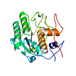
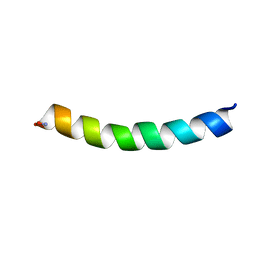
1UPH
 
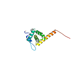 | | HIV-1 Myristoylated Matrix | | 分子名称: | GAG POLYPROTEIN | | 著者 | Tang, C, Loeliger, E, Luncsford, P, Kinde, I, Beckett, D, Summers, M.F. | | 登録日 | 2003-10-01 | | 公開日 | 2004-01-08 | | 最終更新日 | 2011-07-13 | | 実験手法 | SOLUTION NMR | | 主引用文献 | Entropic Switch Regulates Myristate Exposure in the HIV-1 Matrix Protein
Proc.Natl.Acad.Sci.USA, 101, 2004
|
|
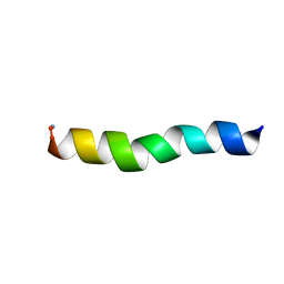
3DE2
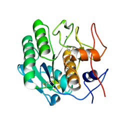 | |
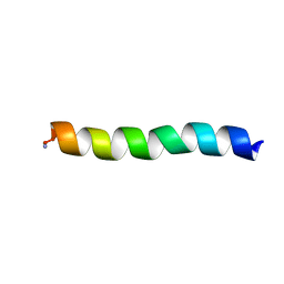
3DE6
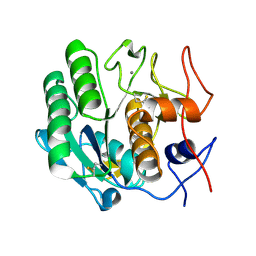 | |
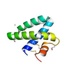
1S6Z
 | | Enhanced Green Fluorescent Protein Containing the Y66L Substitution | | 分子名称: | CHLORIDE ION, green fluorescent protein | | 著者 | Rosenow, M.A, Huffman, H.A, Phail, M.E, Wachter, R.M. | | 登録日 | 2004-01-28 | | 公開日 | 2004-05-04 | | 最終更新日 | 2024-07-10 | | 実験手法 | X-RAY DIFFRACTION (1.5 Å) | | 主引用文献 | The Crystal Structure of the Y66L Variant of Green Fluorescent Protein Supports a Cyclization-Oxidation-Dehydration Mechanism for Chromophore Maturation(,).
Biochemistry, 43, 2004
|
|
2DOB
 | |
3DE7
 | |
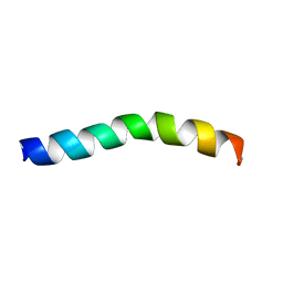
3DE4
 | |
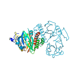
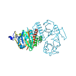
3H55
 | | Crystal Structure of human alpha-N-acetylgalactosaminidase, Complex with Galactose | | 分子名称: | 2-acetamido-2-deoxy-beta-D-glucopyranose, 2-acetamido-2-deoxy-beta-D-glucopyranose-(1-4)-[alpha-L-fucopyranose-(1-6)]2-acetamido-2-deoxy-beta-D-glucopyranose, Alpha-N-acetylgalactosaminidase, ... | | 著者 | Clark, N.E, Garman, S.C. | | 登録日 | 2009-04-21 | | 公開日 | 2009-10-20 | | 最終更新日 | 2023-09-06 | | 実験手法 | X-RAY DIFFRACTION (1.91 Å) | | 主引用文献 | The 1.9 a structure of human alpha-N-acetylgalactosaminidase: The molecular basis of Schindler and Kanzaki diseases
J.Mol.Biol., 393, 2009
|
|
1TOW
 | | Crystal structure of human adipocyte fatty acid binding protein in complex with a carboxylic acid ligand | | 分子名称: | 4-(9H-CARBAZOL-9-YL)BUTANOIC ACID, Fatty acid-binding protein, adipocyte | | 著者 | Lehmann, F, Haile, S, Axen, E, Medina, C, Uppenberg, J, Svensson, S, Lundback, T, Rondahl, L, Barf, T. | | 登録日 | 2004-06-15 | | 公開日 | 2004-08-24 | | 最終更新日 | 2024-03-13 | | 実験手法 | X-RAY DIFFRACTION (2 Å) | | 主引用文献 | Discovery of inhibitors of human adipocyte fatty acid-binding protein, a potential type 2 diabetes target.
Bioorg.Med.Chem.Lett., 14, 2004
|
|
3H53
 | | Crystal Structure of human alpha-N-acetylgalactosaminidase | | 分子名称: | 2-acetamido-2-deoxy-beta-D-glucopyranose, 2-acetamido-2-deoxy-beta-D-glucopyranose-(1-4)-[alpha-L-fucopyranose-(1-6)]2-acetamido-2-deoxy-beta-D-glucopyranose, Alpha-N-acetylgalactosaminidase, ... | | 著者 | Clark, N.E, Garman, S.C. | | 登録日 | 2009-04-21 | | 公開日 | 2009-10-20 | | 最終更新日 | 2023-09-06 | | 実験手法 | X-RAY DIFFRACTION (2.01 Å) | | 主引用文献 | The 1.9 a structure of human alpha-N-acetylgalactosaminidase: The molecular basis of Schindler and Kanzaki diseases
J.Mol.Biol., 393, 2009
|
|
3H54
 | | Crystal Structure of human alpha-N-acetylgalactosaminidase,complex with GalNAc | | 分子名称: | 2-acetamido-2-deoxy-alpha-D-galactopyranose, 2-acetamido-2-deoxy-beta-D-glucopyranose, 2-acetamido-2-deoxy-beta-D-glucopyranose-(1-4)-[alpha-L-fucopyranose-(1-6)]2-acetamido-2-deoxy-beta-D-glucopyranose, ... | | 著者 | Clark, N.E, Garman, S.C. | | 登録日 | 2009-04-21 | | 公開日 | 2009-10-20 | | 最終更新日 | 2023-09-06 | | 実験手法 | X-RAY DIFFRACTION (2.2 Å) | | 主引用文献 | The 1.9 a structure of human alpha-N-acetylgalactosaminidase: The molecular basis of Schindler and Kanzaki diseases
J.Mol.Biol., 393, 2009
|
|
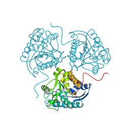
4M6J
 | | Crystal structure of human dihydrofolate reductase (DHFR) bound to NADPH | | 分子名称: | Dihydrofolate reductase, NADPH DIHYDRO-NICOTINAMIDE-ADENINE-DINUCLEOTIDE PHOSPHATE | | 著者 | Bhabha, G, Ekiert, D.C, Wright, P.E, Wilson, I.A. | | 登録日 | 2013-08-09 | | 公開日 | 2013-09-25 | | 最終更新日 | 2023-09-20 | | 実験手法 | X-RAY DIFFRACTION (1.201 Å) | | 主引用文献 | Divergent evolution of protein conformational dynamics in dihydrofolate reductase.
Nat.Struct.Mol.Biol., 20, 2013
|
|
4LTV
 | | Crystal structure of epi-isozizaene synthase from Streptomyces coelicolor A3(2) | | 分子名称: | Epi-isozizaene synthase, SULFATE ION | | 著者 | Li, R, Chou, W, Himmelberger, J.A, Litwin, K, Harris, G, Cane, D.E, Christianson, D.W. | | 登録日 | 2013-07-24 | | 公開日 | 2013-12-18 | | 最終更新日 | 2023-09-20 | | 実験手法 | X-RAY DIFFRACTION (2.403 Å) | | 主引用文献 | Reprogramming the Chemodiversity of Terpenoid Cyclization by Remolding the Active Site Contour of epi-Isozizaene Synthase.
Biochemistry, 53, 2014
|
|
4M4Z
 | |
5UA6
 | | Ocellatin-LB1, solution structure in SDS micelle by NMR spectroscopy | | 分子名称: | Ocellatin-LB1 | | 著者 | Gusmao, K.A.G, dos Santos, D.M, Santos, V.M, Pilo-Veloso, D, de Lima, M.E, Resende, J.M. | | 登録日 | 2016-12-19 | | 公開日 | 2017-03-29 | | 最終更新日 | 2018-04-18 | | 実験手法 | SOLUTION NMR | | 主引用文献 | NMR structures in different membrane environments of three ocellatin peptides isolated from Leptodactylus labyrinthicus.
Peptides, 103, 2018
|
|
5UA7
 | | Ocellatin-LB2, solution structure in SDS micelle by NMR spectroscopy | | 分子名称: | Ocellatin-LB2 | | 著者 | Gusmao, K.A.G, dos Santos, D.M, Santos, V.M, Pilo-Veloso, D, de Lima, M.E, Resende, J.M. | | 登録日 | 2016-12-19 | | 公開日 | 2017-03-29 | | 最終更新日 | 2018-04-18 | | 実験手法 | SOLUTION NMR | | 主引用文献 | NMR structures in different membrane environments of three ocellatin peptides isolated from Leptodactylus labyrinthicus.
Peptides, 103, 2018
|
|
5U9Y
 | | Ocellatin-F1 | | 分子名称: | Ocellatin-K1 | | 著者 | Gusmao, K.A.G, dos Santos, D.M, Santos, V.M, Pilo-Veloso, D, de Lima, M.E, Resende, J.M. | | 登録日 | 2016-12-18 | | 公開日 | 2017-03-01 | | 最終更新日 | 2018-04-18 | | 実験手法 | SOLUTION NMR | | 主引用文献 | NMR structures in different membrane environments of three ocellatin peptides isolated from Leptodactylus labyrinthicus.
Peptides, 103, 2018
|
|
3S7O
 | | Crystal Structure of the Infrared Fluorescent D207H variant of Deinococcus Bacteriophytochrome chromophore binding domain at 1.24 angstrom resolution | | 分子名称: | 3-[2-[(Z)-[3-(2-carboxyethyl)-5-[(Z)-(4-ethenyl-3-methyl-5-oxidanylidene-pyrrol-2-ylidene)methyl]-4-methyl-pyrrol-1-ium -2-ylidene]methyl]-5-[(Z)-[(3E)-3-ethylidene-4-methyl-5-oxidanylidene-pyrrolidin-2-ylidene]methyl]-4-methyl-1H-pyrrol-3- yl]propanoic acid, Bacteriophytochrome, GLYCEROL | | 著者 | Forest, K.T, Auldridge, M.E, Satyshur, K.A, Anstrom, D.M. | | 登録日 | 2011-05-26 | | 公開日 | 2011-12-21 | | 最終更新日 | 2023-09-13 | | 実験手法 | X-RAY DIFFRACTION (1.24 Å) | | 主引用文献 | Structure-guided engineering enhances a phytochrome-based infrared fluorescent protein.
J.Biol.Chem., 287, 2012
|
|
5UA8
 | | Ocellatin-F1, solution structure in SDS micelle by NMR spectroscopy | | 分子名称: | Ocellatin-F1 | | 著者 | Gusmao, K.A.G, dos Santos, D.M, Santos, V.M, Pilo-Veloso, D, de Lima, M.E, Resende, J.M. | | 登録日 | 2016-12-19 | | 公開日 | 2017-03-29 | | 最終更新日 | 2018-04-18 | | 実験手法 | SOLUTION NMR | | 主引用文献 | NMR structures in different membrane environments of three ocellatin peptides isolated from Leptodactylus labyrinthicus.
Peptides, 103, 2018
|
|
3S7N
 | |
3QSQ
 | |
4K2O
 | |
3TF3
 | |
3TFB
 | | Transthyretin natural mutant A25T | | 分子名称: | Transthyretin | | 著者 | Azevedo, E.P.C, Pereira, H.M, Garratt, R.C, Kelly, J.W, Foguel, D, Palhano, F.L. | | 登録日 | 2011-08-15 | | 公開日 | 2011-12-07 | | 最終更新日 | 2023-09-13 | | 実験手法 | X-RAY DIFFRACTION (2.033 Å) | | 主引用文献 | Dissecting the Structure, Thermodynamic Stability, and Aggregation Properties of the A25T Transthyretin (A25T-TTR) Variant Involved in Leptomeningeal Amyloidosis: Identifying Protein Partners That Co-Aggregate during A25T-TTR Fibrillogenesis in Cerebrospinal Fluid.
Biochemistry, 50, 2011
|
|
5UK6
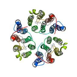 | | Structure of Anabaena Sensory Rhodopsin Determined by Solid State NMR Spectroscopy and DEER | | 分子名称: | Bacteriorhodopsin | | 著者 | Milikisiyants, S, Wang, S, Munro, R.A, Donohue, M, Ward, M.E, Brown, L.S, Smirnova, T.I, Ladizhansky, V, Smirnov, A.I. | | 登録日 | 2017-01-20 | | 公開日 | 2017-05-31 | | 最終更新日 | 2020-01-08 | | 実験手法 | SOLID-STATE NMR | | 主引用文献 | Oligomeric Structure of Anabaena Sensory Rhodopsin in a Lipid Bilayer Environment by Combining Solid-State NMR and Long-range DEER Constraints.
J. Mol. Biol., 429, 2017
|
|


