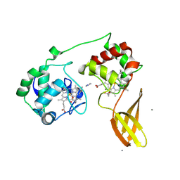
7UY3
 
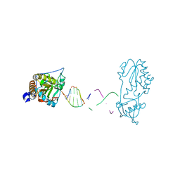
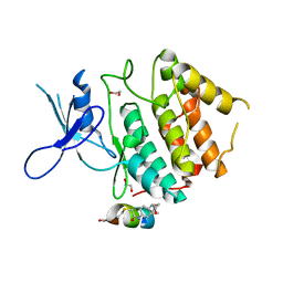
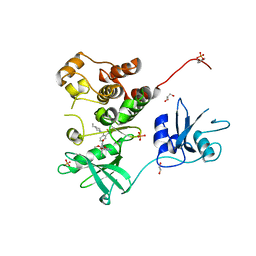 | | Crystal structure of human Fgr tyrosine kinase in complex with TL02-59 | | 分子名称: | 1,2-ETHANEDIOL, 3-[(6,7-dimethoxyquinazolin-4-yl)oxy]-N-{4-[(4-ethylpiperazin-1-yl)methyl]-3-(trifluoromethyl)phenyl}-4-methylbenzamide, GLYCEROL, ... | | 著者 | Du, S, Alvarado, J.J, Smithgall, T.E. | | 登録日 | 2022-05-06 | | 公開日 | 2022-12-28 | | 最終更新日 | 2023-11-15 | | 実験手法 | X-RAY DIFFRACTION (2.99 Å) | | 主引用文献 | ATP-site inhibitors induce unique conformations of the acute myeloid leukemia-associated Src-family kinase, Fgr.
Structure, 30, 2022
|
|
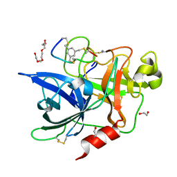
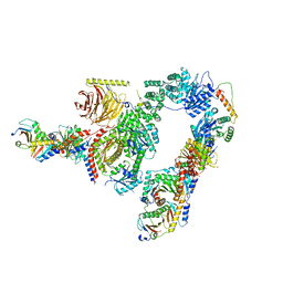
7ZSC
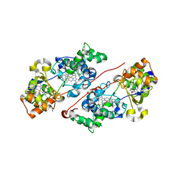
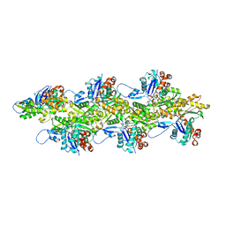
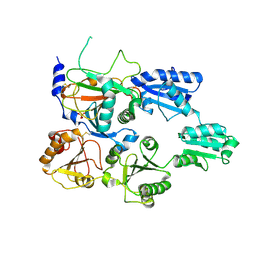 | | Crystal structure of the heterodimeric human C-P4H-II with truncated alpha subunit (C-P4H-II delta281) | | 分子名称: | Prolyl 4-hydroxylase subunit alpha-2, Protein disulfide-isomerase, SULFATE ION | | 著者 | Lebedev, A, Koski, M.K, Wierenga, R.K, Murthy, A.V, Sulu, R. | | 登録日 | 2022-05-06 | | 公開日 | 2022-11-09 | | 最終更新日 | 2024-01-31 | | 実験手法 | X-RAY DIFFRACTION (3.85 Å) | | 主引用文献 | Crystal structure of the collagen prolyl 4-hydroxylase (C-P4H) catalytic domain complexed with PDI: Toward a model of the C-P4H alpha 2 beta 2 tetramer.
J.Biol.Chem., 298, 2022
|
|
7UYO
 | |
7UYN
 | |
7UYP
 | |
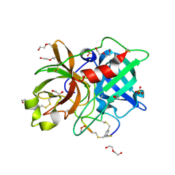
7ZS8
 | | Mixed-valence, active form, of cytochrome c peroxidase from obligate human pathogenic bacterium Neisseria gonorrhoeae at 1.4 Angstrom resolution | | 分子名称: | CALCIUM ION, Cytochrome-c peroxidase, HEME C, ... | | 著者 | Carvalho, A.L, Romao, M.J, Pauleta, S, Nobrega, C. | | 登録日 | 2022-05-06 | | 公開日 | 2023-04-12 | | 最終更新日 | 2024-02-07 | | 実験手法 | X-RAY DIFFRACTION (1.4 Å) | | 主引用文献 | Structural Characterization of Neisseria gonorrhoeae Bacterial Peroxidase-Insights into the Catalytic Cycle of Bacterial Peroxidases.
Int J Mol Sci, 24, 2023
|
|
7ZS0
 | |
7ZS1
 | |
7ZS2
 | |
7ZRR
 | | Crystal structure of human Urokinase-type plasminogen activator in complex with bicycle peptide inhibitor UK965 | | 分子名称: | 1,2-ETHANEDIOL, 1,3,5-tris(bromomethyl)benzene, AMINO GROUP, ... | | 著者 | Caregnato, A, Angela, P, Mazzoccato, Y, Frasson, N, Angelini, A, Cendron, L. | | 登録日 | 2022-05-05 | | 公開日 | 2023-11-15 | | 実験手法 | X-RAY DIFFRACTION (1.64 Å) | | 主引用文献 | Crystal structure of human Urokinase-type plasminogen activator in complex with bicycle peptide inhibitor UK965
To Be Published
|
|
7ZRT
 | | Crystal structure of human Urokinase-type plasminogen activator in complex with bicycle peptide inhibitor UK970 | | 分子名称: | 1,2-ETHANEDIOL, 1,3,5-tris(bromomethyl)benzene, DI(HYDROXYETHYL)ETHER, ... | | 著者 | Caregnato, A, Angela, P, Mazzoccato, Y, Frasson, N, Angelini, A, Cendron, L. | | 登録日 | 2022-05-05 | | 公開日 | 2023-11-15 | | 実験手法 | X-RAY DIFFRACTION (1.8 Å) | | 主引用文献 | Crystal structure of human Urokinase-type plasminogen activator in complex with bicycle peptide inhibitor UK970
To Be Published
|
|
7UXH
 | |
7UXC
 | |
7UXK
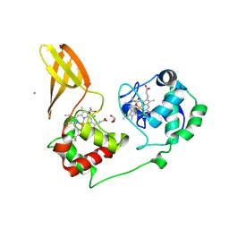
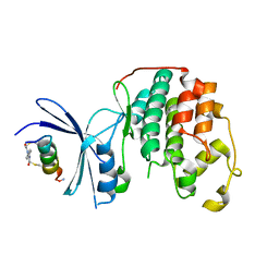
 | | Structure of CDK2 in complex with FP24322, a Helicon Polypeptide | | 分子名称: | 1,2-ETHANEDIOL, Cyclin-dependent kinase 2, FP24322, ... | | 著者 | Li, K, Agarwal, S, Tokareva, O, Thomson, T, Wahl, S, Verdine, G, McGee, J. | | 登録日 | 2022-05-05 | | 公開日 | 2022-12-28 | | 最終更新日 | 2023-10-25 | | 実験手法 | X-RAY DIFFRACTION (2.63 Å) | | 主引用文献 | De novo mapping of alpha-helix recognition sites on protein surfaces using unbiased libraries.
Proc.Natl.Acad.Sci.USA, 119, 2022
|
|
7UXI
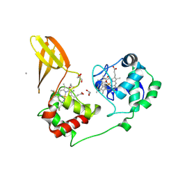
 | | Structure of CDK2 in complex with FP19711, a Helicon Polypeptide | | 分子名称: | AMINO GROUP, Cyclin-dependent kinase 2, FP19711, ... | | 著者 | Li, K, Agarwal, S, Tokareva, O, Thomson, T, Wahl, S, Verdine, G, McGee, J. | | 登録日 | 2022-05-05 | | 公開日 | 2022-12-28 | | 最終更新日 | 2023-10-25 | | 実験手法 | X-RAY DIFFRACTION (2.07 Å) | | 主引用文献 | De novo mapping of alpha-helix recognition sites on protein surfaces using unbiased libraries.
Proc.Natl.Acad.Sci.USA, 119, 2022
|
|
7UXF
 | | Cryogenic electron microscopy 3D map of F-actin | | 分子名称: | ADENOSINE-5'-DIPHOSPHATE, Actin, alpha skeletal muscle, ... | | 著者 | Rangarajan, E.S, Smith, E.W, Izard, T. | | 登録日 | 2022-05-05 | | 公開日 | 2023-03-08 | | 最終更新日 | 2023-03-29 | | 実験手法 | ELECTRON MICROSCOPY (2.7 Å) | | 主引用文献 | Distinct inter-domain interactions of dimeric versus monomeric alpha-catenin link cell junctions to filaments.
Commun Biol, 6, 2023
|
|
7UXG
 | | Crystal structure of putative serine protease YdgD from Escherichia coli | | 分子名称: | Serine protease | | 著者 | Stogios, P.J, Michalska, K, Skarina, T, Di Leo, R, Savchenko, A, Joachimiak, A, Satchell, K.J.F, Center for Structural Genomics of Infectious Diseases (CSGID) | | 登録日 | 2022-05-05 | | 公開日 | 2022-05-18 | | 最終更新日 | 2024-04-03 | | 実験手法 | X-RAY DIFFRACTION (2.24 Å) | | 主引用文献 | Crystal structure of putative serine protease YdgD from Escherichia coli
To Be Published
|
|
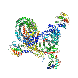
7UX1
 | | EcMscK in an Open Conformation | | 分子名称: | Mechanosensitive channel MscK | | 著者 | Mount, J.W, Yuan, P. | | 登録日 | 2022-05-04 | | 公開日 | 2022-11-23 | | 最終更新日 | 2024-06-12 | | 実験手法 | ELECTRON MICROSCOPY (3.48 Å) | | 主引用文献 | Structural basis for mechanotransduction in a potassium-dependent mechanosensitive ion channel.
Nat Commun, 13, 2022
|
|
7UX2
 | |
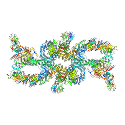
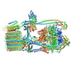
7UX3
 | | Asymmetric unit of AP-1, Arf1, Nef lattice on MHC-I lipopeptide incorporated narrow membrane tubes | | 分子名称: | ADP-ribosylation factor 1, AP-1 complex subunit beta-1, AP-1 complex subunit gamma-1, ... | | 著者 | Hooy, R.M, Hurley, J.H. | | 登録日 | 2022-05-04 | | 公開日 | 2023-05-10 | | 最終更新日 | 2024-06-12 | | 実験手法 | ELECTRON MICROSCOPY (9.6 Å) | | 主引用文献 | Self-assembly and structure of a clathrin-independent AP-1:Arf1 tubular membrane coat.
Sci Adv, 8, 2022
|
|
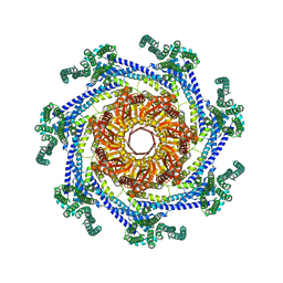
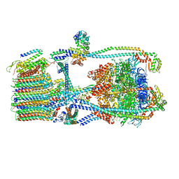
7UW9
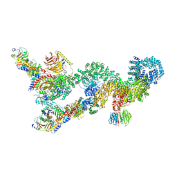
 | | Citrus V-ATPase State 1, H in contact with subunit a | | 分子名称: | V-type proton ATPase catalytic subunit A, V-type proton ATPase subunit AP1 fragment, V-type proton ATPase subunit AP2 fragment, ... | | 著者 | Keon, K.A, Abdelaziz, R.A, Schulze, W.X, Schumacher, K, Rubinstein, J.L. | | 登録日 | 2022-05-03 | | 公開日 | 2022-07-06 | | 最終更新日 | 2024-06-12 | | 実験手法 | ELECTRON MICROSCOPY (4.2 Å) | | 主引用文献 | Structure of V-ATPase from citrus fruit.
Structure, 30, 2022
|
|
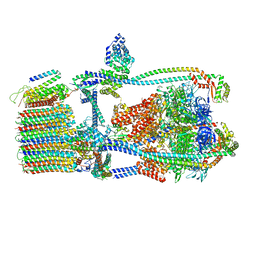
7UWB
 | | Citrus V-ATPase State 2, Highest-Resolution Class | | 分子名称: | V-type proton ATPase catalytic subunit A, V-type proton ATPase subunit AP1 fragment, V-type proton ATPase subunit AP2 fragment, ... | | 著者 | Keon, K.A, Abdelaziz, R.A, Schulze, W.X, Schumacher, K, Rubinstein, J.L. | | 登録日 | 2022-05-03 | | 公開日 | 2022-07-06 | | 最終更新日 | 2024-01-17 | | 実験手法 | ELECTRON MICROSCOPY (3.9 Å) | | 主引用文献 | Structure of V-ATPase from citrus fruit.
Structure, 30, 2022
|
|
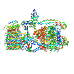
7UWA
 | | Citrus V-ATPase State 1, H in contact with subunits AB | | 分子名称: | V-type proton ATPase catalytic subunit A, V-type proton ATPase subunit AP1 fragment, V-type proton ATPase subunit AP2 fragment, ... | | 著者 | Abdelaziz, R.A, Keon, K.A, Schulze, W.X, Schumacher, K, Rubinstein, J.L. | | 登録日 | 2022-05-03 | | 公開日 | 2022-07-06 | | 最終更新日 | 2024-06-12 | | 実験手法 | ELECTRON MICROSCOPY (4.3 Å) | | 主引用文献 | Structure of V-ATPase from citrus fruit.
Structure, 30, 2022
|
|
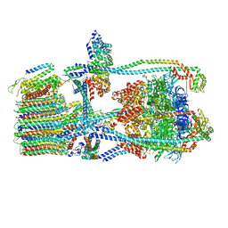
7UWD
 | | Citrus V-ATPase State 2, H in contact with subunits AB | | 分子名称: | V-type proton ATPase catalytic subunit A, V-type proton ATPase subunit, V-type proton ATPase subunit AP1 fragment, ... | | 著者 | Keon, K.A, Abdelaziz, R.A, Schulze, W.X, Schumacher, K, Rubinstein, J.L. | | 登録日 | 2022-05-03 | | 公開日 | 2022-07-06 | | 最終更新日 | 2024-01-17 | | 実験手法 | ELECTRON MICROSCOPY (4.1 Å) | | 主引用文献 | Structure of V-ATPase from citrus fruit.
Structure, 30, 2022
|
|
7UWC
 | | Citrus V-ATPase State 2, H in contact with subunit a | | 分子名称: | V-type proton ATPase catalytic subunit A, V-type proton ATPase subunit AP1 fragment, V-type proton ATPase subunit AP2 fragment, ... | | 著者 | Keon, K.A, Abdelaziz, R.A, Schulze, W.X, Schumacher, K, Rubinstein, J.L. | | 登録日 | 2022-05-03 | | 公開日 | 2022-07-06 | | 最終更新日 | 2024-01-17 | | 実験手法 | ELECTRON MICROSCOPY (4 Å) | | 主引用文献 | Structure of V-ATPase from citrus fruit.
Structure, 30, 2022
|
|


