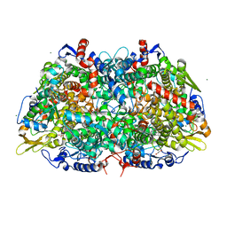
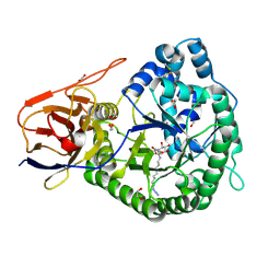
7SUC
 
 | |
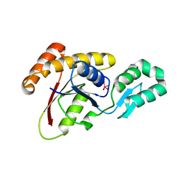
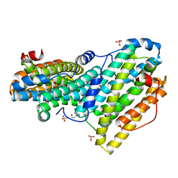
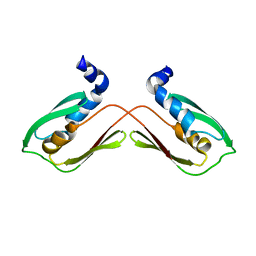
5JFB
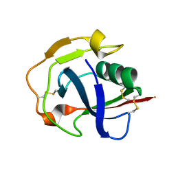 | | Crystal structure of the scavenger receptor cysteine-rich domain 5 (SRCR5) from porcine CD163 | | 分子名称: | Scavenger receptor cysteine-rich type 1 protein M130 | | 著者 | Ma, H, Jiang, L, Qiao, S, Zhang, G, Li, R. | | 登録日 | 2016-04-19 | | 公開日 | 2017-03-22 | | 最終更新日 | 2023-11-08 | | 実験手法 | X-RAY DIFFRACTION (2 Å) | | 主引用文献 | The Crystal Structure of the Fifth Scavenger Receptor Cysteine-Rich Domain of Porcine CD163 Reveals an Important Residue Involved in Porcine Reproductive and Respiratory Syndrome Virus Infection
J. Virol., 91, 2017
|
|
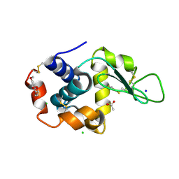
6W67
 | |
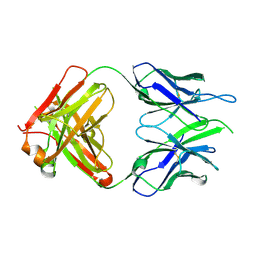
6X3O
 | | Co-structure of BTK kinase domain with L-005191930 inhibitor | | 分子名称: | 4-[8-azanyl-3-[(2~{S})-1-[4-(dimethylamino)butanoyl]pyrrolidin-2-yl]imidazo[1,5-a]pyrazin-1-yl]-~{N}-(1,3-thiazol-2-yl)benzamide, 4-{8-amino-3-[(6R,8aS)-3-oxo-3,5,6,7,8,8a-hexahydroindolizin-6-yl]imidazo[1,5-a]pyrazin-1-yl}-3-methoxy-N-[4-(trifluoromethyl)pyridin-2-yl]benzamide, Tyrosine-protein kinase BTK | | 著者 | Fischmann, T.O. | | 登録日 | 2020-05-21 | | 公開日 | 2020-07-22 | | 最終更新日 | 2024-03-06 | | 実験手法 | X-RAY DIFFRACTION (1.9 Å) | | 主引用文献 | Potent, non-covalent reversible BTK inhibitors with 8-amino-imidazo[1,5-a]pyrazine core featuring 3-position bicyclic ring substitutes.
Bioorg.Med.Chem.Lett., 30, 2020
|
|
5LA7
 | | Crystal structure of human proheparanase, in complex with glucuronic acid configured aziridine probe JJB355 | | 分子名称: | (1~{S},2~{R},3~{S},4~{S},5~{S},6~{R})-2-(8-azidooctylamino)-3,4,5,6-tetrakis(oxidanyl)cyclohexane-1-carboxylic acid, 1,2-ETHANEDIOL, 2-acetamido-2-deoxy-beta-D-glucopyranose, ... | | 著者 | Wu, L, Jin, Y, Davies, G.J. | | 登録日 | 2016-06-13 | | 公開日 | 2017-05-31 | | 最終更新日 | 2024-01-10 | | 実験手法 | X-RAY DIFFRACTION (1.94 Å) | | 主引用文献 | Activity-based probes for functional interrogation of retaining beta-glucuronidases.
Nat. Chem. Biol., 13, 2017
|
|
3OMX
 | | Crystal structure of Ssu72 with vanadate complex | | 分子名称: | CG14216, VANADATE ION | | 著者 | Zhang, Y, Zhang, M, Zhang, Y. | | 登録日 | 2010-08-27 | | 公開日 | 2011-01-19 | | 最終更新日 | 2023-09-06 | | 実験手法 | X-RAY DIFFRACTION (2.3366 Å) | | 主引用文献 | Crystal structure of Ssu72, an essential eukaryotic phosphatase specific for the C-terminal domain of RNA polymerase II, in complex with a transition state analogue.
Biochem.J., 434, 2011
|
|
5KKJ
 | | 2.0-Angstrom In situ Mylar structure of hen egg-white lysozyme (HEWL) at 293 K | | 分子名称: | 3,6,9,12,15,18,21,24-OCTAOXAHEXACOSAN-1-OL, ACETIC ACID, CHLORIDE ION, ... | | 著者 | Broecker, J, Ernst, O.P. | | 登録日 | 2016-06-21 | | 公開日 | 2017-02-15 | | 最終更新日 | 2023-09-27 | | 実験手法 | X-RAY DIFFRACTION (2.001 Å) | | 主引用文献 | A Versatile System for High-Throughput In Situ X-ray Screening and Data Collection of Soluble and Membrane-Protein Crystals.
Cryst Growth Des, 16, 2016
|
|
6VOR
 | |
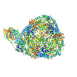
8QOA
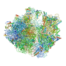 | | Structure of SecM-stalled Escherichia coli 70S ribosome | | 分子名称: | 16S rRNA, 23S rRNA, 30S ribosomal protein S16, ... | | 著者 | Gersteuer, F, Morici, M, Wilson, D.N. | | 登録日 | 2023-09-28 | | 公開日 | 2024-03-20 | | 最終更新日 | 2024-04-24 | | 実験手法 | ELECTRON MICROSCOPY (2 Å) | | 主引用文献 | The SecM arrest peptide traps a pre-peptide bond formation state of the ribosome.
Nat Commun, 15, 2024
|
|
8QCQ
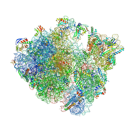 | | B. subtilis ApdA-stalled ribosomal complex | | 分子名称: | 16S rRNA, 23S rRNA, 30S ribosomal protein S10, ... | | 著者 | Morici, M, Wilson, D.N. | | 登録日 | 2023-08-28 | | 公開日 | 2024-03-20 | | 最終更新日 | 2024-04-03 | | 実験手法 | ELECTRON MICROSCOPY (2.3 Å) | | 主引用文献 | RAPP-containing arrest peptides induce translational stalling by short circuiting the ribosomal peptidyltransferase activity.
Nat Commun, 15, 2024
|
|
8QBT
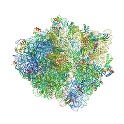 | | E. coli ApdP-stalled ribosomal complex | | 分子名称: | 16S rRNA, 23S rRNA, 30S ribosomal protein S11, ... | | 著者 | Morici, M, Wilson, D.N. | | 登録日 | 2023-08-25 | | 公開日 | 2024-03-20 | | 最終更新日 | 2024-04-03 | | 実験手法 | ELECTRON MICROSCOPY (2.2 Å) | | 主引用文献 | RAPP-containing arrest peptides induce translational stalling by short circuiting the ribosomal peptidyltransferase activity.
Nat Commun, 15, 2024
|
|
6QO8
 | |
6QO9
 | |
7UK4
 | | KS-AT di-domain of mycobacterial Pks13 with endogenous KS ligand bound | | 分子名称: | Polyketide synthase PKS13, UNKNOWN LIGAND | | 著者 | Kim, S.K, Dickinson, M.S, Finer-Moore, J.S, Rosenberg, O.S, Stroud, R.M. | | 登録日 | 2022-03-31 | | 公開日 | 2023-02-15 | | 最終更新日 | 2023-03-29 | | 実験手法 | ELECTRON MICROSCOPY (1.94 Å) | | 主引用文献 | Structure and dynamics of the essential endogenous mycobacterial polyketide synthase Pks13.
Nat.Struct.Mol.Biol., 30, 2023
|
|
8TPJ
 | | Top cylinder bound to OCP from high-resolution phycobilisome quenched by OCP (local refinement) | | 分子名称: | Allophycocyanin alpha chain, Allophycocyanin beta chain, Orange carotenoid-binding protein, ... | | 著者 | Sauer, P.V, Sutter, M, Cupellini, L. | | 登録日 | 2023-08-04 | | 公開日 | 2024-04-17 | | 実験手法 | ELECTRON MICROSCOPY (2.1 Å) | | 主引用文献 | Structural and quantum chemical basis for OCP-mediated quenching of phycobilisomes.
Sci Adv, 10, 2024
|
|
6QO7
 | |
6W66
 | | The structure of the F64A, S172A mutant Keap1-BTB domain in complex with SKP1-FBXL17 | | 分子名称: | F-box/LRR-repeat protein 17, Kelch-like ECH-associated protein 1, S-phase kinase-associated protein 1 | | 著者 | Mena, E.L, Gee, C.L, Kuriyan, J, Rape, M. | | 登録日 | 2020-03-16 | | 公開日 | 2020-08-19 | | 最終更新日 | 2023-10-18 | | 実験手法 | X-RAY DIFFRACTION (3.21 Å) | | 主引用文献 | Structural basis for dimerization quality control.
Nature, 586, 2020
|
|
6QO5
 | |
6QOB
 | |
5EUP
 | |
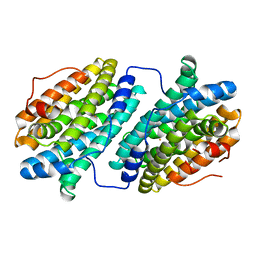
3PXN
 | | Crystal structure of the Drosophila kinesin family member Kin10/NOD in complex with divalent manganese and ADP | | 分子名称: | ADENOSINE-5'-DIPHOSPHATE, Kinesin-like protein Nod, MANGANESE (II) ION | | 著者 | Cochran, J.C, Zhao, Y.C, Wilcox, D.E, Kull, F.J. | | 登録日 | 2010-12-10 | | 公開日 | 2011-12-21 | | 最終更新日 | 2023-09-13 | | 実験手法 | X-RAY DIFFRACTION (2.6 Å) | | 主引用文献 | A metal switch for controlling the activity of molecular motor proteins.
Nat.Struct.Mol.Biol., 19, 2012
|
|
6WA1
 | |
7U0Y
 | |
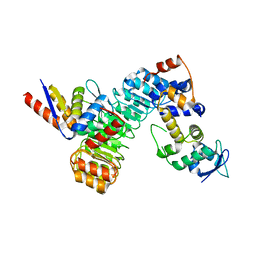
4YMZ
 | | DHAP bound Leptospira Interrogans Triosephosphate Isomerase (LiTIM) | | 分子名称: | 1,2-ETHANEDIOL, 1,3-DIHYDROXYACETONEPHOSPHATE, SULFATE ION, ... | | 著者 | Pareek, V, Balaram, P, Murthy, M.R.N. | | 登録日 | 2015-03-08 | | 公開日 | 2016-03-09 | | 最終更新日 | 2023-11-08 | | 実験手法 | X-RAY DIFFRACTION (1.87 Å) | | 主引用文献 | Connecting Active-Site Loop Conformations and Catalysis in Triosephosphate Isomerase: Insights from a Rare Variation at Residue 96 in the Plasmodial Enzyme
Chembiochem, 17, 2016
|
|
5L77
 | | A glycoside hydrolase mutant with an unreacted activity based probe bound | | 分子名称: | (1~{R},2~{S},3~{R},4~{S},5~{S},6~{R})-7-[8-[(azanylidene-{4}-azanylidene)amino]octyl]-3,4,5-tris(oxidanyl)-7-azabicyclo[4.1.0]heptane-2-carboxylic acid, CHLORIDE ION, GLYCEROL, ... | | 著者 | Jin, Y, Wu, L, Jiang, J.B, Overkleeft, H.S, Davies, G.J. | | 登録日 | 2016-06-02 | | 公開日 | 2017-05-31 | | 最終更新日 | 2024-01-10 | | 実験手法 | X-RAY DIFFRACTION (1.24 Å) | | 主引用文献 | Activity-based probes for functional interrogation of retaining beta-glucuronidases.
Nat. Chem. Biol., 13, 2017
|
|


