Loading
PDBj
MenuPDBj@FacebookPDBj@TwitterPDBj@YouTubewwPDB FoundationwwPDB
RCSB PDBPDBeBMRBAdv. SearchSearch help
PDB: 13 results

6H53
DownloadVisualize
BU of 6h53 by Molmil
Crystal structure of Mycobacterium tuberculosis phosphatidylinositol phosphate synthase (PgsA1) in apo form
Descriptor: CDP-diacylglycerol--inositol 3-phosphatidyltransferase, SULFATE ION, UNKNOWN BRANCHED FRAGMENT OF PHOSPHOLIPID
Authors:Grave, K, Hogbom, M.
Deposit date:2018-07-23
Release date:2019-05-15
Last modified:2024-01-17
Method:X-RAY DIFFRACTION (2.9 Å)
Cite:Structure ofMycobacterium tuberculosisphosphatidylinositol phosphate synthase reveals mechanism of substrate binding and metal catalysis.
Commun Biol, 2, 2019
6H5A
DownloadVisualize
BU of 6h5a by Molmil
Crystal structure of Mycobacterium tuberculosis phosphatidylinositol phosphate synthase (PgsA1) in complex with manganese and citrate
Descriptor: (2R)-2,3-dihydroxypropyl (9Z)-octadec-9-enoate, CDP-diacylglycerol--inositol 3-phosphatidyltransferase, CITRATE ANION, ...
Authors:Grave, K, Hogbom, M.
Deposit date:2018-07-24
Release date:2019-05-15
Last modified:2024-01-17
Method:X-RAY DIFFRACTION (1.88 Å)
Cite:Structure ofMycobacterium tuberculosisphosphatidylinositol phosphate synthase reveals mechanism of substrate binding and metal catalysis.
Commun Biol, 2, 2019
6H59
DownloadVisualize
BU of 6h59 by Molmil
Crystal structure of Mycobacterium tuberculosis phosphatidylinositol phosphate synthase (PgsA1) with CDP-DAG bound
Descriptor: (2S)-2,3-dihydroxypropyl (9Z)-octadec-9-enoate, CDP-diacylglycerol--inositol 3-phosphatidyltransferase, MAGNESIUM ION, ...
Authors:Grave, K, Hogbom, M.
Deposit date:2018-07-24
Release date:2019-05-15
Last modified:2024-01-17
Method:X-RAY DIFFRACTION (1.8 Å)
Cite:Structure ofMycobacterium tuberculosisphosphatidylinositol phosphate synthase reveals mechanism of substrate binding and metal catalysis.
Commun Biol, 2, 2019
6QO8
DownloadVisualize
BU of 6qo8 by Molmil
Crystal structure of ribonucleotide reductase NrdF from Bacillus anthracis anaerobically soaked with ferrous ions
Descriptor: 2-[BIS-(2-HYDROXY-ETHYL)-AMINO]-2-HYDROXYMETHYL-PROPANE-1,3-DIOL, FE (II) ION, Ribonucleoside-diphosphate reductase subunit beta, ...
Authors:Grave, K, Hogbom, M.
Deposit date:2019-02-12
Release date:2019-08-21
Last modified:2024-01-24
Method:X-RAY DIFFRACTION (1.31921768 Å)
Cite:Redox-induced structural changes in the di-iron and di-manganese forms of Bacillus anthracis ribonucleotide reductase subunit NrdF suggest a mechanism for gating of radical access.
J.Biol.Inorg.Chem., 24, 2019
6QO9
DownloadVisualize
BU of 6qo9 by Molmil
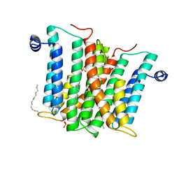
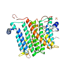
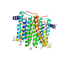
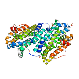
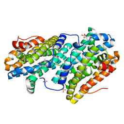
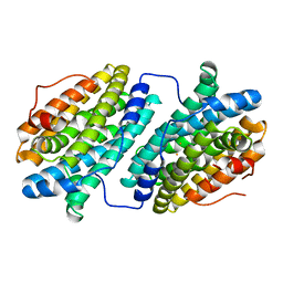
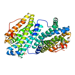
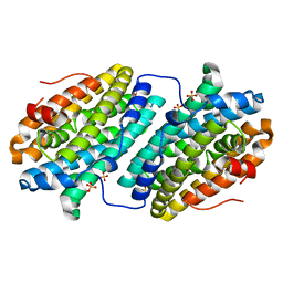
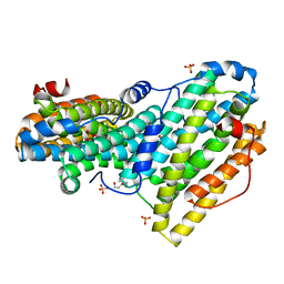
Crystal structure of ribonucleotide reductase NrdF from Bacillus anthracis soaked with manganese ions
Descriptor: 2-[BIS-(2-HYDROXY-ETHYL)-AMINO]-2-HYDROXYMETHYL-PROPANE-1,3-DIOL, MANGANESE (II) ION, Ribonucleoside-diphosphate reductase subunit beta, ...
Authors:Grave, K, Hogbom, M.
Deposit date:2019-02-12
Release date:2019-08-21
Last modified:2024-01-24
Method:X-RAY DIFFRACTION (1.299 Å)
Cite:Redox-induced structural changes in the di-iron and di-manganese forms of Bacillus anthracis ribonucleotide reductase subunit NrdF suggest a mechanism for gating of radical access.
J.Biol.Inorg.Chem., 24, 2019
6QOB
DownloadVisualize
BU of 6qob by Molmil
Crystal structure of ribonucleotide reductase NrdF from Bacillus anthracis with partially oxidised di-iron metallocofactor
Descriptor: 2-[BIS-(2-HYDROXY-ETHYL)-AMINO]-2-HYDROXYMETHYL-PROPANE-1,3-DIOL, FE (III) ION, Ribonucleoside-diphosphate reductase subunit beta, ...
Authors:Grave, K, Hogbom, M.
Deposit date:2019-02-12
Release date:2019-08-21
Last modified:2024-01-24
Method:X-RAY DIFFRACTION (1.457 Å)
Cite:Redox-induced structural changes in the di-iron and di-manganese forms of Bacillus anthracis ribonucleotide reductase subunit NrdF suggest a mechanism for gating of radical access.
J.Biol.Inorg.Chem., 24, 2019
6TQY
DownloadVisualize
BU of 6tqy by Molmil
Crystal structure of ribonucleotide reductase NrdF L61G variant from Bacillus anthracis anaerobically soaked with Fe(II) and Mn(II) ions
Descriptor: MANGANESE (II) ION, Ribonucleoside-diphosphate reductase subunit beta, SULFATE ION
Authors:Grave, K, Hogbom, M.
Deposit date:2019-12-17
Release date:2020-04-15
Last modified:2024-01-24
Method:X-RAY DIFFRACTION (1.77 Å)
Cite:The Bacillus anthracis class Ib ribonucleotide reductase subunit NrdF intrinsically selects manganese over iron.
J.Biol.Inorg.Chem., 25, 2020
6TQW
DownloadVisualize
BU of 6tqw by Molmil
Crystal structure of ribonucleotide reductase NrdF from Bacillus anthracis anaerobically soaked with Fe(II) and Mn(II) ions
Descriptor: MANGANESE (II) ION, Ribonucleoside-diphosphate reductase subunit beta, SULFATE ION
Authors:Grave, K, Hogbom, M.
Deposit date:2019-12-17
Release date:2020-04-15
Last modified:2024-01-24
Method:X-RAY DIFFRACTION (1.55 Å)
Cite:The Bacillus anthracis class Ib ribonucleotide reductase subunit NrdF intrinsically selects manganese over iron.
J.Biol.Inorg.Chem., 25, 2020
6TQZ
DownloadVisualize
BU of 6tqz by Molmil
Crystal structure of ribonucleotide reductase NrdF L61G variant from Bacillus anthracis aerobically soaked with Fe(II) and Mn(II) ions
Descriptor: FE (II) ION, MANGANESE (II) ION, Ribonucleoside-diphosphate reductase subunit beta, ...
Authors:Grave, K, Hogbom, M.
Deposit date:2019-12-17
Release date:2020-04-15
Last modified:2024-01-24
Method:X-RAY DIFFRACTION (2.099335 Å)
Cite:The Bacillus anthracis class Ib ribonucleotide reductase subunit NrdF intrinsically selects manganese over iron.
J.Biol.Inorg.Chem., 25, 2020
6TQV
DownloadVisualize
BU of 6tqv by Molmil
Crystal structure of ribonucleotide reductase NrdF from Bacillus anthracis aerobically soaked with Fe(II) and Mn(II) ions
Descriptor: MANGANESE (II) ION, Ribonucleoside-diphosphate reductase subunit beta, SULFATE ION
Authors:Grave, K, Hogbom, M.
Deposit date:2019-12-17
Release date:2020-04-15
Last modified:2024-01-24
Method:X-RAY DIFFRACTION (1.35000968 Å)
Cite:The Bacillus anthracis class Ib ribonucleotide reductase subunit NrdF intrinsically selects manganese over iron.
J.Biol.Inorg.Chem., 25, 2020
6QO7
DownloadVisualize
BU of 6qo7 by Molmil
Crystal structure of ribonucleotide reductase NrdF from Bacillus anthracis aerobically soaked with ferrous ions (photo-reduced)
Descriptor: CHLORIDE ION, FE (II) ION, Ribonucleoside-diphosphate reductase subunit beta
Authors:Grave, K, Hogbom, M.
Deposit date:2019-02-12
Release date:2019-08-21
Last modified:2019-10-02
Method:X-RAY DIFFRACTION (1.628 Å)
Cite:Redox-induced structural changes in the di-iron and di-manganese forms of Bacillus anthracis ribonucleotide reductase subunit NrdF suggest a mechanism for gating of radical access.
J.Biol.Inorg.Chem., 24, 2019
6QO5
DownloadVisualize
BU of 6qo5 by Molmil
Crystal structure of apo (metal-free) ribonucleotide reductase NrdF from Bacillus anthracis
Descriptor: CHLORIDE ION, Ribonucleoside-diphosphate reductase subunit beta, SULFATE ION
Authors:Grave, K, Hogbom, M.
Deposit date:2019-02-12
Release date:2019-08-21
Last modified:2024-01-24
Method:X-RAY DIFFRACTION (1.511 Å)
Cite:Redox-induced structural changes in the di-iron and di-manganese forms of Bacillus anthracis ribonucleotide reductase subunit NrdF suggest a mechanism for gating of radical access.
J.Biol.Inorg.Chem., 24, 2019
6TQX
DownloadVisualize
BU of 6tqx by Molmil
Crystal structure of apo (metal-free) ribonucleotide reductase NrdF L61G variant from Bacillus anthracis
Descriptor: Ribonucleoside-diphosphate reductase subunit beta, SULFATE ION
Authors:Grave, K, Hogbom, M.
Deposit date:2019-12-17
Release date:2020-04-15
Last modified:2024-01-24
Method:X-RAY DIFFRACTION (2.0500176 Å)
Cite:The Bacillus anthracis class Ib ribonucleotide reductase subunit NrdF intrinsically selects manganese over iron.
J.Biol.Inorg.Chem., 25, 2020

218853

PDB entries from 2024-04-24

PDB statisticsPDBj update infoContact PDBjnumon