2H6R
 
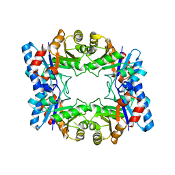 | | Crystal Structure of triosephosphate isomerase (TIM) from Methanocaldococcus jannaschii | | 分子名称: | Triosephosphate isomerase | | 著者 | Gayathri, P, Banerjee, M, Vijayalakshmi, A, Balaram, H, Balaram, P, Murthy, M.R.N. | | 登録日 | 2006-06-01 | | 公開日 | 2007-02-06 | | 最終更新日 | 2023-10-25 | | 実験手法 | X-RAY DIFFRACTION (2.3 Å) | | 主引用文献 | Structure of triosephosphate isomerase (TIM) from Methanocaldococcus jannaschii
Acta Crystallogr.,Sect.D, 63, 2007
|
|
2H6S
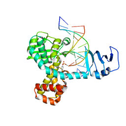
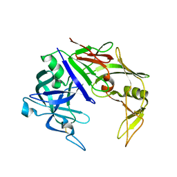 | | Secreted aspartic proteinase (Sap) 3 from Candida albicans | | 分子名称: | Candidapepsin-3, ZINC ION | | 著者 | Ruge, E, Borelli, C, Maskos, K, Huber, R. | | 登録日 | 2006-06-01 | | 公開日 | 2007-06-12 | | 最終更新日 | 2017-10-18 | | 実験手法 | X-RAY DIFFRACTION (2.2 Å) | | 主引用文献 | The crystal structure of the secreted aspartic proteinase 3 from Candida albicans and its complex with pepstatin A.
Proteins, 68, 2007
|
|
2H6T
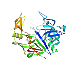 | | Secreted aspartic proteinase (Sap) 3 from Candida albicans complexed with pepstatin A | | 分子名称: | Candidapepsin-3, ZINC ION, pepstatin A | | 著者 | Ruge, E, Borelli, C, Maskos, K, Huber, R. | | 登録日 | 2006-06-01 | | 公開日 | 2007-06-12 | | 最終更新日 | 2017-10-18 | | 実験手法 | X-RAY DIFFRACTION (1.9 Å) | | 主引用文献 | The crystal structure of the secreted aspartic proteinase 3 from Candida albicans and its complex with pepstatin A.
Proteins, 68, 2007
|
|
2H6U
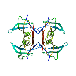 | | Crystal structure of 5-hydroxyisourate hydrolase (formerly known as TRP, transthyretin related protein) | | 分子名称: | 5-HYDROXYISOURATE HYDROLASE (FORMERLY KNOWN AS TRP, TRANSTHYRETIN RELATED PROTEIN) | | 著者 | Zanotti, G, Cendron, L, Folli, C, Ramazzina, I, Percudani, R, Berni, R. | | 登録日 | 2006-06-01 | | 公開日 | 2006-10-31 | | 最終更新日 | 2023-08-30 | | 実験手法 | X-RAY DIFFRACTION (1.7 Å) | | 主引用文献 | Structure of Zebra fish HIUase: Insights into Evolution of an Enzyme to a Hormone Transporter.
J.Mol.Biol., 363, 2006
|
|
2H6V
 | |
2H6X
 | |
2H6Y
 | |
2H6Z
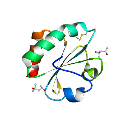
 | | Crystal Structure of Thioredoxin Mutant E44D in Hexagonal (p61) Space Group | | 分子名称: | (4S)-2-METHYL-2,4-PENTANEDIOL, Thioredoxin | | 著者 | Gavira, J.A, Godoy-Ruiz, R, Ibarra-Molero, B, Sanchez-Ruiz, J.M. | | 登録日 | 2006-06-01 | | 公開日 | 2007-05-15 | | 最終更新日 | 2023-08-30 | | 実験手法 | X-RAY DIFFRACTION (2.25 Å) | | 主引用文献 | A stability pattern of protein hydrophobic mutations that reflects evolutionary structural optimization.
Biophys.J., 89, 2005
|
|
2H70
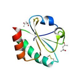
 | | Crystal Structure of Thioredoxin Mutant D9E in Hexagonal (p61) Space Group | | 分子名称: | (4S)-2-METHYL-2,4-PENTANEDIOL, Thioredoxin | | 著者 | Gavira, J.A, Godoy-Ruiz, R, Ibarra-Molero, B, Sanchez-Ruiz, J.M. | | 登録日 | 2006-06-01 | | 公開日 | 2007-05-15 | | 最終更新日 | 2023-08-30 | | 実験手法 | X-RAY DIFFRACTION (2.7 Å) | | 主引用文献 | A stability pattern of protein hydrophobic mutations that reflects evolutionary structural optimization.
Biophys.J., 89, 2005
|
|
2H71
 | |
2H72
 | |
2H73
 | |
2H74
 | |
2H75
 | |
2H76
 | |
2H77
 | | Crystal structure of human TR alpha bound T3 in monoclinic space group | | 分子名称: | 3,5,3'TRIIODOTHYRONINE, THRA protein | | 著者 | Nascimento, A.S, Dias, S.M.G, Nunes, F.M, Aparicio, R, Bleicher, L, Ambrosio, A.L.B, Figueira, A.C.M, Santos, M.A.M, Neto, M.O, Fischer, H, Togashi, H.F.M, Craievich, A.F, Garrat, R.C, Baxter, J.D, Webb, P, Polikarpov, I. | | 登録日 | 2006-06-01 | | 公開日 | 2006-07-25 | | 最終更新日 | 2023-11-15 | | 実験手法 | X-RAY DIFFRACTION (2.33 Å) | | 主引用文献 | Structural rearrangements in the thyroid hormone receptor hinge domain and their putative role in the receptor function.
J.Mol.Biol., 360, 2006
|
|
2H79
 | | Crystal Structure of human TR alpha bound T3 in orthorhombic space group | | 分子名称: | 3,5,3'TRIIODOTHYRONINE, THRA protein | | 著者 | Nascimento, A.S, Dias, S.M.G, Nunes, F.M, Aparicio, R, Bleicher, L, Ambrosio, A.L.B, Figueira, A.C.M, Santos, M.A.M, Neto, M.O, Fischer, H, Togashi, H.F.M, Craievich, A.F, Garrat, R.C, Baxter, J.D, Webb, P, Polikarpov, I. | | 登録日 | 2006-06-01 | | 公開日 | 2006-07-25 | | 最終更新日 | 2023-11-15 | | 実験手法 | X-RAY DIFFRACTION (1.87 Å) | | 主引用文献 | Structural rearrangements in the thyroid hormone receptor hinge domain and their putative role in the receptor function.
J.Mol.Biol., 360, 2006
|
|
2H7A
 | | NMR Structure of the Conserved Protein YcgL from Escherichia coli representing the DUF709 Family Reveals a Novel a/b/a Sandwich Fold | | 分子名称: | Hypothetical protein ycgL | | 著者 | Minailiuc, O.M, Vavelyuk, O, Ekiel, I, Hung, M.-Ni, Cygler, M, Gandhi, S, Montreal-Kingston Bacterial Structural Genomics Initiative (BSGI) | | 登録日 | 2006-06-01 | | 公開日 | 2007-04-17 | | 最終更新日 | 2024-05-01 | | 実験手法 | SOLUTION NMR | | 主引用文献 | NMR structure of YcgL, a conserved protein from Escherichia coli representing the DUF709 family, with a novel alpha/beta/alpha sandwich fold.
Proteins, 66, 2007
|
|
2H7B
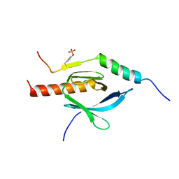
 | | Solution structure of the eTAFH domain from the human leukemia-associated fusion protein AML1-ETO | | 分子名称: | Core-binding factor, ML1-ETO | | 著者 | Plevin, M.J, Zhang, J, Guo, C, Roeder, R.G, Ikura, M. | | 登録日 | 2006-06-01 | | 公開日 | 2006-07-11 | | 最終更新日 | 2024-05-29 | | 実験手法 | SOLUTION NMR | | 主引用文献 | The acute myeloid leukemia fusion protein AML1-ETO targets E proteins via a paired amphipathic helix-like TBP-associated factor homology domain
Proc.Natl.Acad.Sci.USA, 103, 2006
|
|
2H7C
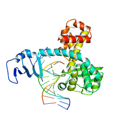
 | | Crystal structure of human carboxylesterase in complex with Coenzyme A | | 分子名称: | 2-acetamido-2-deoxy-beta-D-glucopyranose, 2-acetamido-2-deoxy-beta-D-glucopyranose-(1-4)-2-acetamido-2-deoxy-beta-D-glucopyranose, COENZYME A, ... | | 著者 | Bencharit, S, Edwards, C.C, Morton, C.L, Howard-Williams, E.L, Potter, P.M, Redinbo, M.R. | | 登録日 | 2006-06-02 | | 公開日 | 2006-08-29 | | 最終更新日 | 2023-10-25 | | 実験手法 | X-RAY DIFFRACTION (2 Å) | | 主引用文献 | Multisite promiscuity in the processing of endogenous substrates by human carboxylesterase 1
J.Mol.Biol., 363, 2006
|
|
2H7D
 | |
2H7E
 | |
2H7F
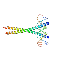
 | | Structure of variola topoisomerase covalently bound to DNA | | 分子名称: | 5'-D(*TP*AP*AP*TP*AP*AP*GP*GP*GP*CP*GP*AP*CP*A)-3', 5'-D(*TP*TP*GP*TP*CP*GP*CP*CP*CP*TP*T)-3', DNA topoisomerase 1 | | 著者 | Perry, K, Hwang, Y, Bushman, F.D, Van Duyne, G.D. | | 登録日 | 2006-06-02 | | 公開日 | 2006-08-15 | | 最終更新日 | 2021-10-20 | | 実験手法 | X-RAY DIFFRACTION (2.7 Å) | | 主引用文献 | Structural basis for specificity in the poxvirus topoisomerase.
Mol.Cell, 23, 2006
|
|
2H7G
 | | Structure of variola topoisomerase non-covalently bound to DNA | | 分子名称: | 5'-D(*TP*AP*AP*TP*AP*AP*GP*GP*GP*CP*GP*AP*CP*A)-3', 5'-D(*TP*TP*GP*TP*CP*GP*CP*CP*CP*TP*TP*A)-3', DNA topoisomerase 1 | | 著者 | Perry, K, Hwang, Y, Bushman, F.D, Van Duyne, G.D. | | 登録日 | 2006-06-02 | | 公開日 | 2006-08-15 | | 最終更新日 | 2024-02-14 | | 実験手法 | X-RAY DIFFRACTION (1.9 Å) | | 主引用文献 | Structural basis for specificity in the poxvirus topoisomerase.
Mol.Cell, 23, 2006
|
|
2H7H
 | |


