2GWE
 
 | |
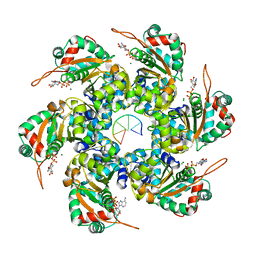
2GWF
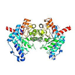
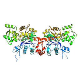
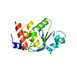
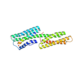
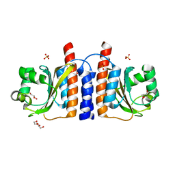
 | | Structure of a USP8-NRDP1 complex | | 分子名称: | RING finger protein 41, Ubiquitin carboxyl-terminal hydrolase 8 | | 著者 | Walker, J.R, Avvakumov, G.V, Xue, S, Newman, E.M, Butler-Cole, C, Finerty Jr, P.J, Weigelt, J, Sundstrom, M, Arrowsmith, C.H, Edwards, A.M, Bochkarev, A, Dhe-Paganon, S, Structural Genomics Consortium (SGC) | | 登録日 | 2006-05-04 | | 公開日 | 2006-06-06 | | 最終更新日 | 2024-02-14 | | 実験手法 | X-RAY DIFFRACTION (2.3 Å) | | 主引用文献 | Amino-terminal Dimerization, NRDP1-Rhodanese Interaction, and Inhibited Catalytic Domain Conformation of the Ubiquitin-specific Protease 8 (USP8).
J.Biol.Chem., 281, 2006
|
|
2GWG
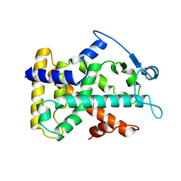
 | | Crystal Structure of 4-Oxalomesaconate Hydratase, LigJ, from Rhodopseudomonas palustris, Northeast Structural Genomics Target RpR66. | | 分子名称: | 4-oxalomesaconate hydratase, ZINC ION | | 著者 | Forouhar, F, Abashidze, M, Jayaraman, S, Cunningham, K, Ciao, M, Ma, L, Xiao, R, Acton, T.B, Montelione, G.T, Hunt, J.F, Tong, L, Northeast Structural Genomics Consortium (NESG) | | 登録日 | 2006-05-04 | | 公開日 | 2006-05-23 | | 最終更新日 | 2017-10-18 | | 実験手法 | X-RAY DIFFRACTION (1.8 Å) | | 主引用文献 | Crystal Structure of 4-Oxalomesaconate Hydratase, LigJ, from Rhodopseudomonas palustris, Northeast Structural Genomics Target RpR66
To be Published
|
|
2GWH
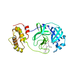
 | | Human Sulfotranferase SULT1C2 in complex with PAP and pentachlorophenol | | 分子名称: | ADENOSINE-3'-5'-DIPHOSPHATE, PENTACHLOROPHENOL, Sulfotransferase 1C2, ... | | 著者 | Tempel, W, Pan, P.W, Dombrovski, L, Allali-Hassani, A, Vedadi, M, Loppnau, P, Weigelt, J, Sundstrom, M, Arrowsmith, C.H, Edwards, A.M, Bochkarev, A, Plotnikov, A.N, Structural Genomics Consortium (SGC) | | 登録日 | 2006-05-04 | | 公開日 | 2006-05-16 | | 最終更新日 | 2023-08-30 | | 実験手法 | X-RAY DIFFRACTION (1.8 Å) | | 主引用文献 | Structural and chemical profiling of the human cytosolic sulfotransferases.
Plos Biol., 5, 2007
|
|
2GWJ
 | |
2GWK
 | |
2GWL
 | |
2GWM
 | |
2GWN
 | | The structure of putative dihydroorotase from Porphyromonas gingivalis. | | 分子名称: | BETA-MERCAPTOETHANOL, CACODYLATE ION, CHLORIDE ION, ... | | 著者 | Cuff, M.E, Borovilos, M, Moy, S, Joachimiak, A, Midwest Center for Structural Genomics (MCSG) | | 登録日 | 2006-05-04 | | 公開日 | 2006-06-06 | | 最終更新日 | 2011-07-13 | | 実験手法 | X-RAY DIFFRACTION (1.85 Å) | | 主引用文献 | The structure of putative dihydroorotase from Porphyromonas gingivalis.
To be Published
|
|
2GWO
 | | crystal structure of TMDP | | 分子名称: | Dual specificity protein phosphatase 13 | | 著者 | Kim, S.J, Ryu, S.E, Kim, J.H. | | 登録日 | 2006-05-05 | | 公開日 | 2007-03-20 | | 最終更新日 | 2024-03-13 | | 実験手法 | X-RAY DIFFRACTION (2.4 Å) | | 主引用文献 | Crystal structure of human TMDP, a testis-specific dual specificity protein phosphatase: implications for substrate specificity
Proteins, 66, 2007
|
|
2GWP
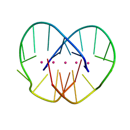
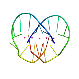
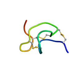
 | | High-resolution solution structure of the salt-bridge defficient mouse defensin (E15D)-Cryptdin4 | | 分子名称: | Defensin-related cryptdin 4 | | 著者 | Rosengren, K.J, Craik, D.J, Vogel, H.J, Daly, N.L, Ouellette, A.J. | | 登録日 | 2006-05-05 | | 公開日 | 2006-07-25 | | 最終更新日 | 2021-10-20 | | 実験手法 | SOLUTION NMR | | 主引用文献 | Structural and functional characterization of the conserved salt bridge in mammalian paneth cell alpha-defensins: solution structures of mouse CRYPTDIN-4 and (E15D)-CRYPTDIN-4.
J.Biol.Chem., 281, 2006
|
|
2GWQ
 | |
2GWR
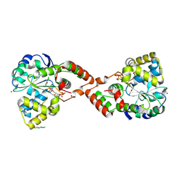
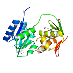 | | Crystal structure of the response regulator protein mtrA from Mycobacterium Tuberculosis | | 分子名称: | CALCIUM ION, DNA-binding response regulator mtrA, GLYCEROL | | 著者 | Friedland, N, Mack, T.R, Yu, M, Bursey, E.H, Hung, L.W, Stock, A.M, Waldo, G.S, Terwilliger, T.C. | | 登録日 | 2006-05-05 | | 公開日 | 2006-05-23 | | 最終更新日 | 2017-10-18 | | 実験手法 | X-RAY DIFFRACTION (2.1 Å) | | 主引用文献 | Domain orientation in the inactive response regulator Mycobacterium tuberculosis MtrA provides a barrier to activation.
Biochemistry, 46, 2007
|
|
2GWS
 | | Crystal Structure of human DNA Polymerase lambda with a G/G mismatch in the primer terminus | | 分子名称: | 1,2-ETHANEDIOL, 5'-D(*CP*GP*GP*CP*AP*GP*CP*GP*CP*AP*C)-3', 5'-D(*GP*TP*GP*CP*GP*G)-3', ... | | 著者 | Garcia-Diaz, M, Picher, A.J, Bebenek, K, Pedersen, L.C, Kunkel, T.A, Blanco, L. | | 登録日 | 2006-05-05 | | 公開日 | 2006-09-05 | | 最終更新日 | 2023-08-30 | | 実験手法 | X-RAY DIFFRACTION (2.4 Å) | | 主引用文献 | Promiscuous mismatch extension by human DNA polymerase lambda.
Nucleic Acids Res., 34, 2006
|
|
2GWW
 | |
2GWX
 | | MOLECULAR RECOGNITION OF FATTY ACIDS BY PEROXISOME PROLIFERATOR-ACTIVATED RECEPTORS | | 分子名称: | PROTEIN (PPAR-DELTA) | | 著者 | Xu, H.E, Lambert, M.H, Montana, V.G, Park, D.J, Blanchard, S, Brown, P, Sternbach, D, Lehmann, J, Bruce, G.W, Willson, T.M, Kliewer, S.A, Milburn, M.V. | | 登録日 | 1999-03-11 | | 公開日 | 2000-03-11 | | 最終更新日 | 2023-12-27 | | 実験手法 | X-RAY DIFFRACTION (2.3 Å) | | 主引用文献 | Molecular recognition of fatty acids by peroxisome proliferator-activated receptors.
Mol.Cell, 3, 1999
|
|
2GX0
 | |
2GX1
 | | Solution structure and alanine scan of a spider toxin that affects the activation of mammalian sodium channels | | 分子名称: | Neurotoxin magi-5 | | 著者 | Sabo, J.K, Corzo, G, Bosmans, F, Billen, B, Villegas, E, Tytgat, J, Norton, R.S. | | 登録日 | 2006-05-08 | | 公開日 | 2006-12-05 | | 最終更新日 | 2022-03-09 | | 実験手法 | SOLUTION NMR | | 主引用文献 | Solution Structure and Alanine Scan of a Spider Toxin That Affects the Activation of Mammalian Voltage-gated Sodium Channels
J.Biol.Chem., 282, 2007
|
|
2GX2
 | | Crystal structural and functional analysis of GFP-like fluorescent protein Dronpa | | 分子名称: | MAGNESIUM ION, fluorescent protein Dronpa | | 著者 | Hwang, K.Y, Nam, K.-H, Park, S.-Y, Sugiyama, K. | | 登録日 | 2006-05-08 | | 公開日 | 2007-05-08 | | 最終更新日 | 2024-10-09 | | 実験手法 | X-RAY DIFFRACTION (1.8 Å) | | 主引用文献 | Structural characterization of the photoswitchable fluorescent protein Dronpa-C62S
Biochem.Biophys.Res.Commun., 354, 2007
|
|
2GX4
 | | Crystal structure of SARS coronavirus 3CL protease inhibitor complex | | 分子名称: | 3C-like proteinase, N-[(BENZYLOXY)CARBONYL]-O-(TERT-BUTYL)-L-THREONYL-3-CYCLOHEXYL-N-[(1S)-2-HYDROXY-1-{[(3S)-2-OXOPYRROLIDIN-3-YL]METHYL}ETHYL]-L-ALANINAMIDE | | 著者 | Hsu, M.F, Wang, A.H.-J. | | 登録日 | 2006-05-08 | | 公開日 | 2007-05-08 | | 最終更新日 | 2020-04-08 | | 実験手法 | X-RAY DIFFRACTION (1.93 Å) | | 主引用文献 | Synthesis, crystal structure, structure-activity relationships, and antiviral activity of a potent SARS coronavirus 3CL protease inhibitor.
J.Med.Chem., 49, 2006
|
|
2GX5
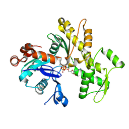
 | | N-terminal GAF domain of transcriptional pleiotropic repressor CodY | | 分子名称: | CYCLIC GUANOSINE MONOPHOSPHATE, GLYCEROL, GTP-sensing transcriptional pleiotropic repressor codY, ... | | 著者 | Wilkinson, A.J, Levdikov, V.M, Blagova, E.V. | | 登録日 | 2006-05-08 | | 公開日 | 2007-04-17 | | 最終更新日 | 2023-08-30 | | 実験手法 | X-RAY DIFFRACTION (1.74 Å) | | 主引用文献 | The structure of CodY, a GTP- and isoleucine-responsive regulator of stationary phase and virulence in gram-positive bacteria.
J.Biol.Chem., 281, 2006
|
|
2GX6
 | |
2GX8
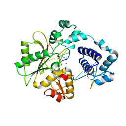
 | | The Crystal Structure of Bacillus cereus protein related to NIF3 | | 分子名称: | 2-AMINO-2-HYDROXYMETHYL-PROPANE-1,3-DIOL, 4-(2-HYDROXYETHYL)-1-PIPERAZINE ETHANESULFONIC ACID, NIF3-related protein, ... | | 著者 | Minasov, G, Brunzelle, J.S, Shuvalova, L, Vorontsov, I.I, Collart, F.R, Joachimiak, A, Anderson, W.F, Midwest Center for Structural Genomics (MCSG) | | 登録日 | 2006-05-08 | | 公開日 | 2006-05-16 | | 最終更新日 | 2024-02-14 | | 実験手法 | X-RAY DIFFRACTION (2.2 Å) | | 主引用文献 | The 2.2 A resolution crystal structure of Bacillus cereus Nif3-family protein YqfO reveals a conserved dimetal-binding motif and a regulatory domain
Protein Sci., 16, 2007
|
|
2GX9
 | |
2GXA
 | |


