6DLR
 
 | |
9ILA
 | |
6C67
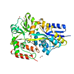
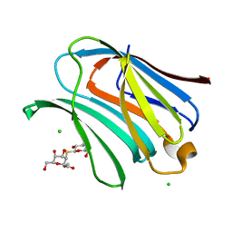
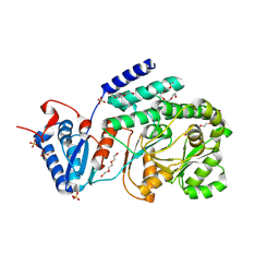
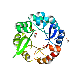
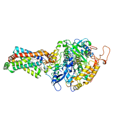
 | | Mycobacterium tuberculosis adenosine kinase bound to iodotubercidin | | 分子名称: | (2R,3R,4S,5R)-2-(4-AMINO-5-IODO-7H-PYRROLO[2,3-D]PYRIMIDIN-7-YL)-5-(HYDROXYMETHYL)TETRAHYDROFURAN-3,4-DIOL, Adenosine kinase, GLYCEROL, ... | | 著者 | Crespo, R.A, TB Structural Genomics Consortium (TBSGC) | | 登録日 | 2018-01-17 | | 公開日 | 2019-05-01 | | 最終更新日 | 2023-10-04 | | 実験手法 | X-RAY DIFFRACTION (2.11 Å) | | 主引用文献 | Structure-Guided Drug Design of 6-Substituted Adenosine Analogues as Potent Inhibitors of Mycobacterium tuberculosis Adenosine Kinase.
J.Med.Chem., 62, 2019
|
|
5MTT
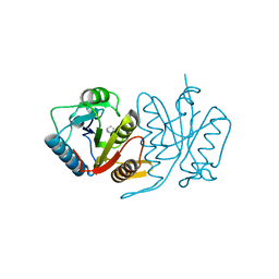
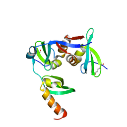
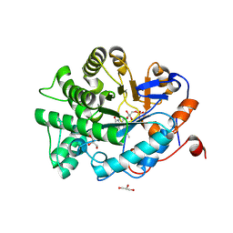
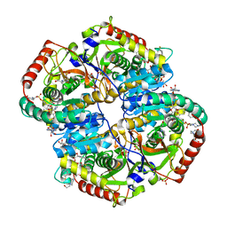
 | | Maltodextrin binding protein MalE1 from L. casei BL23 bound to maltotetraose | | 分子名称: | 1,2-ETHANEDIOL, CHLORIDE ION, MalE1, ... | | 著者 | Homburg, C, Bommer, M, Wuttge, S, Hobe, C, Beck, S, Dobbek, H, Deutscher, J, Licht, A, Schneider, E. | | 登録日 | 2017-01-10 | | 公開日 | 2017-07-05 | | 最終更新日 | 2024-05-08 | | 実験手法 | X-RAY DIFFRACTION (1.12 Å) | | 主引用文献 | Inducer exclusion in Firmicutes: insights into the regulation of a carbohydrate ATP binding cassette transporter from Lactobacillus casei BL23 by the signal transducing protein P-Ser46-HPr.
Mol. Microbiol., 105, 2017
|
|
3GR8
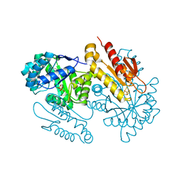
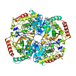
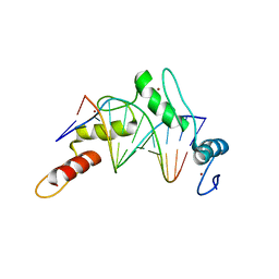
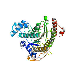
 | | Structure of OYE from Geobacillus kaustophilus, orthorhombic crystal form | | 分子名称: | 2-[BIS-(2-HYDROXY-ETHYL)-AMINO]-2-HYDROXYMETHYL-PROPANE-1,3-DIOL, FLAVIN MONONUCLEOTIDE, NADPH dehydrogenase, ... | | 著者 | Uhl, M.K, Gruber, K. | | 登録日 | 2009-03-25 | | 公開日 | 2010-03-31 | | 最終更新日 | 2023-11-01 | | 実験手法 | X-RAY DIFFRACTION (2.5 Å) | | 主引用文献 | Old Yellow Enzyme-Catalyzed Dehydrogenation of Saturated Ketones
ADV.SYNTH.CATAL., 353, 2011
|
|
8OJI
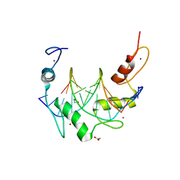
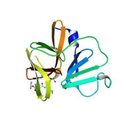
 | | Galectin-3 in complex with Methyl 2,6-anhydro-3-deoxy-3-S-(b-D-galactopyranosyl)-3-thio-D-glycero-L-altro-heptonate | | 分子名称: | 1-thio-beta-D-galactopyranose, CHLORIDE ION, Galectin-3, ... | | 著者 | Tsagkarakou, A.S, Leonidas, D.D. | | 登録日 | 2023-03-24 | | 公開日 | 2023-09-13 | | 最終更新日 | 2023-09-20 | | 実験手法 | X-RAY DIFFRACTION (1.75 Å) | | 主引用文献 | Strong Binding of C -Glycosylic1,2-Thiodisaccharides to Galectin-3─Enthalpy-Driven Affinity Enhancement by Water-Mediated Hydrogen Bonds.
J.Med.Chem., 66, 2023
|
|
3GZD
 | | Human selenocysteine lyase, P1 crystal form | | 分子名称: | (5-HYDROXY-4,6-DIMETHYLPYRIDIN-3-YL)METHYL DIHYDROGEN PHOSPHATE, NITRATE ION, Selenocysteine lyase | | 著者 | Karlberg, T, Hogbom, M, Arrowsmith, C.H, Berglund, H, Bountra, C, Collins, R, Edwards, A.M, Flodin, S, Flores, A, Graslund, S, Hammarstrom, M, Johansson, A, Johansson, I, Kotenyova, T, Moche, M, Nordlund, P, Nyman, T, Persson, C, Sagemark, J, Schutz, P, Siponen, M.I, Thorsell, A.G, Tresaugues, L, Van Den Berg, S, Weigelt, J, Welin, M, Wisniewska, M, Schuler, H, Structural Genomics Consortium (SGC) | | 登録日 | 2009-04-07 | | 公開日 | 2009-04-28 | | 最終更新日 | 2024-11-20 | | 実験手法 | X-RAY DIFFRACTION (1.8 Å) | | 主引用文献 | Biochemical discrimination between selenium and sulfur 1: a single residue provides selenium specificity to human selenocysteine lyase.
Plos One, 7, 2012
|
|
5HWH
 | |
4S3D
 | | IspG in complex with PPi | | 分子名称: | 4-hydroxy-3-methylbut-2-en-1-yl diphosphate synthase, DIPHOSPHATE, FE3-S4 CLUSTER | | 著者 | Quitterer, F, Frank, A, Wang, K, Rao, G, O'Dowd, B, Li, J, Guerra, F, Abdel-Azeim, S, Bacher, A, Eppinger, J, Oldfield, E, Groll, M. | | 登録日 | 2015-01-26 | | 公開日 | 2015-04-29 | | 最終更新日 | 2023-12-06 | | 実験手法 | X-RAY DIFFRACTION (1.8 Å) | | 主引用文献 | Atomic-Resolution Structures of Discrete Stages on the Reaction Coordinate of the [Fe4S4] Enzyme IspG (GcpE).
J.Mol.Biol., 427, 2015
|
|
6B0O
 | |
9C2R
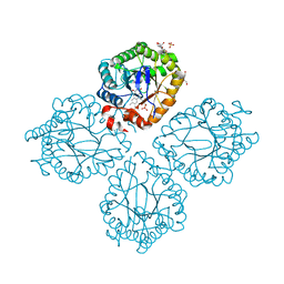
 | | M. tuberculosis PKS13 acyltransferase (AT) domain sulfate free apo form | | 分子名称: | PENTAETHYLENE GLYCOL, Polyketide synthase Pks13, SULFATE ION, ... | | 著者 | Krieger, I.V, Tang, S, Sacchettini, J.C, TB Structural Genomics Consortium (TBSGC) | | 登録日 | 2024-05-31 | | 公開日 | 2025-05-07 | | 最終更新日 | 2025-10-01 | | 実験手法 | X-RAY DIFFRACTION (2.18 Å) | | 主引用文献 | SuFEx-based antitubercular compound irreversibly inhibits Pks13.
Nature, 645, 2025
|
|
6FTO
 | |
4RLS
 | | Lactate Dehydrogenase in complex with inhibitor compound 47 | | 分子名称: | (1R)-5'-[(2-chlorophenyl)sulfanyl]-4'-hydroxy-2,3-dihydrospiro[indene-1,2'-pyran]-6'(3'H)-one, (2S)-2-HYDROXYPROPANOIC ACID, 1,4-DIHYDRONICOTINAMIDE ADENINE DINUCLEOTIDE, ... | | 著者 | Eigenbrot, C, Ultsch, M.H. | | 登録日 | 2014-10-17 | | 公開日 | 2014-11-12 | | 最終更新日 | 2024-02-28 | | 実験手法 | X-RAY DIFFRACTION (1.91 Å) | | 主引用文献 | Identification of 3,6-disubstituted dihydropyrones as inhibitors of human lactate dehydrogenase.
Bioorg.Med.Chem.Lett., 24, 2014
|
|
5UPU
 | | Crystal Structure of the Catalytic Domain of the Inosine Monophosphate Dehydrogenase from Mycobacterium tuberculosis in the presence of TBK6 | | 分子名称: | INOSINIC ACID, Inosine-5'-monophosphate dehydrogenase, ~{N}-(2~{H}-indazol-6-yl)-3,5-dimethyl-1~{H}-pyrazole-4-sulfonamide | | 著者 | Kim, Y, Makowska-Grzyska, M, Maltseva, N, Mulligan, R, Gu, M, Sacchettini, J, Anderson, W.F, Joachimiak, A, Center for Structural Genomics of Infectious Diseases (CSGID) | | 登録日 | 2017-02-04 | | 公開日 | 2017-02-22 | | 最終更新日 | 2023-10-04 | | 実験手法 | X-RAY DIFFRACTION (2.905 Å) | | 主引用文献 | Crystal Structure of the Catalytic Domain of the Inosine Monophosphate Dehydrogenase from Mycobacterium tuberculosis in the presence of TBK6
To Be Published
|
|
5K6Z
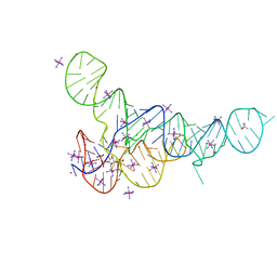
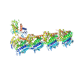
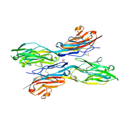 | | Sidekick chimera containing sidekick-2 immunoglobulin domains 1-2 and sidekick-1 immunoglobulin domains 3-4 | | 分子名称: | (4S)-2-METHYL-2,4-PENTANEDIOL, 2-acetamido-2-deoxy-beta-D-glucopyranose, 2-acetamido-2-deoxy-beta-D-glucopyranose-(1-4)-2-acetamido-2-deoxy-beta-D-glucopyranose, ... | | 著者 | Goodman, K.M, Mannepalli, S, Honig, B, Shapiro, L. | | 登録日 | 2016-05-25 | | 公開日 | 2016-09-28 | | 最終更新日 | 2024-11-20 | | 実験手法 | X-RAY DIFFRACTION (2.7 Å) | | 主引用文献 | Molecular basis of sidekick-mediated cell-cell adhesion and specificity.
Elife, 5, 2016
|
|
5AHI
 | | Crystal structure of salmonalla enterica HisA mutant D7N with ProFAR | | 分子名称: | 1-(5-PHOSPHORIBOSYL)-5-[(5-PHOSPHORIBOSYLAMINO) METHYLIDENE AMINO] IMIDAZOLE-4-CARBOXAMIDE ISOMERASE, CHLORIDE ION, GLYCEROL, ... | | 著者 | Soderholm, A, Guo, X, Newton, M.S, Evans, G.B, Nasvall, J, Patrick, W.M, Selmer, M. | | 登録日 | 2015-02-06 | | 公開日 | 2016-03-02 | | 最終更新日 | 2024-05-01 | | 実験手法 | X-RAY DIFFRACTION (1.999 Å) | | 主引用文献 | Structure and Mechanism of Hisa from Salmonella Enterica
To be Published
|
|
9FH7
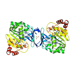
 | | OYE2 from Saccharomyces cerevisiae | | 分子名称: | FLAVIN MONONUCLEOTIDE, GLYCEROL, NADPH dehydrogenase 2 | | 著者 | Opperman, D.J, Paul, C.E. | | 登録日 | 2024-05-26 | | 公開日 | 2024-12-25 | | 実験手法 | X-RAY DIFFRACTION (1.529 Å) | | 主引用文献 | Asymmetric Monoreduction of alpha , beta-Dicarbonyls to alpha-Hydroxy Carbonyls by Ene Reductases.
Acs Catalysis, 14, 2024
|
|
6G7K
 | | Retinal isomerization in bacteriorhodopsin revealed by a femtosecond X-ray laser: 10 ps state structure | | 分子名称: | (2R)-2,3-dihydroxypropyl (9Z)-octadec-9-enoate, 1-[2,6,10.14-TETRAMETHYL-HEXADECAN-16-YL]-2-[2,10,14-TRIMETHYLHEXADECAN-16-YL]GLYCEROL, Bacteriorhodopsin, ... | | 著者 | Nogly, P, Weinert, T, James, D, Cabajo, S, Ozerov, D, Furrer, A, Gashi, D, Borin, V, Skopintsev, P, Jaeger, K, Nass, K, Bath, P, Bosman, R, Koglin, J, Seaberg, M, Lane, T, Kekilli, D, Bruenle, S, Tanaka, T, Wu, W, Milne, C, White, T, Barty, A, Weierstall, U, Panneels, V, Nango, E, Iwata, S, Hunter, M, Schapiro, I, Schertler, G, Neutze, R, Standfuss, J. | | 登録日 | 2018-04-06 | | 公開日 | 2018-06-27 | | 最終更新日 | 2024-10-09 | | 実験手法 | X-RAY DIFFRACTION (1.9 Å) | | 主引用文献 | Retinal isomerization in bacteriorhodopsin captured by a femtosecond x-ray laser.
Science, 361, 2018
|
|
6GF3
 | | Tubulin-Jerantinine B acetate complex | | 分子名称: | 1,2-ETHANEDIOL, 2-(N-MORPHOLINO)-ETHANESULFONIC ACID, CALCIUM ION, ... | | 著者 | Smedley, C.J, Stanley, P.A, Qazzaz, M.E, Prota, A.E, Olieric, N, Collins, H, Eastman, H, Barrow, A.S, Lim, K.-H, Kam, T.-S, Smith, B.J, Duivenvoorden, H.M, Parker, B.S, Bradshaw, T.D, Steinmetz, M.O, Moses, J.E. | | 登録日 | 2018-04-29 | | 公開日 | 2019-05-08 | | 最終更新日 | 2024-01-17 | | 実験手法 | X-RAY DIFFRACTION (2.4 Å) | | 主引用文献 | Sustainable Syntheses of (-)-Jerantinines A & E and Structural Characterisation of the Jerantinine-Tubulin Complex at the Colchicine Binding Site.
Sci Rep, 8, 2018
|
|
6B0P
 | |
4YXD
 | | CRYSTAL STRUCTURE OF PORCINE HEART MITOCHONDRIAL COMPLEX II BOUND WITH flutolanil | | 分子名称: | FE2/S2 (INORGANIC) CLUSTER, FE3-S4 CLUSTER, FLAVIN-ADENINE DINUCLEOTIDE, ... | | 著者 | Harada, S, Shiba, T, Sato, D, Yamamoto, A, Nagahama, M, Yone, A, Inaoka, D.K, Sakamoto, K, Inoue, M, Honma, T, Kita, K. | | 登録日 | 2015-03-23 | | 公開日 | 2016-03-02 | | 最終更新日 | 2023-11-08 | | 実験手法 | X-RAY DIFFRACTION (3 Å) | | 主引用文献 | Structural Insights into the Molecular Design of Flutolanil Derivatives Targeted for Fumarate Respiration of Parasite Mitochondria
Int J Mol Sci, 16, 2015
|
|
6BAD
 | |
9IQX
 | | Cryo-EM structure of the human TRPV4-RhoA in complex with AH001 | | 分子名称: | (1~{R})-1-(3-ethylphenyl)ethane-1,2-diol, GUANOSINE-5'-DIPHOSPHATE, MAGNESIUM ION, ... | | 著者 | Yuan, Z, Ruan, S.S, Li, S.L. | | 登録日 | 2024-07-13 | | 公開日 | 2025-05-28 | | 最終更新日 | 2025-09-03 | | 実験手法 | ELECTRON MICROSCOPY (3.37 Å) | | 主引用文献 | Inactivation of RhoA for Hypertension Treatment Through the TRPV4-RhoA-RhoGDI1 Axis.
Circulation, 152, 2025
|
|
6HSH
 | | Crystal structure of Schistosoma mansoni HDAC8 complexed with Quisinostat | | 分子名称: | 2-[4-[[(1-methylindol-3-yl)methylamino]methyl]piperidin-1-yl]-~{N}-oxidanyl-pyrimidine-5-carboxamide, DIMETHYLFORMAMIDE, GLYCEROL, ... | | 著者 | Shaik, T.B, Marek, M, Romier, C. | | 登録日 | 2018-10-01 | | 公開日 | 2018-10-31 | | 最終更新日 | 2024-01-24 | | 実験手法 | X-RAY DIFFRACTION (1.545 Å) | | 主引用文献 | Characterization of Histone Deacetylase 8 (HDAC8) Selective Inhibition Reveals Specific Active Site Structural and Functional Determinants.
J. Med. Chem., 61, 2018
|
|
6BIB
 | | 1.95 A resolution structure of Norovirus 3CL protease in complex with a triazole-based macrocyclic inhibitor | | 分子名称: | 3C-like protease, benzyl [(9S,12S,15S)-12-(cyclohexylmethyl)-9-(hydroxymethyl)-6,11,14-trioxo-1,5,10,13,18,19-hexaazabicyclo[15.2.1]icosa-17(20),18-dien-15-yl]carbamate | | 著者 | Lovell, S, Battaile, K.P, Mehzabeen, N, Kankanamalage, A.C.G, Weerawarna, P.M, Rathnayake, A.D, Kim, Y, Chang, K.O, Groutas, W.C. | | 登録日 | 2017-11-01 | | 公開日 | 2018-11-07 | | 最終更新日 | 2024-11-20 | | 実験手法 | X-RAY DIFFRACTION (1.95 Å) | | 主引用文献 | Putative structural rearrangements associated with the interaction of macrocyclic inhibitors with norovirus 3CL protease.
Proteins, 87, 2019
|
|


