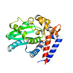
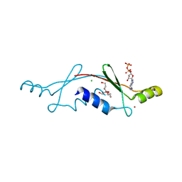
4DL1
 
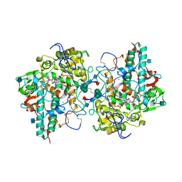 | | Crystal Structure of human Myeloperoxidase with covalent thioxanthine analog | | 分子名称: | 2-acetamido-2-deoxy-beta-D-glucopyranose, 3-[(2R)-2-ethoxypropyl]-2-thioxo-1,2,3,9-tetrahydro-6H-purin-6-one, CALCIUM ION, ... | | 著者 | Vajdos, F, Varghese, A. | | 登録日 | 2012-02-05 | | 公開日 | 2012-03-21 | | 最終更新日 | 2020-07-29 | | 実験手法 | X-RAY DIFFRACTION (2 Å) | | 主引用文献 | Deconstruction of activity-dependent covalent modification of heme in human neutrophil myeloperoxidase by multistage mass spectrometry (MS(4)).
Biochemistry, 51, 2012
|
|
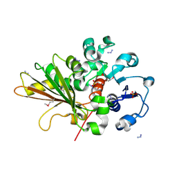
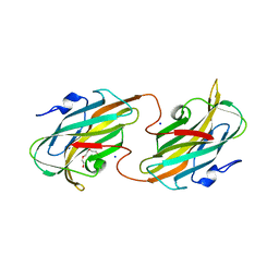
4R8N
 | | Crystal structure of Staphylococcal nuclease variant V23I/V66I/I72V/I92V at cryogenic temperature | | 分子名称: | (4R)-2-METHYLPENTANE-2,4-DIOL, CALCIUM ION, PHOSPHATE ION, ... | | 著者 | Caro, J.A, Flores, E, Schlessman, J.L, Heroux, A, Garcia-Moreno, E.B. | | 登録日 | 2014-09-02 | | 公開日 | 2014-09-17 | | 最終更新日 | 2023-09-20 | | 実験手法 | X-RAY DIFFRACTION (1.65 Å) | | 主引用文献 | Cavities in proteins
To be Published
|
|
4RKB
 | |
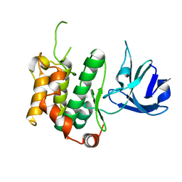
4RUV
 | | Crystal structure of thioredoxin 2 from Staphylococcus aureus NCTC8325 | | 分子名称: | Thioredoxin | | 著者 | Bose, M, Biswas, R, Roychowdhury, A, Bhattacharyya, S, Ghosh, A.K, Das, A.K. | | 登録日 | 2014-11-22 | | 公開日 | 2015-12-09 | | 最終更新日 | 2023-09-20 | | 実験手法 | X-RAY DIFFRACTION (1.93 Å) | | 主引用文献 | Elucidation of the mechanism of disulfide exchange between staphylococcal thioredoxin2 and thioredoxin reductase2: A structural insight.
Biochimie, 160, 2019
|
|
4UG0
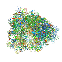 | | STRUCTURE OF THE HUMAN 80S RIBOSOME | | 分子名称: | 18S ribosomal RNA, 28S ribosomal RNA, 40S RIBOSOMAL PROTEIN, ... | | 著者 | Khatter, H, Myasnikov, A.G, Natchiar, S.K, Klaholz, B.P. | | 登録日 | 2015-03-20 | | 公開日 | 2015-06-10 | | 最終更新日 | 2019-12-18 | | 実験手法 | ELECTRON MICROSCOPY (3.6 Å) | | 主引用文献 | Structure of the human 80S ribosome
NATURE, 520, 2015
|
|
5Y8U
 | | Crystal structure of the C276S mutant of MAP2K7 | | 分子名称: | Dual specificity mitogen-activated protein kinase kinase 7 | | 著者 | Kinoshita, T. | | 登録日 | 2017-08-21 | | 公開日 | 2017-10-11 | | 最終更新日 | 2023-11-22 | | 実験手法 | X-RAY DIFFRACTION (2.92 Å) | | 主引用文献 | High-resolution structure discloses the potential for allosteric regulation of mitogen-activated protein kinase kinase 7
Biochem. Biophys. Res. Commun., 493, 2017
|
|
5Y90
 | | MAP2K7 mutant -C218S | | 分子名称: | Dual specificity mitogen-activated protein kinase kinase 7, GLYCEROL | | 著者 | Kinoshita, T, Hashimoto, T, Sogabe, Y, Matsumoto, T, Sawa, M, Fukada, H. | | 登録日 | 2017-08-22 | | 公開日 | 2017-10-11 | | 最終更新日 | 2023-11-22 | | 実験手法 | X-RAY DIFFRACTION (1.3 Å) | | 主引用文献 | High-resolution structure discloses the potential for allosteric regulation of mitogen-activated protein kinase kinase 7
Biochem. Biophys. Res. Commun., 493, 2017
|
|
6ZR5
 | | Crystal structure of JNK1 in complex with ATF2(19-58) | | 分子名称: | Cyclic AMP-dependent transcription factor ATF-2, MAGNESIUM ION, Mitogen-activated protein kinase 8, ... | | 著者 | Kirsch, K, Zeke, A, Remenyi, A. | | 登録日 | 2020-07-10 | | 公開日 | 2020-11-18 | | 最終更新日 | 2024-01-31 | | 実験手法 | X-RAY DIFFRACTION (2.699 Å) | | 主引用文献 | Co-regulation of the transcription controlling ATF2 phosphoswitch by JNK and p38.
Nat Commun, 11, 2020
|
|
6BE0
 | | AvrA delL154 with IP6, CoA | | 分子名称: | AvrA, COENZYME A, INOSITOL HEXAKISPHOSPHATE | | 著者 | Labriola, J.M, Nagar, B. | | 登録日 | 2017-10-24 | | 公開日 | 2018-08-01 | | 最終更新日 | 2023-10-04 | | 実験手法 | X-RAY DIFFRACTION (2.438 Å) | | 主引用文献 | Structural Analysis of the Bacterial Effector AvrA Identifies a Critical Helix Involved in Substrate Recognition.
Biochemistry, 57, 2018
|
|
2OZP
 | |
2PEX
 | |
2PHC
 | | Crystal structure of conserved uncharacterized protein PH0987 from Pyrococcus horikoshii | | 分子名称: | Uncharacterized protein PH0987 | | 著者 | Swindell II, J.T, Chen, L, Zhu, J, Ebihara, A, Shinkai, A, Kuramitsu, S, Yokoyama, S, Fu, Z.-Q, Chrzas, J, Rose, J.P, Wang, B.-C, Southeast Collaboratory for Structural Genomics (SECSG), RIKEN Structural Genomics/Proteomics Initiative (RSGI) | | 登録日 | 2007-04-10 | | 公開日 | 2007-05-08 | | 最終更新日 | 2024-02-21 | | 実験手法 | X-RAY DIFFRACTION (2.29 Å) | | 主引用文献 | Crystal structure of conserved uncharacterized protein PH0987 from Pyrococcus horikoshii.
To be Published
|
|
2P05
 | | Structural Insights into the Evolution of a Non-Biological Protein | | 分子名称: | ADENOSINE-5'-DIPHOSPHATE, CHLORIDE ION, PENTAETHYLENE GLYCOL, ... | | 著者 | Smith, M, Rosenow, M, Wang, M, Allen, J.P, Szostak, J.W, Chaput, J.C. | | 登録日 | 2007-02-28 | | 公開日 | 2007-06-05 | | 最終更新日 | 2024-02-21 | | 実験手法 | X-RAY DIFFRACTION (2.8 Å) | | 主引用文献 | Structural insights into the evolution of a non-biological protein: importance of surface residues in protein fold optimization.
PLoS ONE, 2, 2007
|
|
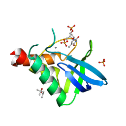
2R5O
 | | Crystal structure of the C-terminal domain of wzt | | 分子名称: | CHLORIDE ION, Putative ATP binding component of ABC-transporter, SODIUM ION, ... | | 著者 | Kimber, M.S, Cuthbertson, L, Whitfield, C. | | 登録日 | 2007-09-04 | | 公開日 | 2007-12-25 | | 最終更新日 | 2024-02-21 | | 実験手法 | X-RAY DIFFRACTION (1.3 Å) | | 主引用文献 | Substrate binding by a bacterial ABC transporter involved in polysaccharide export.
Proc.Natl.Acad.Sci.Usa, 104, 2007
|
|
2RET
 | | The crystal structure of a binary complex of two pseudopilins: EpsI and EpsJ from the Type 2 Secretion System of Vibrio vulnificus | | 分子名称: | CHLORIDE ION, EpsJ, Pseudopilin EpsI, ... | | 著者 | Yanez, M.E, Korotkov, K.V, Abendroth, J, Hol, W.G.J. | | 登録日 | 2007-09-27 | | 公開日 | 2008-02-05 | | 最終更新日 | 2024-02-21 | | 実験手法 | X-RAY DIFFRACTION (2.21 Å) | | 主引用文献 | The crystal structure of a binary complex of two pseudopilins: EpsI and EpsJ from the type 2 secretion system of Vibrio vulnificus.
J.Mol.Biol., 375, 2008
|
|
2QF7
 | | Crystal structure of a complete multifunctional pyruvate carboxylase from Rhizobium etli | | 分子名称: | CHLORIDE ION, COENZYME A, FORMIC ACID, ... | | 著者 | St Maurice, M, Surinya, K.H, Rayment, I. | | 登録日 | 2007-06-27 | | 公開日 | 2007-09-04 | | 最終更新日 | 2012-02-15 | | 実験手法 | X-RAY DIFFRACTION (2 Å) | | 主引用文献 | Domain architecture of pyruvate carboxylase, a biotin-dependent multifunctional enzyme
Science, 317, 2007
|
|
8IFE
 | | Arbekacin-added human 80S ribosome | | 分子名称: | 18S ribosomal RNA, 28S ribosomal RNA, 40S ribosomal protein S10, ... | | 著者 | Tomono, J, Asano, K, Chiashi, T, Tanaka, Y, Yokoyama, T. | | 登録日 | 2023-02-17 | | 公開日 | 2024-02-14 | | 最終更新日 | 2024-06-19 | | 実験手法 | ELECTRON MICROSCOPY (2.57 Å) | | 主引用文献 | Direct visualization of ribosomes in the cell-free system revealed the functional evolution of aminoglycoside.
J.Biochem., 175, 2024
|
|
8IFD
 | | Dibekacin-added human 80S ribosome | | 分子名称: | 18S ribosomal RNA, 28S ribosomal RNA, 40S ribosomal protein S10, ... | | 著者 | Tomono, J, Asano, K, Chiashi, T, Tanaka, Y, Yokoyama, T. | | 登録日 | 2023-02-17 | | 公開日 | 2024-02-14 | | 最終更新日 | 2024-06-19 | | 実験手法 | ELECTRON MICROSCOPY (2.59 Å) | | 主引用文献 | Direct visualization of ribosomes in the cell-free system revealed the functional evolution of aminoglycoside.
J.Biochem., 175, 2024
|
|
2REE
 | |
2O61
 | |
2OK1
 | |
4ME5
 | |
4MH5
 | | Crystal structure of the kainate receptor GluK3 ligand binding domain in complex with (S)-glutamate | | 分子名称: | CHLORIDE ION, GLUTAMIC ACID, GLYCEROL, ... | | 著者 | Venskutonyte, R, Frydenvang, K, Gajhede, M, Kastrup, J.S. | | 登録日 | 2013-08-29 | | 公開日 | 2013-10-16 | | 最終更新日 | 2023-09-20 | | 実験手法 | X-RAY DIFFRACTION (1.65 Å) | | 主引用文献 | Binding site and interlobe interactions of the ionotropic glutamate receptor GluK3 ligand binding domain revealed by high resolution crystal structure in complex with (S)-glutamate.
J.Struct.Biol., 176, 2011
|
|
4MAQ
 | |
6JRX
 | | EGFR T790M/C797S in complex with compound 6i | | 分子名称: | Epidermal growth factor receptor, N-{trans-4-[3-(2-chlorophenyl)-7-{[3-methyl-4-(4-methylpiperazin-1-yl)phenyl]amino}-2-oxo-3,4-dihydropyrimido[4,5-d]pyrimidin-1(2H)-yl]cyclohexyl}propanamide | | 著者 | Zhu, S.J, Yun, C.H. | | 登録日 | 2019-04-06 | | 公開日 | 2020-04-15 | | 最終更新日 | 2024-03-27 | | 実験手法 | X-RAY DIFFRACTION (2.201 Å) | | 主引用文献 | Design, Synthesis, and Structure-Activity Relationships of 1,2,3-Triazole Benzenesulfonamides as New Selective Leucine-Zipper and Sterile-alpha Motif Kinase (ZAK) Inhibitors.
J.Med.Chem., 63, 2020
|
|


