2EVM
 
 | |
2EVN
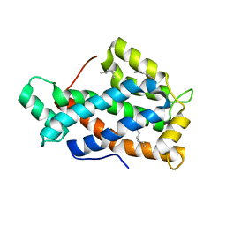
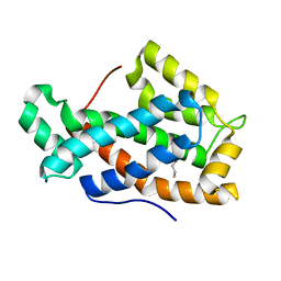
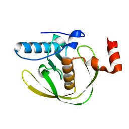
 | | NMR solution structures of At1g77540 | | 分子名称: | Protein At1g77540 | | 著者 | Tyler, R.C, Singh, S, Tonelli, M, Min, M.S, Markley, J.L, Center for Eukaryotic Structural Genomics (CESG) | | 登録日 | 2005-10-31 | | 公開日 | 2005-11-15 | | 最終更新日 | 2024-05-22 | | 実験手法 | SOLUTION NMR | | 主引用文献 | Structure of Arabidopsis thaliana At1g77540 Protein, a Minimal Acetyltransferase from the COG2388 Family.
Biochemistry, 45, 2006
|
|
2EVO
 | |
2EVP
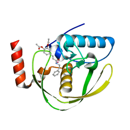
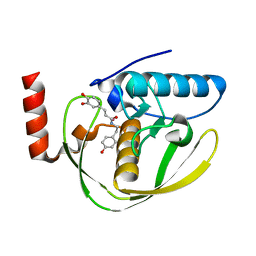
 | | The Structures of Thiolate- and Carboxylate-Ligated Ferric H93G Myoglobin: Models for Cytochrome P450 and for Oxyanion-Bound Heme Proteins | | 分子名称: | BETA-MERCAPTOETHANOL, Myoglobin, PROTOPORPHYRIN IX CONTAINING FE | | 著者 | Qin, J, Perera, R, Lovelace, L.L, Dawson, J.H, Lebioda, L. | | 登録日 | 2005-10-31 | | 公開日 | 2006-03-28 | | 最終更新日 | 2023-08-23 | | 実験手法 | X-RAY DIFFRACTION (1.7 Å) | | 主引用文献 | Structures of Thiolate- and Carboxylate-Ligated Ferric H93G Myoglobin: Models for Cytochrome P450 and for Oxyanion-Bound Heme Proteins(,).
Biochemistry, 45, 2006
|
|
2EVQ
 | |
2EVR
 | |
2EVS
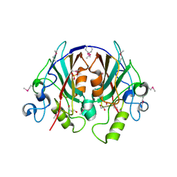
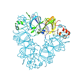
 | | Crystal structure of human Glycolipid Transfer Protein complexed with n-hexyl-beta-D-glucoside | | 分子名称: | DECANE, Glycolipid transfer protein, HEXANE, ... | | 著者 | Malinina, L, Malakhova, M.L, Kanack, A.T, Abagyan, R, Brown, R.E, Patel, D.J. | | 登録日 | 2005-10-31 | | 公開日 | 2006-11-14 | | 最終更新日 | 2023-08-23 | | 実験手法 | X-RAY DIFFRACTION (2.2 Å) | | 主引用文献 | The liganding of glycolipid transfer protein is controlled by glycolipid acyl structure.
Plos Biol., 4, 2006
|
|
2EVT
 | | Crystal structure of D48V mutant of human Glycolipid Transfer Protein | | 分子名称: | Glycolipid transfer protein, HEXANE | | 著者 | Malinina, L, Malakhova, M.L, Teplov, A, Brown, R.E, Patel, D.J. | | 登録日 | 2005-10-31 | | 公開日 | 2005-11-15 | | 最終更新日 | 2023-08-23 | | 実験手法 | X-RAY DIFFRACTION (1.99 Å) | | 主引用文献 | The liganding of glycolipid transfer protein is controlled by glycolipid acyl structure.
Plos Biol., 4, 2006
|
|
2EVU
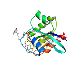
 | | Crystal structure of aquaporin AqpM at 2.3A resolution | | 分子名称: | Aquaporin aqpM, GLYCEROL, octyl beta-D-glucopyranoside | | 著者 | Lee, J.K, Kozono, D, Remis, J, Kitagawa, Y, Agre, P, Stroud, R.M, Center for Structures of Membrane Proteins (CSMP) | | 登録日 | 2005-10-31 | | 公開日 | 2005-12-06 | | 最終更新日 | 2024-02-14 | | 実験手法 | X-RAY DIFFRACTION (2.3 Å) | | 主引用文献 | Structural basis for conductance by the archaeal aquaporin AqpM at 1.68 A.
Proc.Natl.Acad.Sci.Usa, 102, 2005
|
|
2EVV
 | | Crystal Structure of the PEBP-like Protein of Unknown Function HP0218 from Helicobacter pylori | | 分子名称: | GLYCEROL, SULFATE ION, hypothetical protein HP0218 | | 著者 | Kim, Y, Xu, X, Hong, H, Savchenko, A, Edwards, A, Joachimiak, A, Midwest Center for Structural Genomics (MCSG) | | 登録日 | 2005-11-01 | | 公開日 | 2005-12-13 | | 最終更新日 | 2011-07-13 | | 実験手法 | X-RAY DIFFRACTION (2.59 Å) | | 主引用文献 | Crystal Structure of the Hypothetical Protein HP0218 from Helicobacter pylori
To be Published
|
|
2EVW
 | |
2EVX
 | |
2EVY
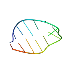 | | GNYA tetranucleotide loops found in poliovirus oriL by in vivo SELEX (un)expectedly form a YNMG-like structure | | 分子名称: | Poliovirus 5'NTR cloverleaf stem loop D mutant | | 著者 | Melchers, W.J.G, Zoll, J, Tessari, M, Bakhmutov, D.V, Gmyl, A.P, Agol, V.I, Heus, H.A. | | 登録日 | 2005-11-01 | | 公開日 | 2006-08-22 | | 最終更新日 | 2024-05-22 | | 実験手法 | SOLUTION NMR | | 主引用文献 | A GCUA tetranucleotide loop found in the poliovirus oriL by in vivo SELEX (un)expectedly forms a YNMG-like structure: Extending the YNMG family with GYYA.
RNA, 12, 2006
|
|
2EVZ
 | |
2EW0
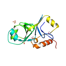 | | X-ray Crystal Structure of Protein Q6FF54 from Acinetobacter sp. ADP1. Northeast Structural Genomics Consortium Target AsR1. | | 分子名称: | SULFATE ION, hypothetical protein ACIAD0353 | | 著者 | Kuzin, A.P, Abashidze, M, Vorobiev, S.M, Forouhar, F, Acton, T, Xiao, R, Northeast Structural Genomics Consortium (NESG) | | 登録日 | 2005-11-01 | | 公開日 | 2005-12-06 | | 最終更新日 | 2011-07-13 | | 実験手法 | X-RAY DIFFRACTION (1.4 Å) | | 主引用文献 | Novel x-ray structure of hypothetical protein Q6FF54 at the resolution 1.4 A. Northeast Structural Genomics target AsR1.
TO BE PUBLISHED
|
|
2EW1
 | | Crystal Structure of Rab30 in complex with a GTP analogue | | 分子名称: | MAGNESIUM ION, PHOSPHOAMINOPHOSPHONIC ACID-GUANYLATE ESTER, Ras-related protein Rab-30 | | 著者 | Wang, J, Shen, Y, Ismail, S, Arrowsmith, C.H, Edwards, A.M, Sundstrom, M, Bochkarev, A, Park, H.W, Structural Genomics Consortium (SGC) | | 登録日 | 2005-11-01 | | 公開日 | 2005-11-08 | | 最終更新日 | 2023-08-23 | | 実験手法 | X-RAY DIFFRACTION (2 Å) | | 主引用文献 | Crystal structure of RAB30 in complex with a GTP analogue
To be Published
|
|
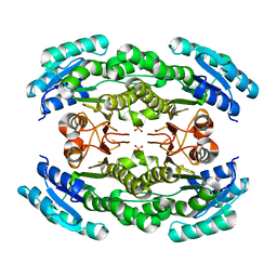
2EW2
 | | Crystal Structure of the Putative 2-Dehydropantoate 2-Reductase from Enterococcus faecalis | | 分子名称: | 2-dehydropantoate 2-reductase, putative, MAGNESIUM ION, ... | | 著者 | Kim, Y, Zhou, M, Moy, S, Clancy, S, Collart, F, Joachimiak, A, Midwest Center for Structural Genomics (MCSG) | | 登録日 | 2005-11-01 | | 公開日 | 2005-12-13 | | 最終更新日 | 2011-07-13 | | 実験手法 | X-RAY DIFFRACTION (2 Å) | | 主引用文献 | Crystal Structure of the Putative 2-Dehydropantoate 2-Reductase from Enterococcus faecalis
To be Published
|
|
2EW3
 | |
2EW4
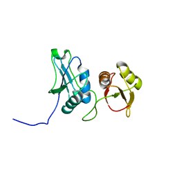
 | | Solution structure of MrIA | | 分子名称: | MrIA | | 著者 | Nilsson, K.P, Lovelace, E.S, Caesar, C.E, Tynngard, N, Alewood, P.F, Johansson, H.M, Sharpe, I.A, Lewis, R.J, Daly, N.L, Craik, D.J. | | 登録日 | 2005-11-02 | | 公開日 | 2005-11-15 | | 最終更新日 | 2020-06-24 | | 実験手法 | SOLUTION NMR | | 主引用文献 | Solution structure of chi-conopeptide MrIA, a modulator of the human norepinephrine transporter
Biopolymers, 80, 2005
|
|
2EW5
 | | Structure of Helicobacter Pylori peptide deformylase in complex with inhibitor | | 分子名称: | 4-{(1E)-3-OXO-3-[(2-PHENYLETHYL)AMINO]PROP-1-EN-1-YL}-1,2-PHENYLENE DIACETATE, COBALT (II) ION, peptide deformylase | | 著者 | Cai, J. | | 登録日 | 2005-11-02 | | 公開日 | 2006-10-24 | | 最終更新日 | 2023-10-25 | | 実験手法 | X-RAY DIFFRACTION (2.2 Å) | | 主引用文献 | Peptide deformylase is a potential target for anti-Helicobacter pylori drugs: reverse docking, enzymatic assay, and X-ray crystallography validation
Protein Sci., 15, 2006
|
|
2EW6
 | | Structure of Helicobacter Pylori peptide deformylase in complex with inhibitor | | 分子名称: | (2E)-3-(3,4-DIHYDROXYPHENYL)-N-[2-(4-HYDROXYPHENYL)ETHYL]ACRYLAMIDE, COBALT (II) ION, peptide deformylase | | 著者 | Cai, J. | | 登録日 | 2005-11-02 | | 公開日 | 2006-10-24 | | 最終更新日 | 2023-10-25 | | 実験手法 | X-RAY DIFFRACTION (2.2 Å) | | 主引用文献 | Peptide deformylase is a potential target for anti-Helicobacter pylori drugs: reverse docking, enzymatic assay, and X-ray crystallography validation
Protein Sci., 15, 2006
|
|
2EW7
 | | Crystal Structure of Helicobacter Pylori peptide deformylase | | 分子名称: | COBALT (II) ION, peptide deformylase | | 著者 | Cai, J. | | 登録日 | 2005-11-02 | | 公開日 | 2006-10-24 | | 最終更新日 | 2023-10-25 | | 実験手法 | X-RAY DIFFRACTION (2.2 Å) | | 主引用文献 | Peptide deformylase is a potential target for anti-Helicobacter pylori drugs: reverse docking, enzymatic assay, and X-ray crystallography validation
Protein Sci., 15, 2006
|
|
2EW8
 | |
2EW9
 | |
2EWA
 | | Dual binding mode of pyridinylimidazole to MAP kinase p38 | | 分子名称: | 4-[5-(4-FLUORO-PHENYL)-2-(4-METHANESULFINYL-PHENYL)-3H-IMIDAZOL-4-YL]-PYRIDINE, Mitogen-activated protein kinase 14 | | 著者 | Delarbre, L, Pouzieux, S, Guilloteau, J.-P, Michot, N. | | 登録日 | 2005-11-02 | | 公開日 | 2006-08-01 | | 最終更新日 | 2024-02-14 | | 実験手法 | X-RAY DIFFRACTION (2.1 Å) | | 主引用文献 | NMR characterization of kinase p38 dynamics in free and ligand-bound forms.
Angew.Chem.Int.Ed.Engl., 45, 2006
|
|


