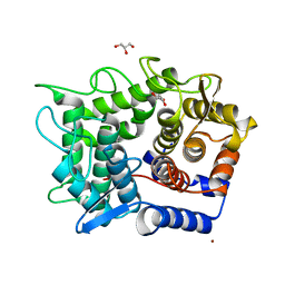
2DQY
 
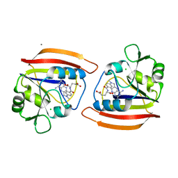
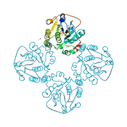
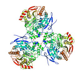 | | Crystal structure of human carboxylesterase in complex with cholate and palmitate | | 分子名称: | 2-acetamido-2-deoxy-beta-D-glucopyranose, CHOLIC ACID, Liver carboxylesterase 1, ... | | 著者 | Bencharit, S, Edwards, C.C, Morton, C.L, Howard-Williams, E.L, Potter, P.M, Redinbo, M.R. | | 登録日 | 2006-06-02 | | 公開日 | 2006-08-29 | | 最終更新日 | 2023-10-25 | | 実験手法 | X-RAY DIFFRACTION (3 Å) | | 主引用文献 | Multisite promiscuity in the processing of endogenous substrates by human carboxylesterase 1
J.Mol.Biol., 363, 2006
|
|
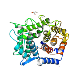
2DQZ
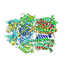
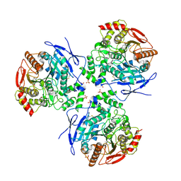 | | Crystal structure of human carboxylesterase in complex with homatropine, coenzyme A, and palmitate | | 分子名称: | 2-acetamido-2-deoxy-beta-D-glucopyranose, COENZYME A, FLUORIDE ION, ... | | 著者 | Bencharit, S, Redinbo, M.R. | | 登録日 | 2006-06-02 | | 公開日 | 2006-08-29 | | 最終更新日 | 2023-10-25 | | 実験手法 | X-RAY DIFFRACTION (2.8 Å) | | 主引用文献 | Multisite promiscuity in the processing of endogenous substrates by human carboxylesterase 1
J.Mol.Biol., 363, 2006
|
|
2DR0
 | |
2DR1
 | |
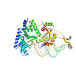
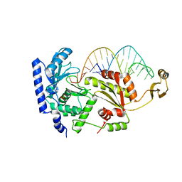
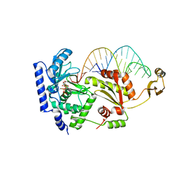
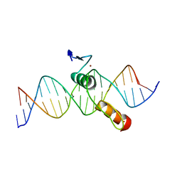
2DR2
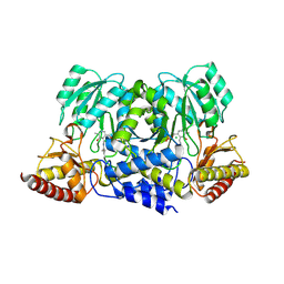
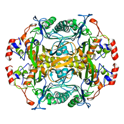
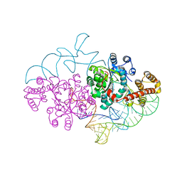 | | Structure of human tryptophanyl-tRNA synthetase in complex with tRNA(Trp) | | 分子名称: | SULFATE ION, TRYPTOPHAN, Tryptophanyl-tRNA synthetase, ... | | 著者 | Shen, N, Guo, L, Yang, B, Jin, Y, Ding, J. | | 登録日 | 2006-06-05 | | 公開日 | 2006-07-11 | | 最終更新日 | 2023-10-25 | | 実験手法 | X-RAY DIFFRACTION (3 Å) | | 主引用文献 | Structure of human tryptophanyl-tRNA synthetase in complex with tRNA(Trp) reveals the molecular basis of tRNA recognition and specificity
Nucleic Acids Res., 34, 2006
|
|
2DR3
 | |
2DR5
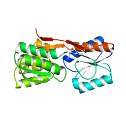
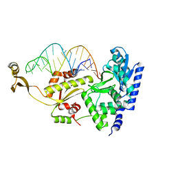 | | Complex structure of CCA adding enzyme with mini-helix lacking CCA | | 分子名称: | CCA-adding enzyme, SULFATE ION, tRNA (32-MER) | | 著者 | Tomita, K, Ishitani, R, Fukai, S, Nureki, O. | | 登録日 | 2006-06-08 | | 公開日 | 2006-10-31 | | 最終更新日 | 2023-10-25 | | 実験手法 | X-RAY DIFFRACTION (2.8 Å) | | 主引用文献 | Complete crystallographic analysis of the dynamics of CCA sequence addition
Nature, 443, 2006
|
|
2DR6
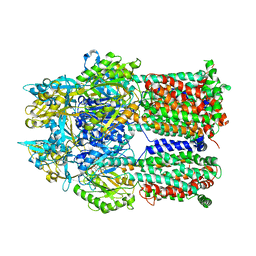 | | Crystal structure of a multidrug transporter reveal a functionally rotating mechanism | | 分子名称: | ACRB, DOXORUBICIN | | 著者 | Murakami, S, Nakashima, R, Yamashita, E, Matsumoto, T. | | 登録日 | 2006-06-08 | | 公開日 | 2006-08-22 | | 最終更新日 | 2024-03-13 | | 実験手法 | X-RAY DIFFRACTION (3.3 Å) | | 主引用文献 | Crystal structures of a multidrug transporter reveal a functionally rotating mechanism
Nature, 443, 2006
|
|
2DR7
 | | Complex structure of CCA-adding enzyme with tRNAminiDC | | 分子名称: | CCA-adding enzyme, SULFATE ION, tRNA (33-MER) | | 著者 | Tomita, K, Ishitani, R, Fukai, S, Nureki, O. | | 登録日 | 2006-06-08 | | 公開日 | 2006-11-14 | | 最終更新日 | 2024-03-13 | | 実験手法 | X-RAY DIFFRACTION (2.8 Å) | | 主引用文献 | Complete crystallographic analysis of the dynamics of CCA sequence addition
Nature, 443, 2006
|
|
2DR8
 | | Complex structure of CCA-adding enzyme with tRNAminiDC and CTP | | 分子名称: | CCA-adding enzyme, CYTIDINE-5'-TRIPHOSPHATE, MAGNESIUM ION, ... | | 著者 | Tomita, K, Ishitani, R, Fukai, S, Nureki, O. | | 登録日 | 2006-06-08 | | 公開日 | 2006-11-14 | | 最終更新日 | 2024-03-13 | | 実験手法 | X-RAY DIFFRACTION (2.5 Å) | | 主引用文献 | Complete crystallographic analysis of the dynamics of CCA sequence addition
Nature, 443, 2006
|
|
2DR9
 | | Complex structure of CCA-adding enzyme with tRNAminiDCC | | 分子名称: | CCA-adding enzyme, SULFATE ION, tRNA (34-MER) | | 著者 | Tomita, K, Ishitani, R, Fukai, S, Nureki, O. | | 登録日 | 2006-06-08 | | 公開日 | 2006-11-14 | | 最終更新日 | 2024-03-13 | | 実験手法 | X-RAY DIFFRACTION (2.8 Å) | | 主引用文献 | Complete crystallographic analysis of the dynamics of CCA sequence addition
Nature, 443, 2006
|
|
2DRA
 | | Complex structure of CCA-adding enzyme with tRNAminiDCC and ATP | | 分子名称: | ADENOSINE-5'-TRIPHOSPHATE, CCA-adding enzyme, MAGNESIUM ION, ... | | 著者 | Tomita, K, Ishitani, R, Fukai, S, Nureki, O. | | 登録日 | 2006-06-08 | | 公開日 | 2006-11-14 | | 最終更新日 | 2024-03-13 | | 実験手法 | X-RAY DIFFRACTION (2.5 Å) | | 主引用文献 | Complete crystallographic analysis of the dynamics of CCA sequence addition
Nature, 443, 2006
|
|
2DRB
 | | Complex structure of CCA-adding enzyme with tRNAminiCCA | | 分子名称: | CCA-adding enzyme, SULFATE ION, tRNA (35-MER) | | 著者 | Tomita, K, Ishitani, R, Fukai, S, Nureki, O. | | 登録日 | 2006-06-08 | | 公開日 | 2006-11-14 | | 最終更新日 | 2024-03-13 | | 実験手法 | X-RAY DIFFRACTION (2.8 Å) | | 主引用文献 | Complete crystallographic analysis of the dynamics of CCA sequence addition
Nature, 443, 2006
|
|
2DRC
 | |
2DRD
 | | Crystal structure of a multidrug transporter reveal a functionally rotating mechanism | | 分子名称: | (4S,4AS,5AR,12AS)-4,7-BIS(DIMETHYLAMINO)-3,10,12,12A-TETRAHYDROXY-1,11-DIOXO-1,4,4A,5,5A,6,11,12A-OCTAHYDROTETRACENE-2- CARBOXAMIDE, ACRB | | 著者 | Murakami, S, Nakashima, R, Yamashita, E, Matsumoto, T. | | 登録日 | 2006-06-08 | | 公開日 | 2006-08-22 | | 最終更新日 | 2024-03-13 | | 実験手法 | X-RAY DIFFRACTION (3.1 Å) | | 主引用文献 | Crystal structures of a multidrug transporter reveal a functionally rotating mechanism
Nature, 443, 2006
|
|
2DRE
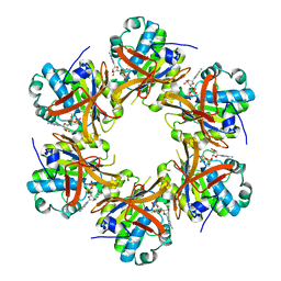
 | | Crystal structure of Water-soluble chlorophyll protein from lepidium virginicum at 2.00 angstrom resolution | | 分子名称: | CHLOROPHYLL A, Water-soluble chlorophyll protein | | 著者 | Horigome, D, Satoh, H, Itoh, N, Mitsunaga, K, Oonishi, I, Nakagawa, A, Uchida, A. | | 登録日 | 2006-06-08 | | 公開日 | 2006-12-26 | | 最終更新日 | 2011-07-13 | | 実験手法 | X-RAY DIFFRACTION (2 Å) | | 主引用文献 | Structural mechanism and photoprotective function of water-soluble chlorophyll-binding protein.
J.Biol.Chem., 282, 2007
|
|
2DRH
 | |
2DRI
 | |
2DRJ
 | |
2DRK
 | |
2DRM
 | |
2DRN
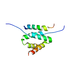 | | Docking and dimerization domain (D/D) of the Type II-alpha regulatory subunity of protein kinase A (PKA) in complex with a peptide from an A-kinase anchoring protein | | 分子名称: | 24-residues peptide from an a-kinase anchoring protein, cAMP-dependent protein kinase type II-alpha regulatory subunit | | 著者 | Newlon, M.G, Roy, M, Morikis, D, Hausken, Z.E, Coghlan, V, Scott, J.D, Jennings, P.A. | | 登録日 | 2006-06-11 | | 公開日 | 2006-08-29 | | 最終更新日 | 2024-05-29 | | 実験手法 | SOLUTION NMR | | 主引用文献 | A novel mechanism of PKA anchoring revealed by solution structures of anchoring complexes.
Embo J., 20, 2001
|
|
2DRO
 | | Crystal structure of reducing-end-xylose releasing exo-oligoxylanase D263C mutant | | 分子名称: | GLYCEROL, NICKEL (II) ION, Xylanase Y | | 著者 | Fushinobu, S, Hidaka, M, Honda, Y, Wakagi, T, Shoun, H, Kitaoka, M. | | 登録日 | 2006-06-12 | | 公開日 | 2006-06-27 | | 最終更新日 | 2023-10-25 | | 実験手法 | X-RAY DIFFRACTION (1.7 Å) | | 主引用文献 | Structural explanation for the acquisition of glycosynthase activity
J.Biochem., 2009
|
|
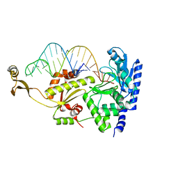
2DRP
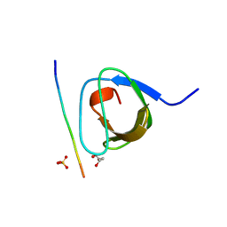
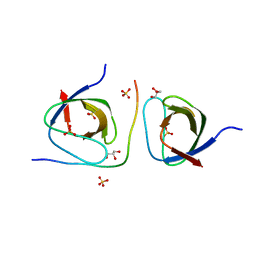
 | | THE CRYSTAL STRUCTURE OF A TWO ZINC-FINGER PEPTIDE REVEALS AN EXTENSION TO THE RULES FOR ZINC-FINGER/DNA RECOGNITION | | 分子名称: | DNA (5'-D(*CP*TP*AP*AP*TP*AP*AP*GP*GP*AP*TP*AP*AP*CP*GP*TP*C P*CP*G)-3'), DNA (5'-D(*TP*CP*GP*GP*AP*CP*GP*TP*TP*AP*TP*CP*CP*TP*TP*AP*T P*TP*A)-3'), PROTEIN (TRAMTRACK DNA-BINDING DOMAIN), ... | | 著者 | Fairall, L, Schwabe, J.W.R, Chapman, L, Finch, J.T, Rhodes, D. | | 登録日 | 1994-06-06 | | 公開日 | 1994-08-31 | | 最終更新日 | 2024-02-14 | | 実験手法 | X-RAY DIFFRACTION (2.8 Å) | | 主引用文献 | The crystal structure of a two zinc-finger peptide reveals an extension to the rules for zinc-finger/DNA recognition.
Nature, 366, 1993
|
|
2DRQ
 | | Crystal structure of reducing-end-xylose releasing exo-oligoxylanase D263G mutant | | 分子名称: | GLYCEROL, NICKEL (II) ION, Xylanase Y | | 著者 | Fushinobu, S, Hidaka, M, Honda, Y, Wakagi, T, Shoun, H, Kitaoka, M. | | 登録日 | 2006-06-12 | | 公開日 | 2006-06-27 | | 最終更新日 | 2023-10-25 | | 実験手法 | X-RAY DIFFRACTION (2.1 Å) | | 主引用文献 | Structural explanation for the acquisition of glycosynthase activity
J.Biochem., 2009
|
|


