4LIT
 
 | |
4EBG
 | |
4R6S
 | |
4RIK
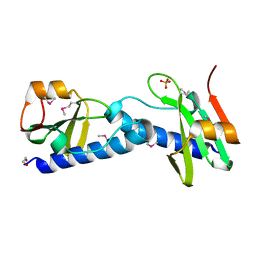
 | | Amyloid forming segment, AVVTGVTAV, from the NAC domain of Parkinson's disease protein alpha-synuclein, residues 69-77 | | 分子名称: | Alpha-synuclein | | 著者 | Guenther, E.L, Sawaya, M.R, Ivanova, M, Eisenberg, D.S. | | 登録日 | 2014-10-06 | | 公開日 | 2015-08-26 | | 最終更新日 | 2024-04-03 | | 実験手法 | X-RAY DIFFRACTION (1.854 Å) | | 主引用文献 | Structure of the toxic core of alpha-synuclein from invisible crystals.
Nature, 525, 2015
|
|
4J6C
 | |
4RIL
 | | Structure of the amyloid forming segment, GAVVTGVTAVA, from the NAC domain of Parkinson's disease protein alpha-synuclein, residues 68-78, determined by electron diffraction | | 分子名称: | Alpha-synuclein | | 著者 | Rodriguez, J.A, Ivanova, M, Sawaya, M.R, Cascio, D, Reyes, F, Shi, D, Johnson, L, Guenther, E, Sangwan, S, Hattne, J, Nannenga, B, Brewster, A.S, Messerschmidt, M, Boutet, S, Sauter, N.K, Gonen, T, Eisenberg, D.S. | | 登録日 | 2014-10-06 | | 公開日 | 2015-08-26 | | 最終更新日 | 2023-09-20 | | 実験手法 | ELECTRON CRYSTALLOGRAPHY (1.43 Å) | | 主引用文献 | Structure of the toxic core of alpha-synuclein from invisible crystals.
Nature, 525, 2015
|
|
4J9I
 | |
3SOO
 | |
4JBA
 | |
4DVH
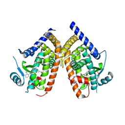
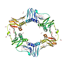
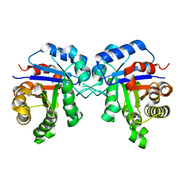
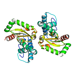 | | Crystal structure of Trypanosoma cruzi mitochondrial iron superoxide dismutase | | 分子名称: | FE (III) ION, Superoxide dismutase | | 著者 | Larrieux, N, Buschiazzo, A. | | 登録日 | 2012-02-23 | | 公開日 | 2013-03-27 | | 最終更新日 | 2023-09-13 | | 実験手法 | X-RAY DIFFRACTION (2.23 Å) | | 主引用文献 | Structural and Molecular Basis of the Peroxynitrite-mediated Nitration and Inactivation of Trypanosoma cruzi Iron-Superoxide Dismutases (Fe-SODs) A and B: DISPARATE SUSCEPTIBILITIES DUE TO THE REPAIR OF TYR35 RADICAL BY CYS83 IN Fe-SODB THROUGH INTRAMOLECULAR ELECTRON TRANSFER.
J.Biol.Chem., 289, 2014
|
|
4JBT
 | |
4DRI
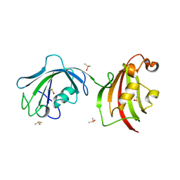
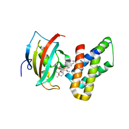 | | Co-crystal structure of the PPIase domain of FKBP51, Rapamycin and the FRB fragment of mTOR | | 分子名称: | Peptidyl-prolyl cis-trans isomerase FKBP5, RAPAMYCIN IMMUNOSUPPRESSANT DRUG, Serine/threonine-protein kinase mTOR | | 著者 | Maerz, A.M, Bracher, A, Hausch, F. | | 登録日 | 2012-02-17 | | 公開日 | 2013-02-06 | | 最終更新日 | 2023-09-13 | | 実験手法 | X-RAY DIFFRACTION (1.45 Å) | | 主引用文献 | Large FK506-Binding Proteins Shape the Pharmacology of Rapamycin.
Mol.Cell.Biol., 33, 2013
|
|
3T0P
 | |
4DIZ
 | |
4DNQ
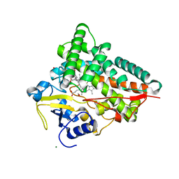
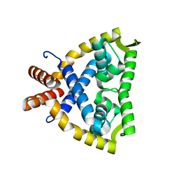
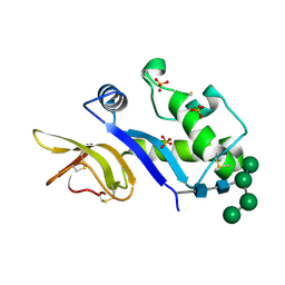
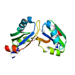
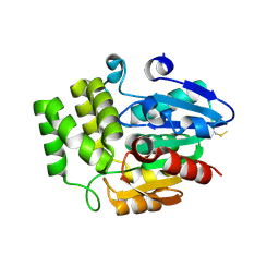 | | Crystal Structure of DAD2 S96A mutant | | 分子名称: | DAD2 | | 著者 | Hamiaux, C. | | 登録日 | 2012-02-08 | | 公開日 | 2012-11-14 | | 最終更新日 | 2013-06-26 | | 実験手法 | X-RAY DIFFRACTION (2.8 Å) | | 主引用文献 | DAD2 Is an alpha/beta Hydrolase likely to Be Involved in the Perception of the Plant Branching Hormone, Strigolactone
Curr.Biol., 22, 2012
|
|
4DYX
 | |
4POC
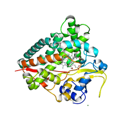
 | | Structure of Triosephosphate Isomerase Wild Type human enzyme. | | 分子名称: | BROMIDE ION, PHOSPHATE ION, POTASSIUM ION, ... | | 著者 | Amrich, C.G, Aslam, A.A, Heroux, A, VanDemark, A.P. | | 登録日 | 2014-02-25 | | 公開日 | 2015-01-14 | | 最終更新日 | 2023-09-20 | | 実験手法 | X-RAY DIFFRACTION (1.601 Å) | | 主引用文献 | Triosephosphate isomerase I170V alters catalytic site, enhances stability and induces pathology in a Drosophila model of TPI deficiency.
Biochim.Biophys.Acta, 1852, 2015
|
|
4DVK
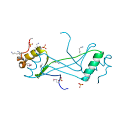
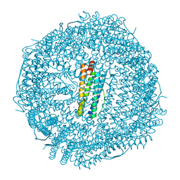
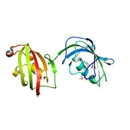
 | | Crystal structure of the glycoprotein Erns from the pestivirus BVDV-1 strain NCP-7 | | 分子名称: | 2-acetamido-2-deoxy-beta-D-glucopyranose, 2-acetamido-2-deoxy-beta-D-glucopyranose-(1-4)-2-acetamido-2-deoxy-beta-D-glucopyranose, E(rns) glycoprotein, ... | | 著者 | Krey, T, Bontems, F, Vonrhein, C, Vaney, M.-C, Bricogne, G, Ruemenapf, T, Rey, F.A. | | 登録日 | 2012-02-23 | | 公開日 | 2012-05-23 | | 最終更新日 | 2020-07-29 | | 実験手法 | X-RAY DIFFRACTION (2.21 Å) | | 主引用文献 | Crystal Structure of the Pestivirus Envelope Glycoprotein E(rns) and Mechanistic Analysis of Its Ribonuclease Activity.
Structure, 20, 2012
|
|
4PUB
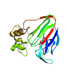
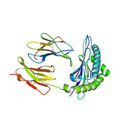
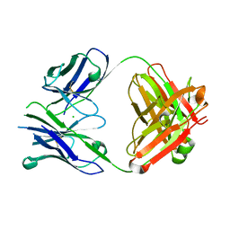 | | Crystal structure of Fab DX-2930 | | 分子名称: | CHLORIDE ION, DX-2930 HEAVY CHAIN, DX-2930 LIGHT CHAIN | | 著者 | Abendroth, J, Edwards, T.E, Nixon, A, Ladner, R. | | 登録日 | 2014-03-12 | | 公開日 | 2014-07-09 | | 最終更新日 | 2023-09-20 | | 実験手法 | X-RAY DIFFRACTION (1.75 Å) | | 主引用文献 | Inhibition of plasma kallikrein by a highly specific active site blocking antibody.
J.Biol.Chem., 289, 2014
|
|
4LAY
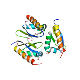
 | | Crystal Structure Analysis of FKBP52, Complex with I63 | | 分子名称: | Peptidyl-prolyl cis-trans isomerase FKBP4, {3-[(1R)-3-(3,4-dimethoxyphenyl)-1-({[(2S)-1-(3,3-dimethyl-2-oxopentanoyl)piperidin-2-yl]carbonyl}oxy)propyl]phenoxy}acetic acid | | 著者 | Bracher, A, Kozany, C, Haehle, A, Wild, P, Zacharias, M, Hausch, F. | | 登録日 | 2013-06-20 | | 公開日 | 2013-08-21 | | 最終更新日 | 2023-09-20 | | 実験手法 | X-RAY DIFFRACTION (1.7 Å) | | 主引用文献 | Crystal Structures of the Free and Ligand-Bound FK1-FK2 Domain Segment of FKBP52 Reveal a Flexible Inter-Domain Hinge.
J.Mol.Biol., 425, 2013
|
|
4PV9
 | | Crystal Structure of H2Kb-Q600V complex | | 分子名称: | ACETATE ION, Beta-2-microglobulin, GLYCEROL, ... | | 著者 | Twist, K.-A, Rossjohn, J, Gras, S. | | 登録日 | 2014-03-15 | | 公開日 | 2014-04-23 | | 最終更新日 | 2023-12-06 | | 実験手法 | X-RAY DIFFRACTION (2 Å) | | 主引用文献 | Structural and functional correlates of enhanced antiviral immunity generated by heteroclitic CD8 T cell epitopes.
J.Immunol., 192, 2014
|
|
4LAW
 | | Crystal Structure Analysis of FKBP52, Crystal Form III | | 分子名称: | DIMETHYL SULFOXIDE, Peptidyl-prolyl cis-trans isomerase FKBP4 | | 著者 | Bracher, A, Kozany, C, Haehle, A, Wild, P, Zacharias, M, Hausch, F. | | 登録日 | 2013-06-20 | | 公開日 | 2013-08-21 | | 最終更新日 | 2023-09-20 | | 実験手法 | X-RAY DIFFRACTION (2.4 Å) | | 主引用文献 | Crystal Structures of the Free and Ligand-Bound FK1-FK2 Domain Segment of FKBP52 Reveal a Flexible Inter-Domain Hinge.
J.Mol.Biol., 425, 2013
|
|
4LIR
 | |
4LIJ
 | |
3R4I
 | |


