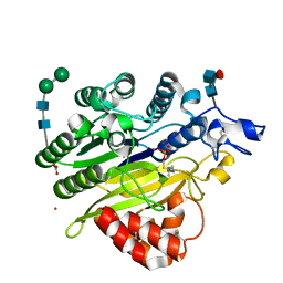
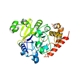
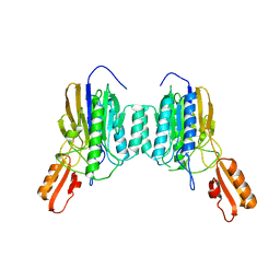
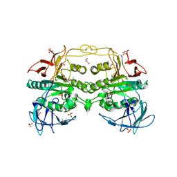
5I85
 
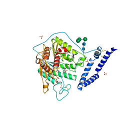 | | aSMase with zinc and phosphocholine | | 分子名称: | 2-acetamido-2-deoxy-beta-D-glucopyranose, 2-acetamido-2-deoxy-beta-D-glucopyranose-(1-4)-2-acetamido-2-deoxy-beta-D-glucopyranose, PHOSPHOCHOLINE, ... | | 著者 | Zhou, Y.F, Wei, R.R. | | 登録日 | 2016-02-18 | | 公開日 | 2016-09-07 | | 最終更新日 | 2023-09-27 | | 実験手法 | X-RAY DIFFRACTION (2.5 Å) | | 主引用文献 | Human acid sphingomyelinase structures provide insight to molecular basis of Niemann-Pick disease.
Nat Commun, 7, 2016
|
|
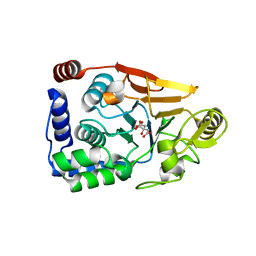
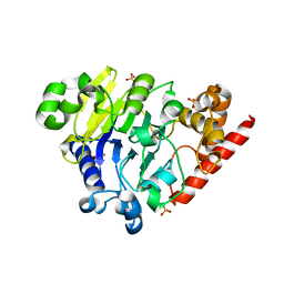
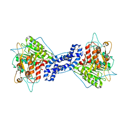
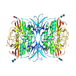
5I81
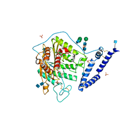 | | aSMase with zinc | | 分子名称: | 2-acetamido-2-deoxy-beta-D-glucopyranose, 2-acetamido-2-deoxy-beta-D-glucopyranose-(1-4)-2-acetamido-2-deoxy-beta-D-glucopyranose, SULFATE ION, ... | | 著者 | Zhou, Y.F, Wei, R.R. | | 登録日 | 2016-02-18 | | 公開日 | 2016-09-07 | | 最終更新日 | 2021-03-24 | | 実験手法 | X-RAY DIFFRACTION (2.25 Å) | | 主引用文献 | Human acid sphingomyelinase structures provide insight to molecular basis of Niemann-Pick disease.
Nat Commun, 7, 2016
|
|
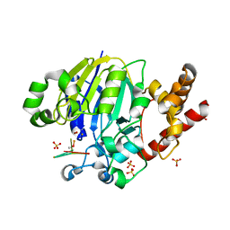
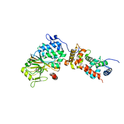
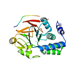
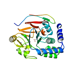
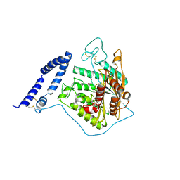
4MOV
 | | 1.45 A Resolution Crystal Structure of Protein Phosphatase 1 | | 分子名称: | CHLORIDE ION, MANGANESE (II) ION, PHOSPHATE ION, ... | | 著者 | Choy, M.S, Peti, W, Page, R. | | 登録日 | 2013-09-12 | | 公開日 | 2014-03-26 | | 最終更新日 | 2023-09-20 | | 実験手法 | X-RAY DIFFRACTION (1.4503 Å) | | 主引用文献 | Understanding the antagonism of retinoblastoma protein dephosphorylation by PNUTS provides insights into the PP1 regulatory code.
Proc.Natl.Acad.Sci.USA, 111, 2014
|
|
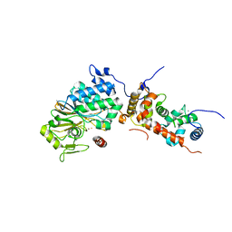
5K78
 | |
5KAS
 | | Murine acid sphingomyelinase-like phosphodiesterase 3b (SMPDL3B) with phosphocholine | | 分子名称: | 2-acetamido-2-deoxy-beta-D-glucopyranose-(1-4)-2-acetamido-2-deoxy-beta-D-glucopyranose, 2-acetamido-2-deoxy-beta-D-glucopyranose-(1-4)-[alpha-L-fucopyranose-(1-6)]2-acetamido-2-deoxy-beta-D-glucopyranose, Acid sphingomyelinase-like phosphodiesterase 3b, ... | | 著者 | Gorelik, A, Illes, K, Heinz, L.X, Superti-Furga, G, Nagar, B. | | 登録日 | 2016-06-02 | | 公開日 | 2016-10-05 | | 最終更新日 | 2020-07-29 | | 実験手法 | X-RAY DIFFRACTION (1.619 Å) | | 主引用文献 | Crystal Structure of the Acid Sphingomyelinase-like Phosphodiesterase SMPDL3B Provides Insights into Determinants of Substrate Specificity.
J.Biol.Chem., 291, 2016
|
|
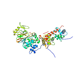
5JPE
 | | Yeast-specific serine/threonine protein phosphatase (PPZ1) of Candida albicans | | 分子名称: | CITRATE ANION, GLYCEROL, Serine/threonine-protein phosphatase | | 著者 | Choy, M.S, Chen, E.H, Peti, W, Page, R. | | 登録日 | 2016-05-03 | | 公開日 | 2016-08-31 | | 最終更新日 | 2023-09-27 | | 実験手法 | X-RAY DIFFRACTION (2.611 Å) | | 主引用文献 | Molecular Insights into the Fungus-Specific Serine/Threonine Protein Phosphatase Z1 in Candida albicans.
Mbio, 7, 2016
|
|
5K77
 | | Dbr1 in complex with 7-mer branched RNA | | 分子名称: | FE (II) ION, HYDROXIDE ION, RNA lariat debranching enzyme, ... | | 著者 | Clark, N.E, Taylor, A.B, Hart, P.J. | | 登録日 | 2016-05-25 | | 公開日 | 2016-12-07 | | 最終更新日 | 2023-09-27 | | 実験手法 | X-RAY DIFFRACTION (2.17 Å) | | 主引用文献 | Metal dependence and branched RNA cocrystal structures of the RNA lariat debranching enzyme Dbr1.
Proc. Natl. Acad. Sci. U.S.A., 113, 2016
|
|
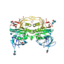
4ORA
 | | Crystal structure of a human calcineurin mutant | | 分子名称: | CALCIUM ION, Calcineurin subunit B type 1, FE (III) ION, ... | | 著者 | Li, S.J, Wang, J, Wu, J.W, Wang, Z.X. | | 登録日 | 2014-02-11 | | 公開日 | 2015-05-20 | | 最終更新日 | 2023-11-08 | | 実験手法 | X-RAY DIFFRACTION (2.747 Å) | | 主引用文献 | Cooperative autoinhibition and multi-level activation mechanisms of calcineurin
To be Published
|
|
5K71
 | | apo Dbr1 | | 分子名称: | RNA lariat debranching enzyme, putative, SULFATE ION | | 著者 | Clark, N.E, Taylor, A.B, Hart, P.J. | | 登録日 | 2016-05-25 | | 公開日 | 2016-12-07 | | 最終更新日 | 2023-09-27 | | 実験手法 | X-RAY DIFFRACTION (2.57 Å) | | 主引用文献 | The RNA lariat debranching enzyme Dbr1: metal dependence and branched RNA co-crystal structures
Proc.Natl.Acad.Sci.USA, 2016
|
|
5KAR
 | | Murine acid sphingomyelinase-like phosphodiesterase 3b (SMPDL3B) | | 分子名称: | 2-acetamido-2-deoxy-beta-D-glucopyranose-(1-4)-2-acetamido-2-deoxy-beta-D-glucopyranose, 2-acetamido-2-deoxy-beta-D-glucopyranose-(1-4)-[alpha-L-fucopyranose-(1-6)]2-acetamido-2-deoxy-beta-D-glucopyranose, Acid sphingomyelinase-like phosphodiesterase 3b, ... | | 著者 | Gorelik, A, Illes, K, Heinz, L.X, Superti-Furga, G, Nagar, B. | | 登録日 | 2016-06-02 | | 公開日 | 2016-10-05 | | 最終更新日 | 2020-07-29 | | 実験手法 | X-RAY DIFFRACTION (1.142 Å) | | 主引用文献 | Crystal Structure of the Acid Sphingomyelinase-like Phosphodiesterase SMPDL3B Provides Insights into Determinants of Substrate Specificity.
J.Biol.Chem., 291, 2016
|
|
5K73
 | |
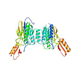
4ORB
 | | Crystal structure of mouse calcineurin | | 分子名称: | CALCIUM ION, Calcineurin subunit B type 1, FE (III) ION, ... | | 著者 | Ma, L, Li, S.J, Wang, J, Wu, J.W, Wang, Z.X. | | 登録日 | 2014-02-11 | | 公開日 | 2015-05-20 | | 最終更新日 | 2023-11-08 | | 実験手法 | X-RAY DIFFRACTION (3.108 Å) | | 主引用文献 | Cooperative autoinhibition and multi-level activation mechanisms of calcineurin
To be Published
|
|
4NZV
 | | DNA Double-Strand Break Repair Pathway Choice Is Directed by Distinct MRE11 Nuclease Activities | | 分子名称: | Exonuclease, putative, MANGANESE (II) ION | | 著者 | Shibata, A, Moiani, D, Arvai, A.S, Perry, J, Harding, S.M, Genois, M, Maity, R, Rossum-Fikkert, S, Kertokalio, A, Romoli, F, Ismail, A, Ismalaj, E, Petricci, E, Neale, M.J, Bristow, R.G, Masson, J, Wyman, C, Jeggo, P.A, Tainer, J.A. | | 登録日 | 2013-12-12 | | 公開日 | 2014-01-08 | | 最終更新日 | 2024-02-28 | | 実験手法 | X-RAY DIFFRACTION (1.901 Å) | | 主引用文献 | DNA Double-Strand Break Repair Pathway Choice Is Directed by Distinct MRE11 Nuclease Activities.
Mol.Cell, 53, 2014
|
|
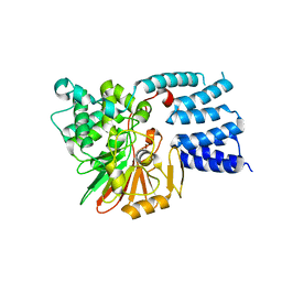
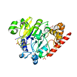
4G9J
 | | Protein Ser/Thr phosphatase-1 in complex with cell-permeable peptide | | 分子名称: | MANGANESE (II) ION, Serine/threonine-protein phosphatase PP1-alpha catalytic subunit, synthetic peptide | | 著者 | Sukackaite, R, Chatterjee, J, Beullens, M, Bollen, M, Koehn, M, Hart, D.J. | | 登録日 | 2012-07-24 | | 公開日 | 2012-09-19 | | 最終更新日 | 2023-09-13 | | 実験手法 | X-RAY DIFFRACTION (3.1 Å) | | 主引用文献 | Development of a Peptide that Selectively Activates Protein Phosphatase-1 in Living Cells.
Angew.Chem.Int.Ed.Engl., 51, 2012
|
|
4ORC
 | | Crystal structure of mammalian calcineurin | | 分子名称: | CALCIUM ION, Calcineurin subunit B type 1, FE (III) ION, ... | | 著者 | Ma, L, Li, S.J, Wang, J, Wu, J.W, Wang, Z.X. | | 登録日 | 2014-02-11 | | 公開日 | 2015-05-20 | | 最終更新日 | 2023-11-08 | | 実験手法 | X-RAY DIFFRACTION (2.7 Å) | | 主引用文献 | Cooperative autoinhibition and multi-level activation mechanisms of calcineurin
To be Published
|
|
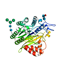
5JG8
 | | Crystal structure of human acid sphingomyelinase | | 分子名称: | 2-acetamido-2-deoxy-beta-D-glucopyranose, 2-acetamido-2-deoxy-beta-D-glucopyranose-(1-3)-2-acetamido-2-deoxy-beta-D-glucopyranose, 2-acetamido-2-deoxy-beta-D-glucopyranose-(1-4)-2-acetamido-2-deoxy-beta-D-glucopyranose, ... | | 著者 | Xiong, Z.J, Prive, G.G. | | 登録日 | 2016-04-19 | | 公開日 | 2016-07-06 | | 最終更新日 | 2023-09-27 | | 実験手法 | X-RAY DIFFRACTION (2.8 Å) | | 主引用文献 | Structure of Human Acid Sphingomyelinase Reveals the Role of the Saposin Domain in Activating Substrate Hydrolysis.
J.Mol.Biol., 428, 2016
|
|
4DT2
 | | Crystal structure of red kidney bean purple acid phosphatase in complex with Maybridge fragment CC27209 | | 分子名称: | (2,2-dimethyl-2,3-dihydro-1-benzofuran-7-yl)methanol, 1,2-ETHANEDIOL, 2-acetamido-2-deoxy-beta-D-glucopyranose, ... | | 著者 | Feder, D, Hussein, W.M, Clayton, D.J, Kan, M, Schenk, G, McGeary, R.P, Guddat, L.W. | | 登録日 | 2012-02-20 | | 公開日 | 2012-09-19 | | 最終更新日 | 2020-07-29 | | 実験手法 | X-RAY DIFFRACTION (2.7 Å) | | 主引用文献 | Identification of purple acid phosphatase inhibitors by fragment-based screening: promising new leads for osteoporosis therapeutics.
Chem.Biol.Drug Des., 80, 2012
|
|
4OR9
 | | Crystal structure of human calcineurin | | 分子名称: | CALCIUM ION, Calcineurin subunit B type 1, FE (III) ION, ... | | 著者 | Li, S.J, Wang, J, Wu, J.W, Wang, Z.X. | | 登録日 | 2014-02-11 | | 公開日 | 2015-05-20 | | 最終更新日 | 2023-11-08 | | 実験手法 | X-RAY DIFFRACTION (2.23 Å) | | 主引用文献 | Cooperative autoinhibition and multi-level activation mechanisms of calcineurin
To be Published
|
|
5J28
 | | Ki67-PP1g (protein phosphatase 1, gamma isoform) holoenzyme complex | | 分子名称: | Antigen KI-67, MALONATE ION, SODIUM ION, ... | | 著者 | Kumar, G.S, Peti, W, Page, R. | | 登録日 | 2016-03-29 | | 公開日 | 2016-10-05 | | 最終更新日 | 2023-09-27 | | 実験手法 | X-RAY DIFFRACTION (2 Å) | | 主引用文献 | The Ki-67 and RepoMan mitotic phosphatases assemble via an identical, yet novel mechanism.
Elife, 5, 2016
|
|
4DSY
 | | Crystal structure of red kidney bean purple acid phosphatase in complex with Maybridge fragment CC24201 | | 分子名称: | 1,2-ETHANEDIOL, 2-acetamido-2-deoxy-beta-D-glucopyranose, 5-phenylpyridine-3-carboxylic acid, ... | | 著者 | Feder, D, Hussein, W.M, Clayton, D.J, Kan, M, Schenk, G, McGeary, R.P, Guddat, L.W. | | 登録日 | 2012-02-20 | | 公開日 | 2012-09-19 | | 最終更新日 | 2020-07-29 | | 実験手法 | X-RAY DIFFRACTION (2.3 Å) | | 主引用文献 | Identification of purple acid phosphatase inhibitors by fragment-based screening: promising new leads for osteoporosis therapeutics.
Chem.Biol.Drug Des., 80, 2012
|
|
5JJT
 | |
4DHL
 | | Crystal structure of red kidney bean purple acid phosphatase in complex with Maybridge fragment MO07123 | | 分子名称: | 1,2-ETHANEDIOL, 2-(4-methylphenyl)-1,3-thiazole-4-carboxylic acid, 2-acetamido-2-deoxy-beta-D-glucopyranose, ... | | 著者 | Feder, D, Clayton, D.J, Hussein, W.M, Schenk, G, McGeary, R, Guddat, L.W. | | 登録日 | 2012-01-29 | | 公開日 | 2012-12-12 | | 最終更新日 | 2020-07-29 | | 実験手法 | X-RAY DIFFRACTION (2.3 Å) | | 主引用文献 | Identification of purple acid phosphatase inhibitors by fragment-based screening: promising new leads for osteoporosis therapeutics.
Chem.Biol.Drug Des., 80, 2012
|
|
4PEH
 | | Dbr1 in complex with synthetic linear RNA | | 分子名称: | GLYCEROL, MANGANESE (II) ION, RNA (5'-R(*CP*UP*AP*(A2P)P*AP*CP*AP*A)-3'), ... | | 著者 | Montemayor, E.J, Katolik, A, Clark, N.E, Taylor, A.B, Schuermann, J.P, Combs, D.J, Johnsson, R, Holloway, S.P, Stevens, S.W, Damha, M.J, Hart, P.J. | | 登録日 | 2014-04-23 | | 公開日 | 2014-08-27 | | 最終更新日 | 2023-12-27 | | 実験手法 | X-RAY DIFFRACTION (2.1 Å) | | 主引用文献 | Structural basis of lariat RNA recognition by the intron debranching enzyme Dbr1.
Nucleic Acids Res., 42, 2014
|
|
4O24
 | | DNA Double-Strand Break Repair Pathway Choice Is Directed by Distinct MRE11 Nuclease Activities | | 分子名称: | (5~{Z})-5-[(4-hydroxyphenyl)methylidene]-3-(2-methylpropyl)-2-sulfanylidene-1,3-thiazolidin-4-one, Exonuclease, putative, ... | | 著者 | Shibata, A, Moiani, D, Arvai, A.S, Perry, J, Harding, S.M, Genois, M, Maity, R, Rossum-Fikkert, S, Kertokalio, A, Romoli, F, Ismail, A, Ismalaj, E, Petricci, E, Neale, M.J, Bristow, R.G, Masson, J, Wyman, C, Jeggo, P.A, Tainer, J.A. | | 登録日 | 2013-12-16 | | 公開日 | 2014-01-15 | | 最終更新日 | 2023-09-20 | | 実験手法 | X-RAY DIFFRACTION (2.3 Å) | | 主引用文献 | DNA Double-Strand Break Repair Pathway Choice Is Directed by Distinct MRE11 Nuclease Activities.
Mol.Cell, 53, 2014
|
|
5I8R
 | | aSMase with zinc | | 分子名称: | 2-acetamido-2-deoxy-beta-D-glucopyranose, 2-acetamido-2-deoxy-beta-D-glucopyranose-(1-4)-2-acetamido-2-deoxy-beta-D-glucopyranose, Sphingomyelin phosphodiesterase, ... | | 著者 | Zhou, Y.F, Wei, R.R. | | 登録日 | 2016-02-19 | | 公開日 | 2016-09-07 | | 最終更新日 | 2023-09-27 | | 実験手法 | X-RAY DIFFRACTION (3.646 Å) | | 主引用文献 | Human acid sphingomyelinase structures provide insight to molecular basis of Niemann-Pick disease.
Nat Commun, 7, 2016
|
|


