2A9N
 
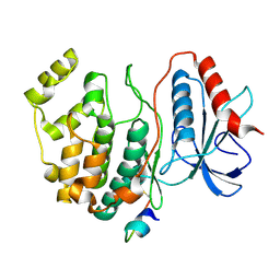
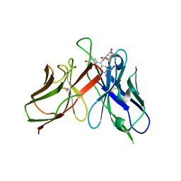 | | A Mutation Designed to Alter Crystal Packing Permits Structural Analysis of a Tight-binding Fluorescein-scFv complex | | 分子名称: | 4-(2,7-DIFLUORO-6-HYDROXY-3-OXO-3H-XANTHEN-9-YL)ISOPHTHALIC ACID, fluorescein-scfv | | 著者 | Cambillau, C, Spinelli, S, Honegger, A, Pluckthun, A. | | 登録日 | 2005-07-12 | | 公開日 | 2005-10-25 | | 最終更新日 | 2024-04-03 | | 実験手法 | X-RAY DIFFRACTION (3 Å) | | 主引用文献 | A mutation designed to alter crystal packing permits structural analysis of a tight-binding fluorescein-scFv complex.
Protein Sci., 14, 2005
|
|
2EUW
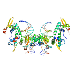
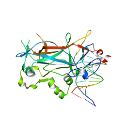
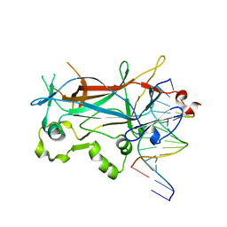 | | Structure of a Ndt80-DNA complex (MSE mutant mA4T) | | 分子名称: | 5'-D(*AP*GP*TP*TP*TP*TP*TP*GP*AP*GP*TP*CP*GP*C)-3', 5'-D(*TP*GP*CP*GP*AP*CP*TP*CP*AP*AP*AP*AP*AP*C)-3', NDT80 protein | | 著者 | Lamoureux, J.S, Glover, J.N. | | 登録日 | 2005-10-30 | | 公開日 | 2006-03-21 | | 最終更新日 | 2023-08-23 | | 実験手法 | X-RAY DIFFRACTION (1.68 Å) | | 主引用文献 | Principles of Protein-DNA Recognition Revealed in the Structural Analysis of Ndt80-MSE DNA Complexes.
Structure, 14, 2006
|
|
2EYV
 | |
2EYW
 | |
2GTY
 | |
2F3Q
 | |
2A11
 | |
2GYR
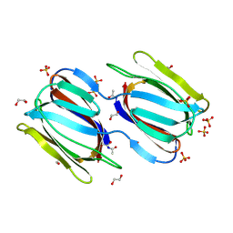
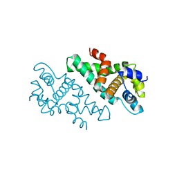
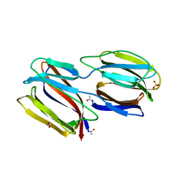
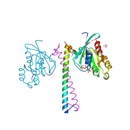
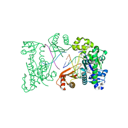
 | | Crystal structure of human artemin | | 分子名称: | Neurotrophic factor artemin, isoform 3 | | 著者 | Wang, X.Q. | | 登録日 | 2006-05-09 | | 公開日 | 2006-06-27 | | 最終更新日 | 2011-07-13 | | 実験手法 | X-RAY DIFFRACTION (2.6 Å) | | 主引用文献 | Structure of Artemin Complexed with Its Receptor GFRalpha3: Convergent Recognition of Glial Cell Line-Derived Neurotrophic Factors.
Structure, 14, 2006
|
|
2F8S
 | | Crystal structure of Aa-Ago with externally-bound siRNA | | 分子名称: | 5'-R(P*AP*GP*AP*CP*AP*GP*CP*AP*UP*AP*UP*AP*UP*GP*CP*UP*GP*UP*CP*UP*UP*U)-3', Argonaute protein | | 著者 | Yuan, Y.R, Chen, H.Y, Patel, D.J. | | 登録日 | 2005-12-03 | | 公開日 | 2006-10-31 | | 最終更新日 | 2023-08-30 | | 実験手法 | X-RAY DIFFRACTION (3 Å) | | 主引用文献 | A Potential Protein-RNA Recognition Event along the RISC-Loading Pathway from the Structure of A. aeolicus Argonaute with Externally Bound siRNA.
Structure, 14, 2006
|
|
2FF3
 | | Crystal structure of Gelsolin domain 1:N-wasp V2 motif hybrid in complex with actin | | 分子名称: | ADENOSINE-5'-TRIPHOSPHATE, Actin, alpha skeletal muscle, ... | | 著者 | Xue, B, Aguda, A.H, Robinson, R.C. | | 登録日 | 2005-12-19 | | 公開日 | 2006-03-21 | | 最終更新日 | 2024-10-09 | | 実験手法 | X-RAY DIFFRACTION (2 Å) | | 主引用文献 | The Structural Basis of Actin Interaction with Multiple WH2/beta-Thymosin Motif-Containing Proteins
Structure, 14, 2006
|
|
2FF6
 | | Crystal structure of Gelsolin domain 1:ciboulot domain 2 hybrid in complex with actin | | 分子名称: | ADENOSINE-5'-TRIPHOSPHATE, Actin, alpha skeletal muscle, ... | | 著者 | Aguda, A.H, Xue, B, Robinson, R.C. | | 登録日 | 2005-12-19 | | 公開日 | 2006-03-21 | | 最終更新日 | 2018-05-23 | | 実験手法 | X-RAY DIFFRACTION (2.05 Å) | | 主引用文献 | The Structural Basis of Actin Interaction with Multiple WH2/beta-Thymosin Motif-Containing Proteins
Structure, 14, 2006
|
|
2A07
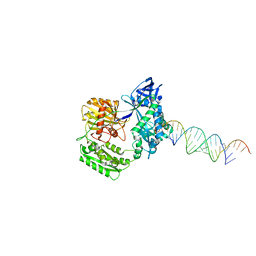
 | | Crystal Structure of Foxp2 bound Specifically to DNA. | | 分子名称: | 5'-D(*AP*AP*CP*TP*AP*TP*GP*AP*AP*AP*CP*AP*AP*AP*TP*TP*TP*TP*CP*CP*T)-3', 5'-D(*TP*TP*AP*GP*GP*AP*AP*AP*AP*TP*TP*TP*GP*TP*TP*TP*CP*AP*TP*AP*G)-3', Forkhead box protein P2, ... | | 著者 | Stroud, J.C, Wu, Y, Bates, D.L, Han, A, Nowick, K, Paabo, S, Tong, H, Chen, L. | | 登録日 | 2005-06-16 | | 公開日 | 2006-01-31 | | 最終更新日 | 2024-02-14 | | 実験手法 | X-RAY DIFFRACTION (1.9 Å) | | 主引用文献 | Structure of the Forkhead Domain of FOXP2 Bound to DNA.
Structure, 14, 2006
|
|
2GUC
 | |
2GWK
 | |
2GPH
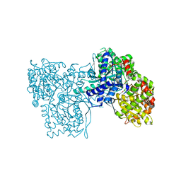
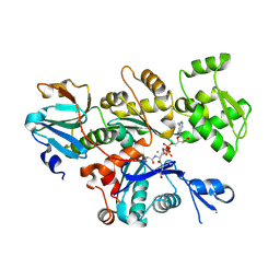
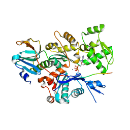
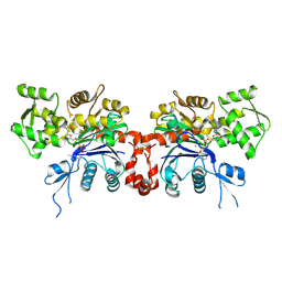
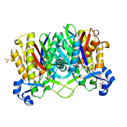
 | | Docking motif interactions in the MAP kinase ERK2 | | 分子名称: | Mitogen-activated protein kinase 1, Tyrosine-protein phosphatase non-receptor type 7 | | 著者 | Zhou, T, Sun, L, Humphreys, J, Goldsmith, E.J. | | 登録日 | 2006-04-17 | | 公開日 | 2006-07-04 | | 最終更新日 | 2023-08-30 | | 実験手法 | X-RAY DIFFRACTION (1.9 Å) | | 主引用文献 | Docking Interactions Induce Exposure of Activation Loop in the MAP Kinase ERK2.
Structure, 14, 2006
|
|
2GZH
 | |
2EXT
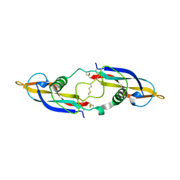
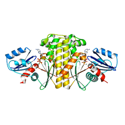
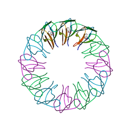 | | TRAP4 (engineered TRAP) | | 分子名称: | TRYPTOPHAN, Transcription attenuation protein mtrB | | 著者 | Heddle, J.G, Yokoyama, T, Yamashita, I, Park, S.Y, Tame, J.R.H. | | 登録日 | 2005-11-08 | | 公開日 | 2006-08-01 | | 最終更新日 | 2024-03-13 | | 実験手法 | X-RAY DIFFRACTION (1.8 Å) | | 主引用文献 | Rounding up: Engineering 12-Membered Rings from the Cyclic 11-Mer TRAP
Structure, 14, 2006
|
|
2FLL
 | | Ternary complex of human DNA polymerase iota with DNA and dTTP | | 分子名称: | DNA polymerase iota, DNA primer strand, DNA template strand, ... | | 著者 | Nair, D.T, Johnson, R.E, Prakash, L, Prakash, S, Aggarwal, A.K. | | 登録日 | 2006-01-06 | | 公開日 | 2006-12-12 | | 最終更新日 | 2023-08-30 | | 実験手法 | X-RAY DIFFRACTION (2.6 Å) | | 主引用文献 | An incoming nucleotide imposes an anti to syn conformational change on the templating purine in the human DNA polymerase-iota active site.
Structure, 14, 2006
|
|
2EUX
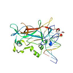
 | | Structure of a Ndt80-DNA complex (MSE VARIANT vA4G) | | 分子名称: | 5'-D(*AP*GP*TP*TP*TP*TP*TP*GP*CP*GP*TP*CP*GP*C)-3', 5'-D(*TP*GP*CP*GP*AP*CP*GP*CP*AP*AP*AP*AP*AP*C)-3', NDT80 protein | | 著者 | Lamoureux, J.S, Glover, J.N. | | 登録日 | 2005-10-30 | | 公開日 | 2006-03-21 | | 最終更新日 | 2023-08-23 | | 実験手法 | X-RAY DIFFRACTION (1.57 Å) | | 主引用文献 | Principles of Protein-DNA Recognition Revealed in the Structural Analysis of Ndt80-MSE DNA Complexes.
Structure, 14, 2006
|
|
2EZ5
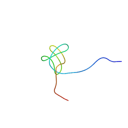
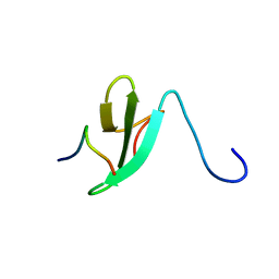 | | Solution Structure of the dNedd4 WW3* Domain- Comm LPSY Peptide Complex | | 分子名称: | Commissureless LPSY Peptide, E3 ubiquitin-protein ligase NEDD4 | | 著者 | Kanelis, V, Bruce, M.C, Skrynnikov, N.R, Rotin, D, Forman-Kay, J.D. | | 登録日 | 2005-11-10 | | 公開日 | 2006-03-28 | | 最終更新日 | 2024-05-22 | | 実験手法 | SOLUTION NMR | | 主引用文献 | Structural Determinants for High-Affinity Binding in a Nedd4 WW3(*) Domain-Comm PY Motif Complex
Structure, 14, 2006
|
|
2EFT
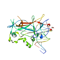
 | | Methanethiol-CYS 112 inhibition complex of E. coli ketoacyl synthase III (FABH) and Coenzyme A (high concentration (1.7mM) soak) | | 分子名称: | 3-oxoacyl-[acyl-carrier-protein] synthase 3, COENZYME A, METHANETHIOL, ... | | 著者 | Alhamadsheh, M.M, Musayev, F, Komissarov, A.A, Sachdeva, S, Wright, H.T, Scarsdale, N, Florova, G, Reynolds, K.A. | | 登録日 | 2007-02-24 | | 公開日 | 2007-06-12 | | 最終更新日 | 2023-10-25 | | 実験手法 | X-RAY DIFFRACTION (2 Å) | | 主引用文献 | Alkyl-CoA Disulfides as Inhibitors and Mechanistic Probes for FabH Enzymes
Chem.Biol., 14, 2007
|
|
2EWS
 | |
2EUV
 | |
2ETW
 | |
2EVJ
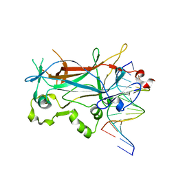 | | Structure of an Ndt80-DNA complex (MSE mutant mA9C) | | 分子名称: | 5'-D(*AP*GP*TP*GP*TP*TP*TP*GP*TP*GP*TP*CP*GP*C)-3', 5'-D(*TP*GP*CP*GP*AP*CP*AP*CP*AP*AP*AP*CP*AP*C)-3', NDT80 protein | | 著者 | Lamoureux, J.S, Glover, J.N. | | 登録日 | 2005-10-31 | | 公開日 | 2006-03-21 | | 最終更新日 | 2023-08-23 | | 実験手法 | X-RAY DIFFRACTION (1.89 Å) | | 主引用文献 | Principles of Protein-DNA Recognition Revealed in the Structural Analysis of Ndt80-MSE DNA Complexes.
Structure, 14, 2006
|
|


