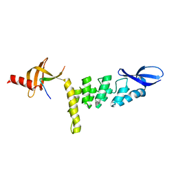
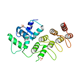
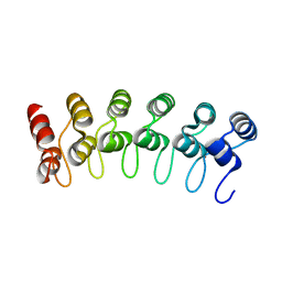
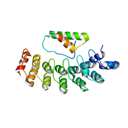
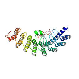
3V31
 
 | | Crystal Structure of the Peptide Bound Complex of the Ankyrin Repeat Domains of Human ANKRA2 | | 分子名称: | Ankyrin repeat family A protein 2, CHLORIDE ION, Histone deacetylase 4, ... | | 著者 | Lam, R, Xu, C, Bian, C.B, Kania, J, Bountra, C, Weigelt, J, Arrowsmith, C.H, Edwards, A.M, Bochkarev, A, Min, J, Structural Genomics Consortium (SGC) | | 登録日 | 2011-12-12 | | 公開日 | 2012-04-04 | | 最終更新日 | 2023-09-13 | | 実験手法 | X-RAY DIFFRACTION (1.57 Å) | | 主引用文献 | Sequence-Specific Recognition of a PxLPxI/L Motif by an Ankyrin Repeat Tumbler Lock.
Sci.Signal., 5, 2012
|
|
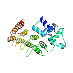
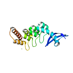
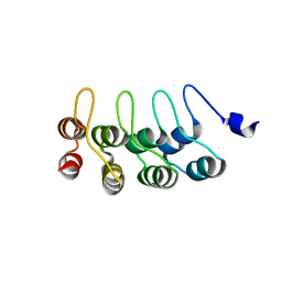
3UXG
 | | Crystal structure of RFXANK | | 分子名称: | DNA-binding protein RFXANK, Histone deacetylase 4, UNKNOWN ATOM OR ION | | 著者 | Tempel, W, Chao, X, Bian, C, Li, Y, Bountra, C, Weigelt, J, Arrowsmith, C.H, Edwards, A.M, Min, J, Structural Genomics Consortium (SGC) | | 登録日 | 2011-12-05 | | 公開日 | 2012-06-13 | | 最終更新日 | 2023-09-13 | | 実験手法 | X-RAY DIFFRACTION (1.85 Å) | | 主引用文献 | Sequence-Specific Recognition of a PxLPxI/L Motif by an Ankyrin Repeat Tumbler Lock.
Sci.Signal., 5, 2012
|
|
3UI2
 | |
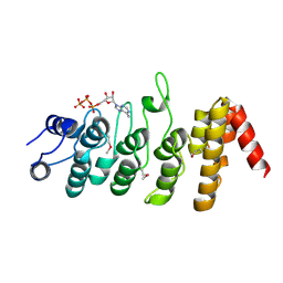
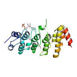
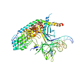
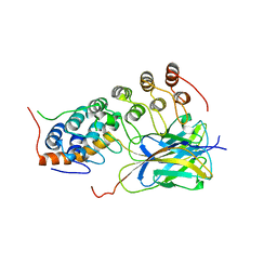
4A63
 | | Crystal structure of the p73-ASPP2 complex at 2.6A resolution | | 分子名称: | ACETATE ION, APOPTOSIS STIMULATING OF P53 PROTEIN 2, TUMOUR PROTEIN 73, ... | | 著者 | Canning, P, Sharpe, T, Krojer, T, Savitsky, P, Cooper, C.D.O, Salah, E, Keates, T, Muniz, J, Vollmar, M, von Delft, F, Weigelt, J, Arrowsmith, C, Bountra, C, Edwards, A, Bullock, A.N. | | 登録日 | 2011-10-31 | | 公開日 | 2011-12-21 | | 最終更新日 | 2023-12-20 | | 実験手法 | X-RAY DIFFRACTION (2.27 Å) | | 主引用文献 | Structural Basis for Aspp2 Recognition by the Tumor Suppressor P73.
J.Mol.Biol., 423, 2012
|
|
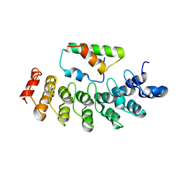
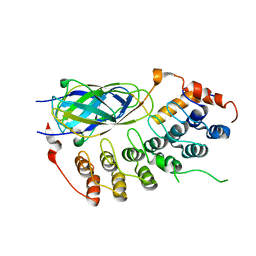
3T9K
 | | Crystal Structure of ACAP1 C-portion mutant S554D fused with integrin beta1 peptide | | 分子名称: | Arf-GAP with coiled-coil, ANK repeat and PH domain-containing protein 1,Peptide from Integrin beta-1, SULFATE ION, ... | | 著者 | Sun, F, Pang, X, Zhang, K, Ma, J, Zhou, Q. | | 登録日 | 2011-08-03 | | 公開日 | 2012-07-18 | | 最終更新日 | 2023-11-01 | | 実験手法 | X-RAY DIFFRACTION (2.3 Å) | | 主引用文献 | Mechanistic insights into regulated cargo binding by ACAP1 protein
J.Biol.Chem., 287, 2012
|
|
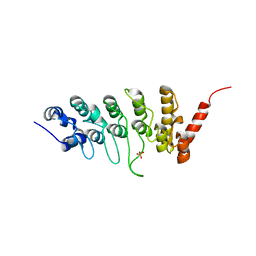
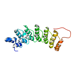
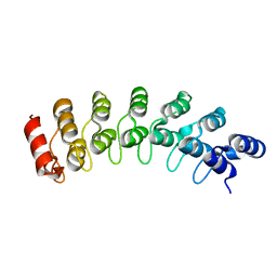
3SO8
 | | Crystal Structure of ANKRA | | 分子名称: | Ankyrin repeat family A protein 2 | | 著者 | Xu, C, Bochkarev, A, Bian, C.B, Min, J, Structural Genomics Consortium (SGC) | | 登録日 | 2011-06-30 | | 公開日 | 2011-10-05 | | 最終更新日 | 2024-02-28 | | 実験手法 | X-RAY DIFFRACTION (1.9 Å) | | 主引用文献 | Sequence-Specific Recognition of a PxLPxI/L Motif by an Ankyrin Repeat Tumbler Lock.
Sci.Signal., 5, 2012
|
|
3JXJ
 | |
3JXI
 | |
3JUE
 | | Crystal Structure of ArfGAP and ANK repeat domain of ACAP1 | | 分子名称: | ARFGAP with coiled-coil, ANK repeat and PH domain-containing protein 1, SULFATE ION, ... | | 著者 | Pang, X, Zhang, K, Ma, J, Zhou, Q, Sun, F. | | 登録日 | 2009-09-15 | | 公開日 | 2010-09-22 | | 最終更新日 | 2024-03-20 | | 実験手法 | X-RAY DIFFRACTION (2.3 Å) | | 主引用文献 | Mechanistic insights into regulated cargo binding by ACAP1 protein
J.Biol.Chem., 287, 2012
|
|
3DEO
 | |
3DEP
 | | Structural basis for specific substrate recognition by the chloroplast signal recognition particle protein cpSRP43 | | 分子名称: | CHLORIDE ION, Signal recognition particle 43 kDa protein, YPGGSFDPLGLA | | 著者 | Holdermann, I, Stengel, K.F, Wild, K, Sinning, I. | | 登録日 | 2008-06-10 | | 公開日 | 2008-08-12 | | 最終更新日 | 2023-11-01 | | 実験手法 | X-RAY DIFFRACTION (2.7 Å) | | 主引用文献 | Structural basis for specific substrate recognition by the chloroplast signal recognition particle protein cpSRP43.
Science, 321, 2008
|
|
3C5R
 | |
2QC9
 | |
2PNN
 | |
2NYJ
 | |
2DZO
 | |
2DZN
 | |
2FO1
 | |
1YMP
 | |
1WG0
 | | Structural comparison of Nas6p protein structures in two different crystal forms | | 分子名称: | Probable 26S proteasome regulatory subunit p28 | | 著者 | Nakamura, Y, Umehara, T, Tanaka, A, Horikoshi, M, Yokoyama, S, Padmanabhan, B, RIKEN Structural Genomics/Proteomics Initiative (RSGI) | | 登録日 | 2004-05-27 | | 公開日 | 2005-06-07 | | 最終更新日 | 2023-10-25 | | 実験手法 | X-RAY DIFFRACTION (2.53 Å) | | 主引用文献 | Structural comparison of Nas6p protein structures in two different crystal forms
To be Published
|
|
1WDY
 | | Crystal structure of ribonuclease | | 分子名称: | 2-5A-dependent ribonuclease, 5'-O-MONOPHOSPHORYLADENYLYL(2'->5')ADENYLYL(2'->5')ADENOSINE | | 著者 | Tanaka, N, Nakanishi, M, Kusakabe, Y, Goto, Y, Kitade, Y, Nakamura, K.T. | | 登録日 | 2004-05-19 | | 公開日 | 2004-10-05 | | 最終更新日 | 2021-11-10 | | 実験手法 | X-RAY DIFFRACTION (1.8 Å) | | 主引用文献 | Structural basis for recognition of 2',5'-linked oligoadenylates by human ribonuclease L
Embo J., 23, 2004
|
|
1OY3
 | | CRYSTAL STRUCTURE OF AN IKBBETA/NF-KB P65 HOMODIMER COMPLEX | | 分子名称: | Transcription factor p65, transcription factor inhibitor I-kappa-B-beta | | 著者 | Malek, S, Huang, D.B, Huxford, T, Ghosh, S, Ghosh, G. | | 登録日 | 2003-04-03 | | 公開日 | 2003-05-20 | | 最終更新日 | 2023-08-16 | | 実験手法 | X-RAY DIFFRACTION (2.05 Å) | | 主引用文献 | X-ray crystal structure of an IkappaBbeta x NF-kappaB p65 homodimer complex.
J.Biol.Chem., 278, 2003
|
|
1N11
 | | D34 REGION OF HUMAN ANKYRIN-R AND LINKER | | 分子名称: | Ankyrin, BROMIDE ION, CHLORIDE ION | | 著者 | Michaely, P, Tomchick, D.R, Machius, M, Anderson, R.G.W. | | 登録日 | 2002-10-16 | | 公開日 | 2002-12-11 | | 最終更新日 | 2024-02-14 | | 実験手法 | X-RAY DIFFRACTION (2.7 Å) | | 主引用文献 | Crystal structure of a 12 ANK repeat stack from human ankyrinR
Embo J., 21, 2002
|
|
1IXV
 | | Crystal Structure Analysis of homolog of oncoprotein gankyrin, an interactor of Rb and CDK4/6 | | 分子名称: | Probable 26S proteasome regulatory subunit p28 | | 著者 | Padmanabhan, B, Adachi, N, Kataoka, K, Horikoshi, M. | | 登録日 | 2002-07-09 | | 公開日 | 2003-12-16 | | 最終更新日 | 2023-12-27 | | 実験手法 | X-RAY DIFFRACTION (2.3 Å) | | 主引用文献 | Crystal structure of the homolog of the oncoprotein gankyrin, an interactor of Rb and CDK4/6
J.BIOL.CHEM., 279, 2004
|
|
1K3Z
 | | X-ray crystal structure of the IkBb/NF-kB p65 homodimer complex | | 分子名称: | Transcription factor p65, transcription factor inhibitor I-kappa-B-beta | | 著者 | Shiva, M, Huang, D.B, Chen, Y, Huxford, T, Ghosh, S, Ghosh, G. | | 登録日 | 2001-10-04 | | 公開日 | 2002-10-04 | | 最終更新日 | 2023-08-16 | | 実験手法 | X-RAY DIFFRACTION (2.5 Å) | | 主引用文献 | X-ray crystal structure of an IkappaBbeta x NF-kappaB p65 homodimer complex.
J.Biol.Chem., 278, 2003
|
|


