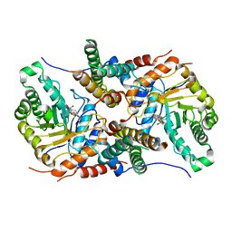
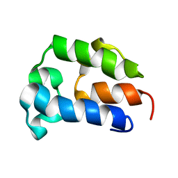
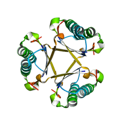
5UHL
 
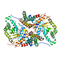 | | Crystal structure of the core catalytic domain of human O-GlcNAcase complexed with Thiamet G | | 分子名称: | (2Z,3aR,5R,6S,7R,7aR)-2-(ethylimino)-5-(hydroxymethyl)hexahydro-3aH-pyrano[3,2-d][1,3]thiazole-6,7-diol, O-GlcNAcase TIM-barrel domain, O-GlcNAcase stalk domain | | 著者 | Klein, D.J, Elsen, N.L. | | 登録日 | 2017-01-11 | | 公開日 | 2017-03-29 | | 最終更新日 | 2024-03-06 | | 実験手法 | X-RAY DIFFRACTION (3.14 Å) | | 主引用文献 | Crystal structure of the core catalytic domain of human O-GlcNAcase and molecular basis of activity and inhibition
To Be Published
|
|
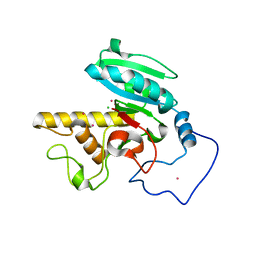
5DDB
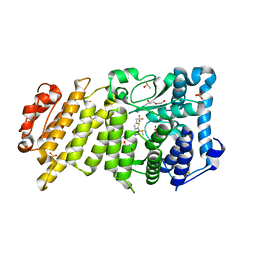 | | Menin in complex with MI-319 | | 分子名称: | 4-{4-[5-(difluoromethyl)-1,3,4-thiadiazol-2-yl]piperazin-1-yl}-6-(2,2,2-trifluoroethyl)thieno[2,3-d]pyrimidine, DI(HYDROXYETHYL)ETHER, DIMETHYL SULFOXIDE, ... | | 著者 | Pollock, J, Dmitry, B, Cierpicki, T, Grembecka, J. | | 登録日 | 2015-08-24 | | 公開日 | 2015-09-09 | | 最終更新日 | 2023-09-27 | | 実験手法 | X-RAY DIFFRACTION (1.54 Å) | | 主引用文献 | Rational Design of Orthogonal Multipolar Interactions with Fluorine in Protein-Ligand Complexes.
J.Med.Chem., 58, 2015
|
|
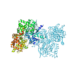
3WA0
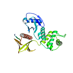 | | Crystal structure of merlin complexed with DCAF1/VprBP | | 分子名称: | Merlin, Protein VPRBP | | 著者 | Mori, T, Gotoh, S, Shirakawa, M, Hakoshima, T. | | 登録日 | 2013-04-20 | | 公開日 | 2014-05-28 | | 最終更新日 | 2023-11-08 | | 実験手法 | X-RAY DIFFRACTION (2.31 Å) | | 主引用文献 | Structural basis of DDB1-and-Cullin 4-associated Factor 1 (DCAF1) recognition by merlin/NF2 and its implication in tumorigenesis by CD44-mediated inhibition of merlin suppression of DCAF1 function.
Genes Cells, 19, 2014
|
|
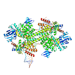
5G2V
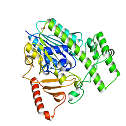 | | Structure of BT4656 in complex with its substrate D-Glucosamine-2-N, 6-O-disulfate. | | 分子名称: | 2-deoxy-6-O-sulfo-2-(sulfoamino)-alpha-D-glucopyranose, CALCIUM ION, N-ACETYLGLUCOSAMINE-6-SULFATASE, ... | | 著者 | Cartmell, A, Lowe, E.C, Basle, A, Crouch, L.I, Czjzek, M, Turnbull, J, Henrissat, B, Terrapon, N, Thomas, S, Murray, H, Firbank, S.J, Bolam, D.N. | | 登録日 | 2016-04-14 | | 公開日 | 2017-05-24 | | 最終更新日 | 2024-05-08 | | 実験手法 | X-RAY DIFFRACTION (1.39 Å) | | 主引用文献 | How members of the human gut microbiota overcome the sulfation problem posed by glycosaminoglycans.
Proc. Natl. Acad. Sci. U.S.A., 114, 2017
|
|
5UHO
 | |
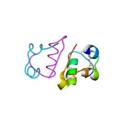
2CTW
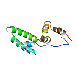 | | Solution structure of J-domain from mouse DnaJ subfamily C menber 5 | | 分子名称: | DnaJ homolog subfamily C member 5 | | 著者 | Kobayashi, N, Tomizawa, T, Koshiba, S, Inoue, M, Kigawa, T, Yokoyama, S, RIKEN Structural Genomics/Proteomics Initiative (RSGI) | | 登録日 | 2005-05-21 | | 公開日 | 2005-11-24 | | 最終更新日 | 2024-05-29 | | 実験手法 | SOLUTION NMR | | 主引用文献 | Solution structure of J-domain from mouse DnaJ subfamily C menber 5
To be Published
|
|
5DEV
 | |
2CUO
 | | Collagen model peptide (PRO-PRO-GLY)9 | | 分子名称: | COLLAGEN MODEL PEPTIDE (PRO-PRO-GLY)9 | | 著者 | Hongo, C, Noguchi, K, Okuyama, K, Tanaka, Y, Nishino, N. | | 登録日 | 2005-05-27 | | 公開日 | 2005-06-14 | | 最終更新日 | 2023-10-25 | | 実験手法 | X-RAY DIFFRACTION (1.33 Å) | | 主引用文献 | Repetitive interactions observed in the crystal structure of a collagen-model peptide, [(Pro-Pro-Gly)9]3
J.Biochem.(Tokyo), 138, 2005
|
|
5CPR
 | | The novel SUV4-20 inhibitor A-196 verifies a role for epigenetics in genomic integrity | | 分子名称: | 6,7-dichloro-N-cyclopentyl-4-(pyridin-4-yl)phthalazin-1-amine, Histone-lysine N-methyltransferase SUV420H1, S-ADENOSYLMETHIONINE, ... | | 著者 | Jakob, C.G, Upadhyay, A.K, Sun, C. | | 登録日 | 2015-07-21 | | 公開日 | 2017-01-25 | | 最終更新日 | 2023-09-27 | | 実験手法 | X-RAY DIFFRACTION (2.22 Å) | | 主引用文献 | The SUV4-20 inhibitor A-196 verifies a role for epigenetics in genomic integrity.
Nat. Chem. Biol., 13, 2017
|
|
2CL3
 | | Crystal structure of human Cleavage and Polyadenylation Specificity Factor 5 (CPSF5) | | 分子名称: | CLEAVAGE AND POLYADENYLATION SPECIFICITY FACTOR 5 | | 著者 | Stenmark, P, Hogbom, M, Arrowsmith, C, Berglund, H, Collins, R, Edwards, A, Ehn, M, Flodin, S, Flores, A, Graslund, S, Hammarstrom, M, Hallberg, B.M, Holmberg Schiavone, L, Kotenyova, T, Magnusdottir, A, Nilsson-Ehle, P, Nyman, T, Ogg, D, Persson, C, Sagemark, J, Sundstrom, M, Thorsell, A.G, Van Den Berg, S, Wallden, K, Weigelt, J, Nordlund, P. | | 登録日 | 2006-04-25 | | 公開日 | 2006-05-04 | | 最終更新日 | 2024-05-08 | | 実験手法 | X-RAY DIFFRACTION (1.9 Å) | | 主引用文献 | The Crystal Structure of Human Cleavage and Polyadenylation Specific Factor-5 Reveals a Dimeric Nudix Protein with a Conserved Catalytic Site.
Proteins, 73, 2008
|
|
5UHU
 | |
2CAX
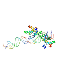 | | STRUCTURAL BASIS FOR COOPERATIVE BINDING OF RIBBON-HELIX-HELIX REPRESSOR OMEGA TO MUTATED DIRECT DNA HEPTAD REPEATS | | 分子名称: | 5'-D(*CP*TP*TP*GP*TP*GP*AP*CP*TP*TP *GP*TP*GP*AP*TP*TP*CP*G)-3', 5'-D(*GP*AP*AP*TP*CP*AP*CP*AP*AP*AP *TP*CP*AP*CP*AP*AP*G)-3', 5'-D(*GP*AP*AP*TP*CP*AP*CP*AP*AP*GP *TP*CP*AP*CP*AP*AP*GP*C)-3', ... | | 著者 | Weihofen, W.A, Cicek, A, Pratto, F, Alonso, J.C, Saenger, W. | | 登録日 | 2005-12-23 | | 公開日 | 2006-03-15 | | 最終更新日 | 2024-05-08 | | 実験手法 | X-RAY DIFFRACTION (2.9 Å) | | 主引用文献 | Structures of Omega Repressors Bound to Direct and Inverted DNA Repeats Explain Modulation of Transcription.
Nucleic Acids Res., 34, 2006
|
|
5G4F
 | | Structure of the ADP-bound VAT complex | | 分子名称: | VCP-LIKE ATPASE | | 著者 | Huang, R, Ripstein, Z.A, Augustyniak, R, Lazniewski, M, Ginalski, K, Kay, L.E, Rubinstein, J.L. | | 登録日 | 2016-05-12 | | 公開日 | 2016-07-27 | | 最終更新日 | 2024-05-08 | | 実験手法 | ELECTRON MICROSCOPY (7 Å) | | 主引用文献 | Unfolding the Mechanism of the Aaa+ Unfoldase Vat by a Combined Cryo-Em, Solution NMR Study.
Proc.Natl.Acad.Sci.USA, 113, 2016
|
|
5DIS
 | | Crystal structure of a CRM1-RanGTP-SPN1 export complex bound to a 113 amino acid FG-repeat containing fragment of Nup214 | | 分子名称: | Exportin-1, GTP-binding nuclear protein Ran, GUANOSINE-5'-TRIPHOSPHATE, ... | | 著者 | Monecke, T, Port, S.A, Dickmanns, A, Kehlenbach, R.H, Ficner, R. | | 登録日 | 2015-09-01 | | 公開日 | 2015-11-04 | | 最終更新日 | 2024-01-10 | | 実験手法 | X-RAY DIFFRACTION (2.85 Å) | | 主引用文献 | Structural and Functional Characterization of CRM1-Nup214 Interactions Reveals Multiple FG-Binding Sites Involved in Nuclear Export.
Cell Rep, 13, 2015
|
|
5GNL
 | | Cytochrome P450 Vdh (CYP107BR1) F106V mutant | | 分子名称: | 2-(2-METHOXYETHOXY)ETHANOL, PROTOPORPHYRIN IX CONTAINING FE, Vitamin D(3) 25-hydroxylase | | 著者 | Yasutake, Y, Tamura, T. | | 登録日 | 2016-07-22 | | 公開日 | 2017-05-17 | | 最終更新日 | 2023-11-08 | | 実験手法 | X-RAY DIFFRACTION (1.95 Å) | | 主引用文献 | Structural insights into the mechanism of the drastic changes in enzymatic activity of the cytochrome P450 vitamin D3 hydroxylase (CYP107BR1) caused by a mutation distant from the active site
Acta Crystallogr F Struct Biol Commun, 73, 2017
|
|
5TTB
 | |
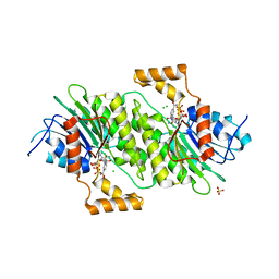
1ZIZ
 | | Crystal Structure of Human Galactosyltransferase (GTB) Complexed with Galactose | | 分子名称: | ABO blood group (transferase A, alpha 1-3-N-acetylgalactosaminyltransferase; transferase B, alpha 1-3-galactosyltransferase), ... | | 著者 | Letts, J.A, Rose, N.L, Fang, Y.R, Barry, C.H, Borisova, S.N, Seto, N.O, Palcic, M.M, Evans, S.V. | | 登録日 | 2005-04-27 | | 公開日 | 2005-12-13 | | 最終更新日 | 2023-08-23 | | 実験手法 | X-RAY DIFFRACTION (1.49 Å) | | 主引用文献 | Differential Recognition of the Type I and II H Antigen Acceptors by the Human ABO(H) Blood Group A and B Glycosyltransferases.
J.Biol.Chem., 281, 2006
|
|
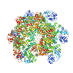
2CHI
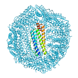 | | Recombinant human H ferritin, K86Q and E27D mutant | | 分子名称: | CALCIUM ION, FERRITIN HEAVY CHAIN, GLYCEROL, ... | | 著者 | Toussaint, L, Crichton, R.R, Declercq, J.P. | | 登録日 | 2006-03-15 | | 公開日 | 2006-12-13 | | 最終更新日 | 2023-12-13 | | 実験手法 | X-RAY DIFFRACTION (1.6 Å) | | 主引用文献 | High-Resolution X-Ray Structures of Human Apoferritin H-Chain Mutants Correlated with Their Activity and Metal-Binding Sites.
J.Mol.Biol., 365, 2007
|
|
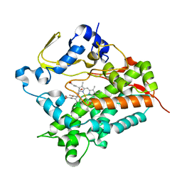
1Z6Z
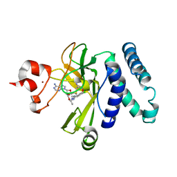
 | | Crystal Structure of Human Sepiapterin Reductase in complex with NADP+ | | 分子名称: | CHLORIDE ION, NADP NICOTINAMIDE-ADENINE-DINUCLEOTIDE PHOSPHATE, SULFATE ION, ... | | 著者 | Ugochukwu, E, Kavanagh, K, Ng, S, Arrowsmith, C, Edwards, A, Sundstrom, M, von Delft, F, Oppermann, U, Structural Genomics Consortium (SGC) | | 登録日 | 2005-03-23 | | 公開日 | 2005-04-05 | | 最終更新日 | 2023-08-23 | | 実験手法 | X-RAY DIFFRACTION (2.5 Å) | | 主引用文献 | Crystal Structure of Human Sepiapterin Reductase
To be Published
|
|
5CLO
 | |
2C8R
 | | insuline(60sec) and UV laser excited fluorescence | | 分子名称: | INSULIN A CHAIN, INSULIN B CHAIN | | 著者 | Vernede, X, Lavault, B, Ohana, J, Nurizzo, D, Joly, J, Jacquamet, L, Felisaz, F, Cipriani, F, Bourgeois, D. | | 登録日 | 2005-12-06 | | 公開日 | 2006-03-08 | | 最終更新日 | 2023-12-13 | | 実験手法 | X-RAY DIFFRACTION (1.5 Å) | | 主引用文献 | Uv Laser-Excited Fluorescence as a Tool for the Visualization of Protein Crystals Mounted in Loops.
Acta Crystallogr.,Sect.D, 62, 2006
|
|
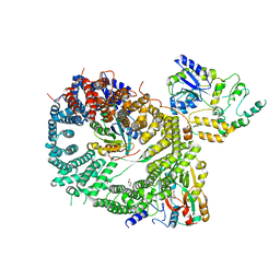
1Z8D
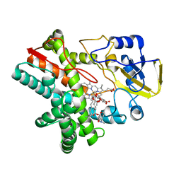
 | | Crystal Structure of Human Muscle Glycogen Phosphorylase a with AMP and Glucose | | 分子名称: | ADENINE, ADENOSINE MONOPHOSPHATE, Glycogen phosphorylase, ... | | 著者 | Lukacs, C.M, Oikonomakos, N.G, Crowther, R.L, Hong, L.N, Kammlott, R.U, Levin, W, Li, S, Liu, C.M, Lucas-McGady, D, Pietranico, S, Reik, L. | | 登録日 | 2005-03-30 | | 公開日 | 2006-03-21 | | 最終更新日 | 2023-11-15 | | 実験手法 | X-RAY DIFFRACTION (2.3 Å) | | 主引用文献 | The crystal structure of human muscle glycogen phosphorylase a with bound glucose and AMP: An intermediate conformation with T-state and R-state features.
Proteins, 63, 2006
|
|
5GIO
 | | Crystal structure of box C/D RNP with 12 nt guide regions and 13 nt substrates | | 分子名称: | 50S ribosomal protein L7Ae, C/D RNA, C/D box methylation guide ribonucleoprotein complex aNOP56 subunit, ... | | 著者 | Yang, Z, Lin, J, Ye, K. | | 登録日 | 2016-06-24 | | 公開日 | 2016-09-14 | | 最終更新日 | 2023-11-08 | | 実験手法 | X-RAY DIFFRACTION (3.604 Å) | | 主引用文献 | Box C/D guide RNAs recognize a maximum of 10 nt of substrates
Proc.Natl.Acad.Sci.USA, 113, 2016
|
|
2CTT
 | | Solution structure of zinc finger domain from human DnaJ subfamily A menber 3 | | 分子名称: | DnaJ homolog subfamily A member 3, ZINC ION | | 著者 | Kobayashi, N, Tochio, N, Saito, K, Koshiba, S, Inoue, M, Kigawa, T, Yokoyama, S, RIKEN Structural Genomics/Proteomics Initiative (RSGI) | | 登録日 | 2005-05-24 | | 公開日 | 2005-11-24 | | 最終更新日 | 2024-05-29 | | 実験手法 | SOLUTION NMR | | 主引用文献 | Solution structure of zinc finger domain from human DnaJ subfamily A menber 3
To be Published
|
|
5UIF
 | | Crystal Structure of Native Ps01740 | | 分子名称: | Ps01740 | | 著者 | LeVieux, J, Baas, B.J, Zhang, Y.J, Whitman, C.P. | | 登録日 | 2017-01-13 | | 公開日 | 2017-11-22 | | 最終更新日 | 2023-10-04 | | 実験手法 | X-RAY DIFFRACTION (2.57 Å) | | 主引用文献 | Kinetic and structural characterization of a cis-3-Chloroacrylic acid dehalogenase homologue in Pseudomonas sp. UW4: A potential step between subgroups in the tautomerase superfamily.
Arch. Biochem. Biophys., 636, 2017
|
|


