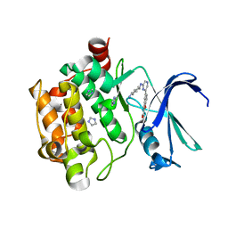
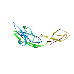
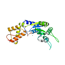
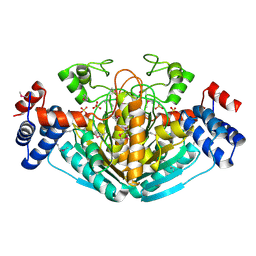
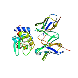
1OD7
 
 | | N-terminal of Sialoadhesin in complex with Me-a-9-N-(naphthyl-2-carbonyl)-amino-9-deoxy-Neu5Ac (NAP compound) | | 分子名称: | ME-A-9-N-(NAPHTHYL-2-CARBONYL)-AMINO-9-DEOXY-NEU5AC, SIALOADHESIN | | 著者 | Zaccai, N.R, Maenaka, K, Maenaka, T, Crocker, P.R, Brossmer, R, Kelm, S, Jones, E.Y. | | 登録日 | 2003-02-14 | | 公開日 | 2003-05-16 | | 最終更新日 | 2023-12-13 | | 実験手法 | X-RAY DIFFRACTION (3 Å) | | 主引用文献 | Structure-Guided Design of Sialic Acid-Based Siglec Inhibitors and Crystallographic Analysis in Complex with Sialoadhesin
Structure, 11, 2003
|
|
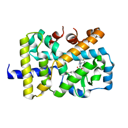
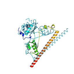
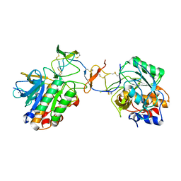
3R04
 | | The discovery of novel benzofuran-2-carboxylic acids as potent Pim-1 inhibitors | | 分子名称: | 5-{6-[(trans-4-aminocyclohexyl)amino]pyrazin-2-yl}-1-benzofuran-2-carboxylic acid, IMIDAZOLE, Proto-oncogene serine/threonine-protein kinase pim-1 | | 著者 | Xiang, Y, Hirth, B, Asmussen, G, Biemann, H.-P, Good, A, Fitzgerald, M, Gladysheva, T, Jancsics, K, Liu, J, Metz, M, Papoulis, A, Skerlj, R, Stepp, D.J, Wei, R.R. | | 登録日 | 2011-03-07 | | 公開日 | 2011-05-11 | | 最終更新日 | 2024-02-21 | | 実験手法 | X-RAY DIFFRACTION (1.7 Å) | | 主引用文献 | The discovery of novel benzofuran-2-carboxylic acids as potent Pim-1 inhibitors.
Bioorg.Med.Chem.Lett., 21, 2011
|
|
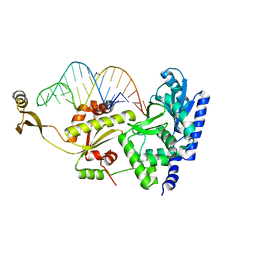
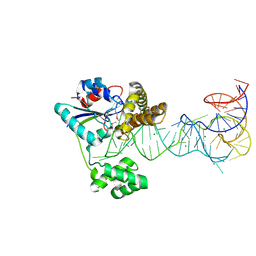
1K9M
 | | Co-crystal structure of tylosin bound to the 50S ribosomal subunit of Haloarcula marismortui | | 分子名称: | 23S RRNA, 5S RRNA, CADMIUM ION, ... | | 著者 | Hansen, J.L, Ippolito, J.A, Ban, N, Nissen, P, Moore, P.B, Steitz, T.A. | | 登録日 | 2001-10-29 | | 公開日 | 2002-07-19 | | 最終更新日 | 2023-08-16 | | 実験手法 | X-RAY DIFFRACTION (3 Å) | | 主引用文献 | The structures of four macrolide antibiotics bound to the large ribosomal subunit.
Mol.Cell, 10, 2002
|
|
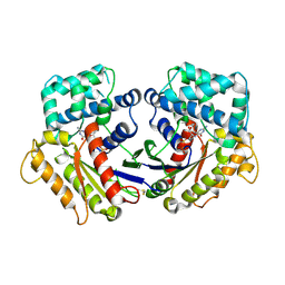
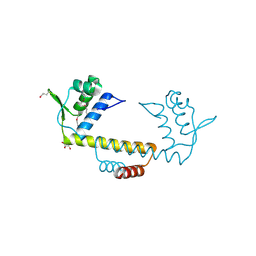
3AA9
 | | Crystal Structure Analysis of the Mutant CutA1 (E61V) from E. coli | | 分子名称: | Divalent-cation tolerance protein cutA | | 著者 | Matsuura, Y, Tanaka, T, Bagautdinov, B, Kunishima, N, Yutani, K. | | 登録日 | 2009-11-12 | | 公開日 | 2010-08-11 | | 最終更新日 | 2023-11-01 | | 実験手法 | X-RAY DIFFRACTION (2.3 Å) | | 主引用文献 | Remarkable improvement in the heat stability of CutA1 from Escherichia coli by rational protein design
J.Biochem., 148, 2010
|
|
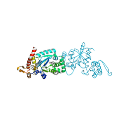
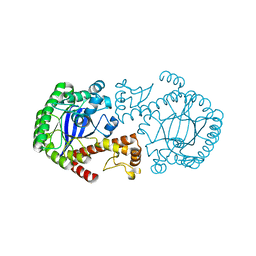
2ZDX
 | | Inhibitor-bound structures of human pyruvate dehydrogenase kinase 4 | | 分子名称: | 4-[4-(4-methoxyphenyl)-5-methyl-1H-pyrazol-3-yl]benzene-1,3-diol, Pyruvate dehydrogenase kinase isozyme 4 | | 著者 | Kawamoto, M, Shiromizu, I, Kukimoto-niino, M, Tokmakov, A, Terada, T, Shirouzu, M, Matsusue, T, Yokoyama, S. | | 登録日 | 2007-11-30 | | 公開日 | 2008-12-09 | | 最終更新日 | 2023-11-01 | | 実験手法 | X-RAY DIFFRACTION (2.54 Å) | | 主引用文献 | Inhibitor-bound structures of human pyruvate dehydrogenase kinase 4.
Acta Crystallogr.,Sect.D, 67, 2011
|
|
2ZG1
 | |
5VQK
 | |
2ZHA
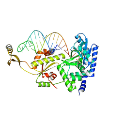 | |
2ZH9
 | | Complex structure of AFCCA with tRNAminiDU | | 分子名称: | CCA-adding enzyme, SULFATE ION, tRNA (33-MER) | | 著者 | Toh, Y, Tomita, K. | | 登録日 | 2008-02-01 | | 公開日 | 2008-08-05 | | 最終更新日 | 2024-03-13 | | 実験手法 | X-RAY DIFFRACTION (2.9 Å) | | 主引用文献 | Molecular basis for maintenance of fidelity during the CCA-adding reaction by a CCA-adding enzyme
Embo J., 27, 2008
|
|
2VII
 | | PspF1-275-Mg-AMP | | 分子名称: | ADENOSINE MONOPHOSPHATE, MAGNESIUM ION, PSP OPERON TRANSCRIPTIONAL ACTIVATOR | | 著者 | Joly, N, Rappas, M, Buck, M, Zhang, X. | | 登録日 | 2007-12-04 | | 公開日 | 2008-01-22 | | 最終更新日 | 2023-12-13 | | 実験手法 | X-RAY DIFFRACTION (2.85 Å) | | 主引用文献 | Trapping of a Transcription Complex Using a New Nucleotide Analogue: AMP Aluminium Fluoride
J.Mol.Biol., 375, 2008
|
|
6NCQ
 | |
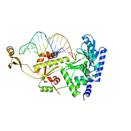
2ZH1
 | | Complex structure of AFCCA with tRNAminiDA | | 分子名称: | CCA-adding enzyme, RNA (33-MER), SULFATE ION | | 著者 | Toh, Y, Tomita, K. | | 登録日 | 2008-02-01 | | 公開日 | 2008-08-05 | | 最終更新日 | 2023-11-01 | | 実験手法 | X-RAY DIFFRACTION (2.8 Å) | | 主引用文献 | Molecular basis for maintenance of fidelity during the CCA-adding reaction by a CCA-adding enzyme
Embo J., 27, 2008
|
|
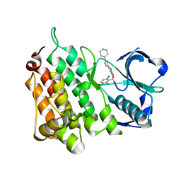
2ZJZ
 | | Structure of the K349P mutant of Gi alpha 1 subunit bound to GDP | | 分子名称: | GUANOSINE-5'-DIPHOSPHATE, Guanine nucleotide-binding protein G(i), alpha-1 subunit | | 著者 | Morikawa, T, Muroya, A, Sugio, S, Wakamatsu, K, Kohno, T. | | 登録日 | 2008-03-11 | | 公開日 | 2009-03-24 | | 最終更新日 | 2023-11-01 | | 実験手法 | X-RAY DIFFRACTION (2.6 Å) | | 主引用文献 | How GPCRs activate G proteins: Structural changes from C-terminal tail to GDP binding pocket
To be Published
|
|
2ZM5
 | | Crystal structure of tRNA modification enzyme MiaA in the complex with tRNA(Phe) | | 分子名称: | MAGNESIUM ION, tRNA delta(2)-isopentenylpyrophosphate transferase, tRNA(Phe) | | 著者 | Sakai, J, Yao, M, Chimnaronk, S, Tanaka, I. | | 登録日 | 2008-04-11 | | 公開日 | 2009-04-14 | | 最終更新日 | 2011-07-13 | | 実験手法 | X-RAY DIFFRACTION (2.55 Å) | | 主引用文献 | Snapshots of dynamics in synthesizing N(6)-isopentenyladenosine at the tRNA anticodon
Biochemistry, 48, 2009
|
|
2ZP1
 | |
3NEU
 | |
2YFX
 | | Structure of L1196M Mutant Anaplastic Lymphoma Kinase in Complex with Crizotinib | | 分子名称: | 3-[(1R)-1-(2,6-dichloro-3-fluorophenyl)ethoxy]-5-(1-piperidin-4-yl-1H-pyrazol-4-yl)pyridin-2-amine, TYROSINE-PROTEIN KINASE RECEPTOR | | 著者 | McTigue, M, Deng, Y, Liu, W, Brooun, A. | | 登録日 | 2011-04-08 | | 公開日 | 2011-05-04 | | 最終更新日 | 2023-12-20 | | 実験手法 | X-RAY DIFFRACTION (1.7 Å) | | 主引用文献 | Design of Potent and Selective Inhibitors to Overcome Clinical Anaplastic Lymphoma Kinase Mutations Resistant to Crizotinib.
J.Med.Chem., 57, 2014
|
|
1NXU
 | | CRYSTAL STRUCTURE OF E. COLI HYPOTHETICAL OXIDOREDUCTASE YIAK NORTHEAST STRUCTURAL GENOMICS CONSORTIUM TARGET ER82. | | 分子名称: | Hypothetical oxidoreductase yiaK, SULFATE ION | | 著者 | Forouhar, F, Lee, I, Benach, J, Kulkarni, K, Xiao, R, Acton, T.B, Shastry, R, Rost, B, Montelione, G.T, Tong, L, Northeast Structural Genomics Consortium (NESG) | | 登録日 | 2003-02-11 | | 公開日 | 2003-03-11 | | 最終更新日 | 2011-07-13 | | 実験手法 | X-RAY DIFFRACTION (1.8 Å) | | 主引用文献 | A Novel NAD-binding Protein Revealed by the Crystal Structure of 2,3-Diketo-L-gulonate Reductase (YiaK).
J.Biol.Chem., 279, 2004
|
|
3OOP
 | | The structure of a protein with unknown function from Listeria innocua Clip11262 | | 分子名称: | Lin2960 protein | | 著者 | Fan, Y, Li, H, Zhou, Y, Gu, M, Joachimiak, A, Midwest Center for Structural Genomics (MCSG) | | 登録日 | 2010-08-31 | | 公開日 | 2010-09-22 | | 最終更新日 | 2017-11-08 | | 実験手法 | X-RAY DIFFRACTION (1.78 Å) | | 主引用文献 | The structure of a protein with unknown function from Listeria innocua Clip11262
To be Published
|
|
2ZZN
 | | The complex structure of aTrm5 and tRNACys | | 分子名称: | MAGNESIUM ION, RNA (71-MER), S-ADENOSYLMETHIONINE, ... | | 著者 | Goto-Ito, S, Ito, T, Yokoyama, S. | | 登録日 | 2009-02-19 | | 公開日 | 2009-09-15 | | 最終更新日 | 2023-11-01 | | 実験手法 | X-RAY DIFFRACTION (2.95 Å) | | 主引用文献 | Tertiary structure checkpoint at anticodon loop modification in tRNA functional maturation.
Nat.Struct.Mol.Biol., 16, 2009
|
|
3A3A
 | |
3A67
 | | Crystal Structure of HyHEL-10 Fv mutant LN31D complexed with hen egg white lysozyme | | 分子名称: | IG VH, anti-lysozyme, Lysozyme C, ... | | 著者 | Yokota, A, Tsumoto, K, Shiroishi, M, Nakanishi, T, Kondo, H, Kumagai, I. | | 登録日 | 2009-08-24 | | 公開日 | 2009-12-22 | | 最終更新日 | 2023-11-01 | | 実験手法 | X-RAY DIFFRACTION (1.8 Å) | | 主引用文献 | Contribution of asparagine residues to the stabilization of a proteinaceous antigen-antibody complex, HyHEL-10-hen egg white lysozyme
J.Biol.Chem., 285, 2010
|
|
3A6C
 | | Crystal Structure of HyHEL-10 Fv mutant LN92D complexed with hen egg white lysozyme | | 分子名称: | IG VH, anti-lysozyme, Lysozyme C, ... | | 著者 | Yokota, A, Tsumoto, K, Shiroishi, M, Nakanishi, T, Kondo, H, Kumagai, I. | | 登録日 | 2009-08-28 | | 公開日 | 2009-12-22 | | 最終更新日 | 2023-11-01 | | 実験手法 | X-RAY DIFFRACTION (1.8 Å) | | 主引用文献 | Contribution of asparagine residues to the stabilization of a proteinaceous antigen-antibody complex, HyHEL-10-hen egg white lysozyme
J.Biol.Chem., 285, 2010
|
|
1OYV
 | | Crystal structure of tomato inhibitor-II in a ternary complex with subtilisin Carlsberg | | 分子名称: | CALCIUM ION, Subtilisin Carlsberg, Wound-induced proteinase inhibitor-II | | 著者 | Barrette-Ng, I.H, Ng, K.K, Cherney, M.M, Pearce, G, Ryan, C.A, James, M.N. | | 登録日 | 2003-04-07 | | 公開日 | 2003-07-15 | | 最終更新日 | 2023-08-16 | | 実験手法 | X-RAY DIFFRACTION (2.5 Å) | | 主引用文献 | Structural basis of inhibition revealed by a 1:2 complex of the two-headed tomato inhibitor-II and subtilisin Carlsberg
J.Biol.Chem., 278, 2003
|
|
1P0D
 | | CRYSTAL STRUCTURE OF ZYMOMONAS MOBILIS tRNA-GUANINE TRANSGLYCOSYLASE (TGT) CRYSTALLISED AT PH 5.5 | | 分子名称: | Queuine tRNA-ribosyltransferase, ZINC ION | | 著者 | Brenk, R, Stubbs, M.T, Heine, A, Reuter, K, Klebe, G. | | 登録日 | 2003-04-10 | | 公開日 | 2003-09-30 | | 最終更新日 | 2023-08-16 | | 実験手法 | X-RAY DIFFRACTION (1.9 Å) | | 主引用文献 | Flexible adaptations in the structure of the tRNA-modifying enzyme
tRNA-guanine transglycosylase and their implications for substrate selectivity,
reaction mechanism and structure-based drug design
Chembiochem, 4, 2003
|
|


