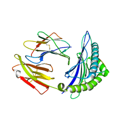
2WR7
 
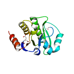
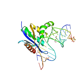
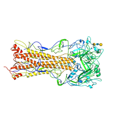 | | the structure of influenza H2 human singapore hemagglutinin with human receptor | | 分子名称: | HEMAGGLUTININ, N-acetyl-alpha-neuraminic acid-(2-6)-beta-D-galactopyranose-(1-4)-2-acetamido-2-deoxy-beta-D-glucopyranose-(1-3)-beta-D-galactopyranose | | 著者 | Liu, J, Stevens, D.J, Haire, L.F, Walker, P.A, Coombs, P.J, Russell, R.J, Gamblin, S.J, Skehel, J.J. | | 登録日 | 2009-08-30 | | 公開日 | 2009-09-29 | | 最終更新日 | 2020-07-29 | | 実験手法 | X-RAY DIFFRACTION (2.5 Å) | | 主引用文献 | From the Cover: Structures of Receptor Complexes Formed by Hemagglutinins from the Asian Influenza Pandemic of 1957.
Proc.Natl.Acad.Sci.USA, 106, 2009
|
|
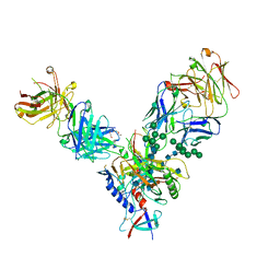
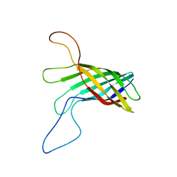
6XYW
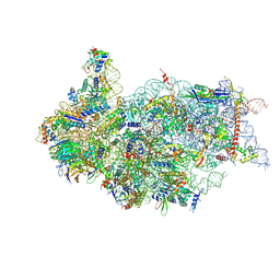
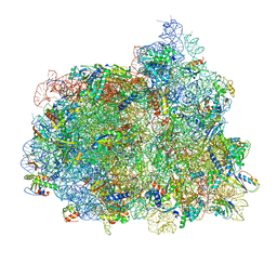
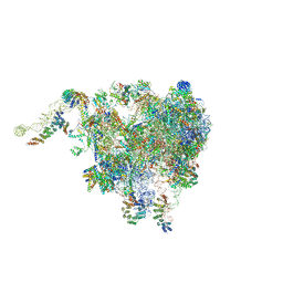 | | Structure of the plant mitochondrial ribosome | | 分子名称: | 28S ribosomal S34 protein, 3-hydroxyisobutyryl-CoA hydrolase-like protein 2, mitochondrial, ... | | 著者 | Soufari, H, Waltz, F, Bochler, A, Giege, P, Hashem, Y. | | 登録日 | 2020-01-31 | | 公開日 | 2020-04-15 | | 最終更新日 | 2020-04-29 | | 実験手法 | ELECTRON MICROSCOPY (3.86 Å) | | 主引用文献 | Cryo-EM structure of the RNA-rich plant mitochondrial ribosome.
Nat.Plants, 6, 2020
|
|
5QN7
 | |
8UT5
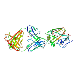
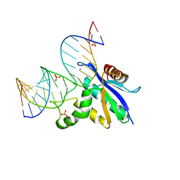
 | | CryoEM structure of A/Michigan/45/2015 H1 in complex with flu HA central stem VH1-18 antibody UCA6_N55T | | 分子名称: | 2-acetamido-2-deoxy-beta-D-glucopyranose, Hemagglutinin HA1 chain, Hemagglutinin HA2 chain, ... | | 著者 | Huang, J, Han, J, Ward, A.B. | | 登録日 | 2023-10-30 | | 公開日 | 2024-05-08 | | 最終更新日 | 2024-05-29 | | 実験手法 | ELECTRON MICROSCOPY (3.5 Å) | | 主引用文献 | Eliciting a single amino acid change by vaccination generates antibody protection against group 1 and group 2 influenza A viruses.
Immunity, 57, 2024
|
|
3CM3
 | |
8H1T
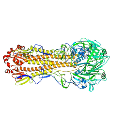
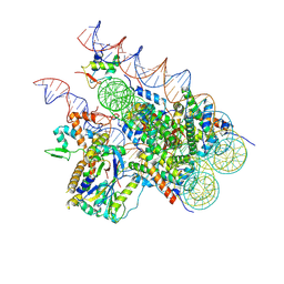 | | Cryo-EM structure of BAP1-ASXL1 bound to chromatosome | | 分子名称: | DNA (187-MER), Histone H1.4, Histone H2A type 1-D, ... | | 著者 | Ge, W, Yu, C, Xu, R.M. | | 登録日 | 2022-10-04 | | 公開日 | 2023-02-01 | | 最終更新日 | 2024-07-03 | | 実験手法 | ELECTRON MICROSCOPY (3 Å) | | 主引用文献 | Basis of the H2AK119 specificity of the Polycomb repressive deubiquitinase.
Nature, 616, 2023
|
|
2K6I
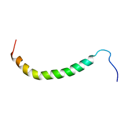 | | The domain features of the peripheral stalk subunit H of the methanogenic A1AO ATP synthase and the NMR solution structure of H1-47 | | 分子名称: | Uncharacterized protein MJ0223 | | 著者 | Biukovic, N, Gayen, S, Pervushin, K, Gruber, G, Biukovic, G. | | 登録日 | 2008-07-09 | | 公開日 | 2009-07-21 | | 最終更新日 | 2024-05-08 | | 実験手法 | SOLUTION NMR | | 主引用文献 | Domain features of the peripheral stalk subunit H of the methanogenic A1AO ATP synthase and the NMR solution structure of H(1-47).
Biophys.J., 97, 2009
|
|
4U3G
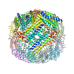 | | Crystal structure of Escherichia coli bacterioferritin mutant D132F | | 分子名称: | Bacterioferritin, SULFATE ION | | 著者 | Wong, S.G, Grigg, J.C, Le Brun, N.E, Moore, G.R, Murphy, M.E.P, Mauk, A.G. | | 登録日 | 2014-07-21 | | 公開日 | 2014-12-24 | | 最終更新日 | 2023-09-27 | | 実験手法 | X-RAY DIFFRACTION (2 Å) | | 主引用文献 | The B-type Channel Is a Major Route for Iron Entry into the Ferroxidase Center and Central Cavity of Bacterioferritin.
J.Biol.Chem., 290, 2015
|
|
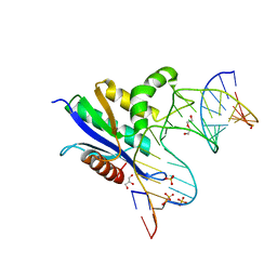
4X1E
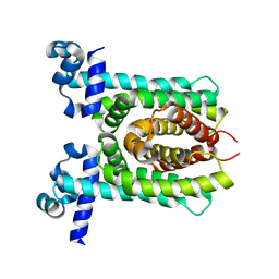 | | Crystal structure of unliganded E. coli transcriptional regulator RutR, W167A mutant | | 分子名称: | HTH-type transcriptional regulator RutR | | 著者 | Nguyen Le Minh, P, de Cima, S, Bervoets, I, Maes, D, Rubio, V, Charlier, D. | | 登録日 | 2014-11-24 | | 公開日 | 2015-01-21 | | 最終更新日 | 2024-01-10 | | 実験手法 | X-RAY DIFFRACTION (2.4 Å) | | 主引用文献 | Ligand binding specificity of RutR, a member of the TetR family of transcription regulators in Escherichia coli.
Febs Open Bio, 5, 2015
|
|
5WWU
 | | Crystal Structure of HLA-A*2402 in complex with 2009 pandemic influenza A(H1N1) virus and avian influenza A(H5N1) virus-derived peptide H1-25 | | 分子名称: | Beta-2-microglobulin, HLA class I histocompatibility antigen, A-24 alpha chain, ... | | 著者 | Zhao, M, Liu, K, Chai, Y, Qi, J, Liu, J, Gao, G.F. | | 登録日 | 2017-01-05 | | 公開日 | 2018-01-17 | | 最終更新日 | 2019-07-31 | | 実験手法 | X-RAY DIFFRACTION (2.794 Å) | | 主引用文献 | Heterosubtypic Protections against Human-Infecting Avian Influenza Viruses Correlate to Biased Cross-T-Cell Responses.
Mbio, 9, 2018
|
|
6VRD
 | | Crystal structure of RNase H/RNA/PS-ASO complex at an atomic level | | 分子名称: | DI(HYDROXYETHYL)ETHER, GLYCEROL, RNA (5'-R(*(OMC)P*(N7X)P*(T39)P*(C5L)P*(A2M))-D(P*(SC)P*(PST)P*(SC)P*(AS)P*(SC)P*(SC)P*(SC)P*(AS)P*(SC)P*(PST))-R(P*(6OO)P*(RFJ)P*(6OO)P*(6OO)P*(6NW))-3'), ... | | 著者 | Cho, Y.-J, Butler, D. | | 登録日 | 2020-02-07 | | 公開日 | 2021-02-10 | | 最終更新日 | 2023-11-15 | | 実験手法 | X-RAY DIFFRACTION (1.299 Å) | | 主引用文献 | Crystal structure of RNase H/RNA/PS-ASO complex at an atomic level
To Be Published
|
|
4JM2
 | |
4JVV
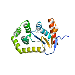
 | | Crystal structure of the evolved variant of the computationally designed serine hydrolase, OSH55.4_H1, covalently bound with diisopropyl fluorophosphate (DFP), Northeast Structural Genomics Consortium (NESG) Target OR273 | | 分子名称: | evolved variant of the computationally designed serine hydrolase | | 著者 | Kuzin, A, Lew, S, Rajagopalan, S, Seetharaman, J, Tong, S, Everett, J.K, Acton, T.B, Baker, D, Montelione, G.T, Tong, L, Hunt, J.F, Northeast Structural Genomics Consortium (NESG) | | 登録日 | 2013-03-26 | | 公開日 | 2013-04-24 | | 最終更新日 | 2023-12-06 | | 実験手法 | X-RAY DIFFRACTION (2.288 Å) | | 主引用文献 | Design of activated serine-containing catalytic triads with atomic-level accuracy.
Nat.Chem.Biol., 10, 2014
|
|
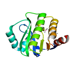
4JCA
 | | Crystal Structure of the apo form of the evolved variant of the computationally designed serine hydrolase, OSH55.4_H1. Northeast Structural Genomics Consortium (NESG) Target OR273 | | 分子名称: | CITRIC ACID, RUBIDIUM ION, serine hydrolase | | 著者 | Kuzin, A.P, Lew, S, Rajagopalan, S, Seetharaman, J, Tong, S, Everett, J.K, Acton, T.B, Baker, D, Montelione, G.T, Tong, L, Hunt, J.F, Northeast Structural Genomics Consortium (NESG) | | 登録日 | 2013-02-21 | | 公開日 | 2013-03-20 | | 最終更新日 | 2023-09-20 | | 実験手法 | X-RAY DIFFRACTION (2.411 Å) | | 主引用文献 | Design of activated serine-containing catalytic triads with atomic-level accuracy.
Nat.Chem.Biol., 10, 2014
|
|
2LHF
 | |
5W0D
 | |
7KNA
 | | Localized reconstruction of the H1 A/Michigan/45/2015 ectodomain displayed at the surface of I53_dn5 nanoparticle | | 分子名称: | 2-acetamido-2-deoxy-beta-D-glucopyranose, 2-acetamido-2-deoxy-beta-D-glucopyranose-(1-4)-2-acetamido-2-deoxy-beta-D-glucopyranose, Hemagglutinin,I53_dn5 | | 著者 | Acton, O.J, Park, Y.J, Veesler, D, Seattle Structural Genomics Center for Infectious Disease (SSGCID) | | 登録日 | 2020-11-04 | | 公開日 | 2021-03-31 | | 最終更新日 | 2024-05-01 | | 実験手法 | ELECTRON MICROSCOPY (3.3 Å) | | 主引用文献 | Quadrivalent influenza nanoparticle vaccines induce broad protection.
Nature, 592, 2021
|
|
8SWB
 | | RNase H complex with streopure ASO and RNA | | 分子名称: | DI(HYDROXYETHYL)ETHER, DNA (5'-R(*(OMC)P*(N7X)P*(T39)P*(C5L)P*(A2M))-D(P*(SC)P*(PST)P*(SC)P*AP*(SC)P*(SC)P*(RC)P*(AS)P*(SC)P*(PST))-R(P*(6OO)P*(RFJ)P*(6OO)P*(6OO)P*(6NW))-3'), GLYCEROL, ... | | 著者 | Cho, Y.-J, Iwamoto, N. | | 登録日 | 2023-05-18 | | 公開日 | 2024-05-22 | | 実験手法 | X-RAY DIFFRACTION (2 Å) | | 主引用文献 | Crystal structure of RNase H/RNA/PS-ASO complex at an atomic level
To Be Published
|
|
8SWC
 | |
6ZXG
 | | Cryo-EM structure of a late human pre-40S ribosomal subunit - State H1 | | 分子名称: | 40S ribosomal protein S10, 40S ribosomal protein S11, 40S ribosomal protein S12, ... | | 著者 | Ameismeier, M, Zemp, I, van den Heuvel, J, Thoms, M, Berninghausen, O, Kutay, U, Beckmann, R. | | 登録日 | 2020-07-29 | | 公開日 | 2020-12-02 | | 最終更新日 | 2024-04-24 | | 実験手法 | ELECTRON MICROSCOPY (2.6 Å) | | 主引用文献 | Structural basis for the final steps of human 40S ribosome maturation.
Nature, 587, 2020
|
|
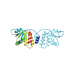
7ODK
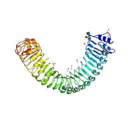 | | Plant peptide hormone receptor H1 | | 分子名称: | 1,2-ETHANEDIOL, 2-acetamido-2-deoxy-beta-D-glucopyranose, 2-acetamido-2-deoxy-beta-D-glucopyranose-(1-4)-2-acetamido-2-deoxy-beta-D-glucopyranose, ... | | 著者 | Roman, A.O, Jimenez-Sandoval, P, Santiago, J. | | 登録日 | 2021-04-29 | | 公開日 | 2022-02-16 | | 最終更新日 | 2024-01-31 | | 実験手法 | X-RAY DIFFRACTION (1.83 Å) | | 主引用文献 | HSL1 and BAM1/2 impact epidermal cell development by sensing distinct signaling peptides.
Nat Commun, 13, 2022
|
|
5TPM
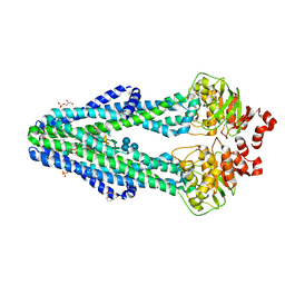
 | | 2.8 Angstrom Crystal Structure of the C-terminal Dimerization Domain of Transcriptional Regulator PdhR from Escherichia coli. | | 分子名称: | Pyruvate dehydrogenase complex repressor | | 著者 | Minasov, G, Wawrzak, Z, Sandoval, J, Skarina, T, Grimshaw, S, Kwon, K, Savchenko, A, Anderson, W.F, Center for Structural Genomics of Infectious Diseases (CSGID) | | 登録日 | 2016-10-20 | | 公開日 | 2016-11-02 | | 実験手法 | X-RAY DIFFRACTION (2.8 Å) | | 主引用文献 | 2.8 Angstrom Crystal Structure of the C-terminal Dimerization Domain of Transcriptional Regulator PdhR from Escherichia coli.
To Be Published
|
|
7PJX
 | | Structure of the 70S-EF-G-GDP ribosome complex with tRNAs in hybrid state 1 (H1-EF-G-GDP) | | 分子名称: | 16S ribosomal RNA, 23S ribosomal RNA, 30S ribosomal protein S10, ... | | 著者 | Petrychenko, V, Peng, B.Z, Schwarzer, A.C, Peske, F, Rodnina, M.V, Fischer, N. | | 登録日 | 2021-08-24 | | 公開日 | 2021-10-20 | | 最終更新日 | 2024-04-24 | | 実験手法 | ELECTRON MICROSCOPY (6.5 Å) | | 主引用文献 | Structural mechanism of GTPase-powered ribosome-tRNA movement.
Nat Commun, 12, 2021
|
|
4R7L
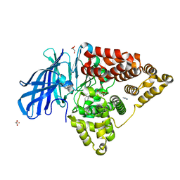 | | Structure of Human Leukotriene A4 Hydrolase in complex with inhibitor H1 | | 分子名称: | ACETATE ION, GLYCEROL, IMIDAZOLE, ... | | 著者 | Ouyang, P, Cui, K, Lu, W, Huang, J. | | 登録日 | 2014-08-27 | | 公開日 | 2015-11-25 | | 最終更新日 | 2024-03-20 | | 実験手法 | X-RAY DIFFRACTION (1.66 Å) | | 主引用文献 | Human Leukotriene A4 Hydrolase in complex with SAHA
To be Published
|
|
6BPP
 | | E. coli MsbA in complex with LPS and inhibitor G092 | | 分子名称: | (2E)-3-{6-[(1S)-1-(3-amino-2,6-dichlorophenyl)ethoxy]-4-cyclopropylquinolin-3-yl}prop-2-enoic acid, 1,2-Distearoyl-sn-glycerophosphoethanolamine, 3-HYDROXY-TETRADECANOIC ACID, ... | | 著者 | Ho, H, Koth, C.M, Payandeh, J. | | 登録日 | 2017-11-24 | | 公開日 | 2018-05-02 | | 最終更新日 | 2024-03-13 | | 実験手法 | X-RAY DIFFRACTION (2.92 Å) | | 主引用文献 | Structural basis for dual-mode inhibition of the ABC transporter MsbA.
Nature, 557, 2018
|
|


