2GH7
 
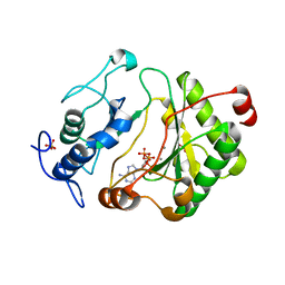
 | | Epi-biotin complex with core streptavidin | | 分子名称: | BIOTIN, EPI-BIOTIN, GLYCEROL, ... | | 著者 | Le Trong, I, Aubert, D.G.L, Thomas, N.R, Stenkamp, R.E. | | 登録日 | 2006-03-26 | | 公開日 | 2006-04-18 | | 最終更新日 | 2024-02-14 | | 実験手法 | X-RAY DIFFRACTION (1 Å) | | 主引用文献 | The high-resolution structure of (+)-epi-biotin bound to streptavidin.
Acta Crystallogr.,Sect.D, 62, 2006
|
|
2R0T
 | |
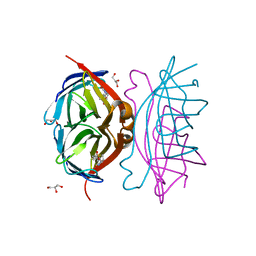
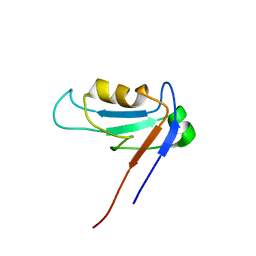
2ATM
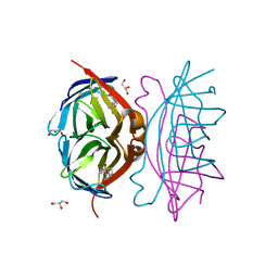
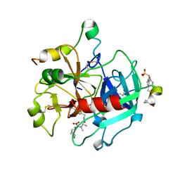
 | | Crystal structure of the recombinant allergen Ves v 2 | | 分子名称: | 2-(N-MORPHOLINO)-ETHANESULFONIC ACID, Hyaluronoglucosaminidase, SULFATE ION | | 著者 | Skov, L.K, Seppala, U, Coen, J.J.F, Crickmore, N, King, T.P, Monsalve, R, Kastrup, J.S, Spangfort, M.D, Gajhede, M. | | 登録日 | 2005-08-25 | | 公開日 | 2006-05-23 | | 最終更新日 | 2023-10-25 | | 実験手法 | X-RAY DIFFRACTION (2 Å) | | 主引用文献 | Structure of recombinant Ves v 2 at 2.0 Angstrom resolution: structural analysis of an allergenic hyaluronidase from wasp venom.
Acta Crystallogr.,Sect.D, 62, 2006
|
|
2DES
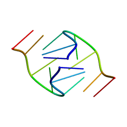
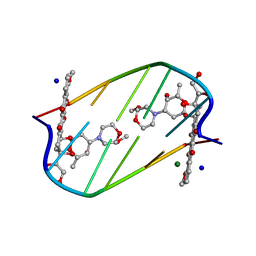 | | INTERACTIONS BETWEEN MORPHOLINYL ANTHRACYCLINES AND DNA: THE CRYSTAL STRUCTURE OF A MORPHOLINO DOXORUBICIN BOUND TO D(CGTACG) | | 分子名称: | 3'-DESAMINO-3'-(2-METHOXY-4-MORPHOLINYL)-DOXORUBICIN, DNA (5'-D(*CP*GP*TP*AP*CP*G)-3'), MAGNESIUM ION, ... | | 著者 | Cirilli, M, Bachechi, F, Ughetto, G, Colonna, F.P, Capobianco, M.L. | | 登録日 | 1993-03-16 | | 公開日 | 1993-07-15 | | 最終更新日 | 2024-02-14 | | 実験手法 | X-RAY DIFFRACTION (1.5 Å) | | 主引用文献 | Interactions between morpholinyl anthracyclines and DNA. The crystal structure of a morpholino doxorubicin bound to d(CGTACG).
J.Mol.Biol., 230, 1993
|
|
1ZOK
 | |
2N94
 | |
2EKP
 | |
2CF9
 | | Complex of recombinant human thrombin with an inhibitor | | 分子名称: | 4-[(1R,3AS,4R,8AS,8BR)-1-ISOPROPYL-2-(4-METHOXYBENZYL)-3-OXODECAHYDROPYRROLO[3,4-A]PYRROLIZIN-4-YL]BENZENECARBOXIMIDAMIDE, CALCIUM ION, HIRUDIN IIIA, ... | | 著者 | Schweizer, E, Hoffmann-Roeder, A, Olsen, J.A, Obst-Sander, U, Wagner, B, Kansy, M, Banner, D.W, Diederich, F. | | 登録日 | 2006-02-17 | | 公開日 | 2006-06-14 | | 最終更新日 | 2023-12-13 | | 実験手法 | X-RAY DIFFRACTION (1.79 Å) | | 主引用文献 | Multipolar Interactions in the D Pocket of Thrombin: Large Differences between Tricyclic Imide and Lactam Inhibitors.
Org.Biomol.Chem., 4, 2006
|
|
2CF8
 | | Complex of recombinant human thrombin with an inhibitor | | 分子名称: | 4- [(1R,3AS,4R,8AS,8BR)- 2- (4-CHLOROBENZYL)- 1- ISOPROPYL- 3- OXODECAHYDROPYRROLO[3,4- A]PYRROLIZIN- 4- YL]BENZENECARBOXIMIDAMIDE, CALCIUM ION, HIRUDIN IIIA, ... | | 著者 | Schweizer, E, Hoffmann-Roeder, A, Olsen, J.A, Obst-Sander, U, Wagner, B, Kansy, M, Banner, D.W, Diederich, F. | | 登録日 | 2006-02-17 | | 公開日 | 2006-06-14 | | 最終更新日 | 2023-12-13 | | 実験手法 | X-RAY DIFFRACTION (1.3 Å) | | 主引用文献 | Multipolar Interactions in the D Pocket of Thrombin: Large Differences between Tricyclic Imide and Lactam Inhibitors.
Org.Biomol.Chem., 4, 2006
|
|
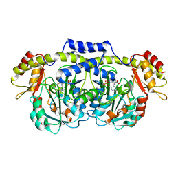
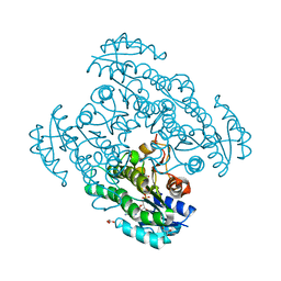
2CBJ
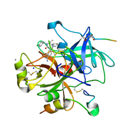
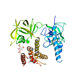
 | | Structure of the Clostridium perfringens NagJ family 84 glycoside hydrolase, a homologue of human O-GlcNAcase in complex with PUGNAc | | 分子名称: | CHLORIDE ION, HYALURONIDASE, O-(2-ACETAMIDO-2-DEOXY D-GLUCOPYRANOSYLIDENE) AMINO-N-PHENYLCARBAMATE | | 著者 | Rao, F.V, Dorfmueller, H.C, Villa, F, Allwood, M, Eggleston, I.M, van Aalten, D.M.F. | | 登録日 | 2006-01-05 | | 公開日 | 2006-02-13 | | 最終更新日 | 2023-12-13 | | 実験手法 | X-RAY DIFFRACTION (2.35 Å) | | 主引用文献 | Structural insights into the mechanism and inhibition of eukaryotic O-GlcNAc hydrolysis.
EMBO J., 25, 2006
|
|
2FG4
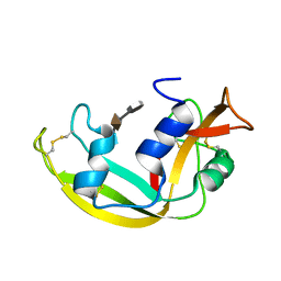
 | | Structure of Human Ferritin L Chain | | 分子名称: | CADMIUM ION, Ferritin light chain | | 著者 | Wang, Z, Li, C, Ellenburg, M, Ruble, J, Ho, J.X, Carter, D.C. | | 登録日 | 2005-12-21 | | 公開日 | 2006-07-04 | | 最終更新日 | 2023-08-30 | | 実験手法 | X-RAY DIFFRACTION (2.1 Å) | | 主引用文献 | Structure of human ferritin L chain.
ACTA CRYSTALLOGR.,SECT.D, 62, 2006
|
|
1Y9Q
 | |
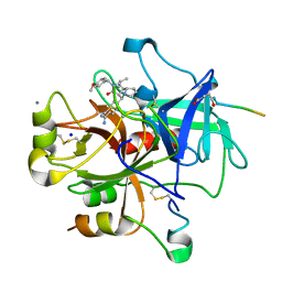
2F5V
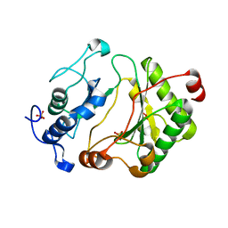
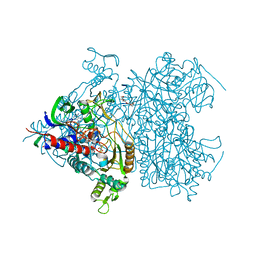 | | Reaction geometry and thermostability mutant of pyranose 2-oxidase from the white-rot fungus Peniophora sp. | | 分子名称: | DI(HYDROXYETHYL)ETHER, FLAVIN-ADENINE DINUCLEOTIDE, Pyranose 2-oxidase, ... | | 著者 | Bannwarth, M, Bastian, S, Heckmann-Pohl, D, Giffhorn, F, Schulz, G.E. | | 登録日 | 2005-11-28 | | 公開日 | 2006-06-13 | | 最終更新日 | 2020-07-29 | | 実験手法 | X-RAY DIFFRACTION (1.41 Å) | | 主引用文献 | Reaction Geometry and Thermostable Variant of Pyranose 2-Oxidase from the White-Rot Fungus Peniophora sp.
Biochemistry, 45, 2006
|
|
2FAR
 | | Crystal Structure of Pseudomonas aeruginosa LigD polymerase domain with dATP and Manganese | | 分子名称: | 2'-DEOXYADENOSINE 5'-TRIPHOSPHATE, MANGANESE (II) ION, SULFATE ION, ... | | 著者 | Zhu, H, Nandakumar, J, Aniukwu, J, Wang, L.K, Glickman, M.S, Lima, C.D, Shuman, S. | | 登録日 | 2005-12-07 | | 公開日 | 2006-05-23 | | 最終更新日 | 2023-08-30 | | 実験手法 | X-RAY DIFFRACTION (1.9 Å) | | 主引用文献 | Atomic structure and nonhomologous end-joining function of the polymerase component of bacterial DNA ligase D
Proc.Natl.Acad.Sci.USA, 103, 2006
|
|
2F01
 | | Epi-biotin complex with core streptavidin | | 分子名称: | BIOTIN, EPI-BIOTIN, GLYCEROL, ... | | 著者 | Le Trong, I, Aubert, D.G, Thomas, N.R, Stenkamp, R.E. | | 登録日 | 2005-11-10 | | 公開日 | 2005-11-29 | | 最終更新日 | 2023-08-23 | | 実験手法 | X-RAY DIFFRACTION (0.85 Å) | | 主引用文献 | The high-resolution structure of (+)-epi-biotin bound to streptavidin.
Acta Crystallogr.,Sect.D, 62, 2006
|
|
2FAQ
 | | Crystal Structure of Pseudomonas aeruginosa LigD polymerase domain with ATP and Manganese | | 分子名称: | ADENOSINE-5'-TRIPHOSPHATE, MANGANESE (II) ION, SULFATE ION, ... | | 著者 | Zhu, H, Nandakumar, J, Aniukwu, J, Wang, L.K, Glickman, M.S, Lima, C.D, Shuman, S. | | 登録日 | 2005-12-07 | | 公開日 | 2006-05-23 | | 最終更新日 | 2023-08-30 | | 実験手法 | X-RAY DIFFRACTION (1.9 Å) | | 主引用文献 | Atomic structure and nonhomologous end-joining function of the polymerase component of bacterial DNA ligase D
Proc.Natl.Acad.Sci.USA, 103, 2006
|
|
2FAO
 | | Crystal Structure of Pseudomonas aeruginosa LigD polymerase domain | | 分子名称: | SULFATE ION, probable ATP-dependent DNA ligase | | 著者 | Zhu, H, Nandakumar, J, Aniukwu, J, Wang, L.K, Glickman, M.S, Lima, C.D, Shuman, S. | | 登録日 | 2005-12-07 | | 公開日 | 2006-05-23 | | 最終更新日 | 2024-02-14 | | 実験手法 | X-RAY DIFFRACTION (1.5 Å) | | 主引用文献 | Atomic structure and nonhomologous end-joining function of the polymerase component of bacterial DNA ligase D
Proc.Natl.Acad.Sci.USA, 103, 2006
|
|
3UJ0
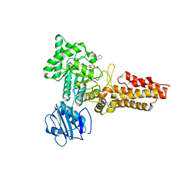
 | | Crystal structure of the inositol 1,4,5-trisphosphate receptor with ligand bound form. | | 分子名称: | D-MYO-INOSITOL-1,4,5-TRIPHOSPHATE, Inositol 1,4,5-trisphosphate receptor type 1 | | 著者 | Ikura, M, Seo, M.D, Ishiyama, N, Stathopulos, P. | | 登録日 | 2011-11-07 | | 公開日 | 2012-02-15 | | 最終更新日 | 2024-02-28 | | 実験手法 | X-RAY DIFFRACTION (3.6 Å) | | 主引用文献 | Structural and functional conservation of key domains in InsP3 and ryanodine receptors.
Nature, 483, 2012
|
|
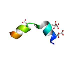
2LVZ
 | | Solution structure of a Eosinophil Cationic Protein-trisaccharide heparin mimetic complex | | 分子名称: | 2-deoxy-6-O-sulfo-2-(sulfoamino)-alpha-D-glucopyranose-(1-4)-2-O-sulfo-alpha-L-idopyranuronic acid-(1-4)-propan-2-yl 2-deoxy-6-O-sulfo-2-(sulfoamino)-alpha-D-glucopyranoside, Eosinophil cationic protein | | 著者 | Garcia Mayoral, M, Canales, A, Diaz, D, Lopez Prados, J, Moussaoui, M, de Paz, J, Angulo, J, Nieto, P, Jimenez Barbero, J, Boix, E, Bruix, M. | | 登録日 | 2012-07-17 | | 公開日 | 2013-07-31 | | 最終更新日 | 2020-07-29 | | 実験手法 | SOLUTION NMR | | 主引用文献 | Insights into the glycosaminoglycan-mediated cytotoxic mechanism of eosinophil cationic protein revealed by NMR.
Acs Chem.Biol., 8, 2013
|
|
2A2X
 | | Orally Active Thrombin Inhibitors in Complex with Thrombin Inh12 | | 分子名称: | N-(CARBOXYMETHYL)-3-CYCLOHEXYL-D-ALANYL-N-({6-[AMINO(IMINO)METHYL]PYRIDIN-3-YL}METHYL)-N~2~-METHYL-L-ALANINAMIDE, Thrombin heavy chain, Thrombin light chain, ... | | 著者 | Lange, U.E.W, Baucke, D, Hornberger, W, Mack, H, Seitz, W, Hoeffken, H.W. | | 登録日 | 2005-06-23 | | 公開日 | 2006-11-14 | | 最終更新日 | 2023-08-23 | | 実験手法 | X-RAY DIFFRACTION (2.44 Å) | | 主引用文献 | Orally active thrombin inhibitors. Part 2: optimization of the P2-moiety
BIOORG.MED.CHEM.LETT., 16, 2006
|
|
2P0D
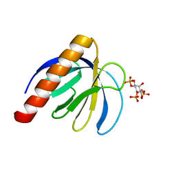 | | ArhGAP9 PH domain in complex with Ins(1,4,5)P3 | | 分子名称: | D-MYO-INOSITOL-1,4,5-TRIPHOSPHATE, Rho GTPase-activating protein 9 | | 著者 | Ceccarelli, D.F.J, Blasutig, I, Goudreault, M, Ruston, J, Pawson, T, Sicheri, F. | | 登録日 | 2007-02-28 | | 公開日 | 2007-03-27 | | 最終更新日 | 2024-02-21 | | 実験手法 | X-RAY DIFFRACTION (1.811 Å) | | 主引用文献 | Non-canonical Interaction of Phosphoinositides with Pleckstrin Homology Domains of Tiam1 and ArhGAP9.
J.Biol.Chem., 282, 2007
|
|
1B3P
 | | 5'-D(*GP*GP*AP*GP*GP*AP*T)-3' | | 分子名称: | DNA (5'-D(*GP*GP*AP*GP*GP*AP*T)-3') | | 著者 | Kettani, A, Bouaziz, S, Skripkin, E, Majumdar, A, Wang, W, Jones, R.A, Patel, D.J. | | 登録日 | 1998-12-14 | | 公開日 | 1999-08-31 | | 最終更新日 | 2023-12-27 | | 実験手法 | SOLUTION NMR | | 主引用文献 | Interlocked mismatch-aligned arrowhead DNA motifs.
Structure Fold.Des., 7, 1999
|
|
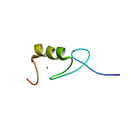
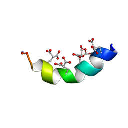
2MZK
 | | The solution structure of the Magnesium-bound Conantokin RLB | | 分子名称: | Conantokin-R1-B | | 著者 | Kunda, S, Yuan, Y, Balsara, R.D, Zajicek, J, Castellino, F.J. | | 登録日 | 2015-02-14 | | 公開日 | 2015-06-17 | | 最終更新日 | 2015-08-05 | | 実験手法 | SOLUTION NMR | | 主引用文献 | Hydroxyproline-induced Helical Disruption in Conantokin Rl-B Affects Subunit-selective Antagonistic Activities toward Ion Channels of N-Methyl-d-aspartate Receptors.
J.Biol.Chem., 290, 2015
|
|
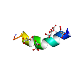
2MYZ
 | | The Solution Structure of the Magnesium-bound Conantokin-R1B Mutant | | 分子名称: | Conantokin-R1-B | | 著者 | Kunda, S, Yuan, Y, Balsara, R.D, Zajicek, J, Castellino, F.J. | | 登録日 | 2015-02-04 | | 公開日 | 2015-06-17 | | 最終更新日 | 2023-06-14 | | 実験手法 | SOLUTION NMR | | 主引用文献 | Hydroxyproline-induced Helical Disruption in Conantokin Rl-B Affects Subunit-selective Antagonistic Activities toward Ion Channels of N-Methyl-d-aspartate Receptors.
J.Biol.Chem., 290, 2015
|
|
2MZM
 | | The Solution Structure of the Magnesium-bound Conantokin-G | | 分子名称: | Conantokin-R1-B | | 著者 | Kunda, S, Yuan, Y, Balsara, R.D, Zajicek, J, Castellino, F.J. | | 登録日 | 2015-02-14 | | 公開日 | 2015-06-17 | | 最終更新日 | 2023-06-14 | | 実験手法 | SOLUTION NMR | | 主引用文献 | Hydroxyproline-induced Helical Disruption in Conantokin Rl-B Affects Subunit-selective Antagonistic Activities toward Ion Channels of N-Methyl-d-aspartate Receptors.
J.Biol.Chem., 290, 2015
|
|


