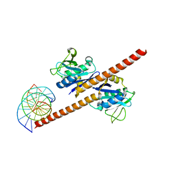
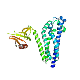
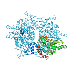
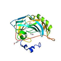
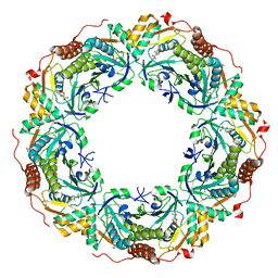
2H01
 
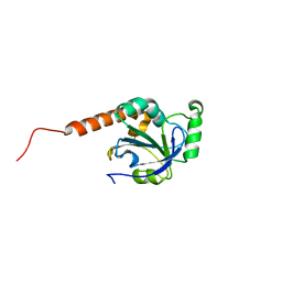 | | PY00414- Plasmodium yoelii thioredoxin peroxidase I | | 分子名称: | 2-CYS PEROXIREDOXIN | | 著者 | Artz, J, Qiu, W, Min, J.R, Dong, A, Lew, J, Melone, M, Alam, Z, Weigelt, J, Sundstrom, M, Edwards, A.M, Arrowsmith, C.H, Bochkarev, A, Hui, R, Structural Genomics Consortium (SGC) | | 登録日 | 2006-05-12 | | 公開日 | 2006-05-30 | | 最終更新日 | 2023-08-30 | | 実験手法 | X-RAY DIFFRACTION (2.3 Å) | | 主引用文献 | Crystal structure of PY00414 - a Plasmodium yoelii thioredoxin peroxidase I
To be Published
|
|
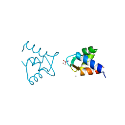
2H07
 | |
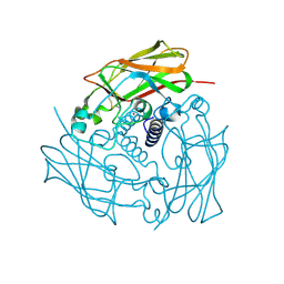
5BKA
 | |
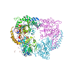
7EOE
 | |
7EOF
 | | Structure of CCDC25-DNA complex | | 分子名称: | Coiled-coil domain-containing protein 25, DNA (5'-D(*CP*AP*GP*AP*TP*CP*AP*CP*TP*AP*GP*TP*AP*GP*AP*T)-3'), DNA (5'-D(*GP*AP*TP*CP*TP*AP*CP*TP*AP*GP*TP*GP*AP*TP*CP*T)-3') | | 著者 | Liu, Y.R. | | 登録日 | 2021-04-22 | | 公開日 | 2022-11-02 | | 最終更新日 | 2024-04-03 | | 実験手法 | X-RAY DIFFRACTION (2.733 Å) | | 主引用文献 | Structure of CCDC25-DNA complex
To Be Published
|
|
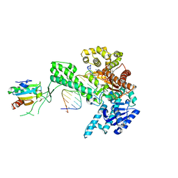
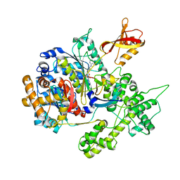
1R03
 | | crystal structure of a human mitochondrial ferritin | | 分子名称: | MAGNESIUM ION, mitochondrial ferritin | | 著者 | Corsi, B, Santambrogio, P, Arosio, P, Levi, S, Langlois d'Estaintot, B, Granier, T, Gallois, B, Chevallier, J.M, Precigoux, G. | | 登録日 | 2003-09-19 | | 公開日 | 2004-06-29 | | 最終更新日 | 2023-08-23 | | 実験手法 | X-RAY DIFFRACTION (1.7 Å) | | 主引用文献 | Crystal Structure and Biochemical Properties of the Human Mitochondrial Ferritin and its Mutant Ser144Ala
J.Mol.Biol., 340, 2004
|
|
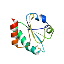
3UF1
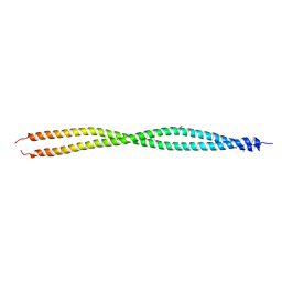 | | Crystal Structure of Vimentin (fragment 144-251) from Homo sapiens, Northeast Structural Genomics Consortium Target HR4796B | | 分子名称: | Vimentin | | 著者 | Kuzin, A, Abashidze, M, Vorobiev, S.M, Patel, P, Xiao, R, Ciccosanti, C, Shastry, R, Everett, J.K, Nair, R, Acton, T.B, Rost, B, Montelione, G.T, Tong, L, Hunt, J.F, Northeast Structural Genomics Consortium (NESG) | | 登録日 | 2011-10-31 | | 公開日 | 2011-11-30 | | 最終更新日 | 2024-11-06 | | 実験手法 | X-RAY DIFFRACTION (2.81 Å) | | 主引用文献 | The structure of vimentin linker 1 and rod 1B domains characterized by site-directed spin-labeling electron paramagnetic resonance (SDSL-EPR) and X-ray crystallography.
J.Biol.Chem., 287, 2012
|
|
2CBE
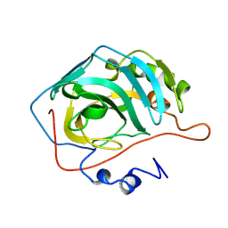 | |
3UMA
 | | Crystal structure of a hypothetical peroxiredoxin protein frm Sinorhizobium meliloti | | 分子名称: | Hypothetical peroxiredoxin protein, SULFATE ION | | 著者 | Eswaramoorthy, S, Chamala, S, Evans, B, Foti, R, Gizzi, A, Hillerich, B, Kar, A, LaFleur, J, Seidel, R, Villigas, G, Zencheck, W, Almo, S.C, Swaminathan, S, New York Structural Genomics Research Consortium (NYSGRC) | | 登録日 | 2011-11-12 | | 公開日 | 2011-11-23 | | 最終更新日 | 2024-10-09 | | 実験手法 | X-RAY DIFFRACTION (2.2 Å) | | 主引用文献 | Crystal structure of a hypothetical peroxiredoxin protein from Sinorhizobium meliloti
To be Published
|
|
1JKN
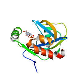 | | Solution Structure of the Nudix Enzyme Diadenosine Tetraphosphate Hydrolase from Lupinus angustifolius Complexed with ATP | | 分子名称: | ADENOSINE-5'-TRIPHOSPHATE, diadenosine 5',5'''-P1,P4-tetraphosphate hydrolase | | 著者 | Fletcher, J.I, Swarbrick, J.D, Maksel, D, Gayler, K.R, Gooley, P.R. | | 登録日 | 2001-07-12 | | 公開日 | 2002-02-27 | | 最終更新日 | 2024-05-22 | | 実験手法 | SOLUTION NMR | | 主引用文献 | The structure of Ap(4)A hydrolase complexed with ATP-MgF(x) reveals the basis of substrate binding.
Structure, 10, 2002
|
|
1RJ2
 | |
4DXT
 | | Human SUN2 (AA 522-717) | | 分子名称: | POTASSIUM ION, SUN domain-containing protein 2 | | 著者 | Sosa, B, Schwartz, T.U. | | 登録日 | 2012-02-28 | | 公開日 | 2012-06-06 | | 最終更新日 | 2024-11-06 | | 実験手法 | X-RAY DIFFRACTION (2.22 Å) | | 主引用文献 | LINC Complexes Form by Binding of Three KASH Peptides to Domain Interfaces of Trimeric SUN Proteins.
Cell(Cambridge,Mass.), 149, 2012
|
|
3UCY
 | |
5CR7
 | |
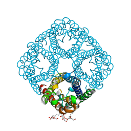
1RLZ
 | | Deoxyhypusine synthase holoenzyme in its high ionic strength, low pH crystal form | | 分子名称: | Deoxyhypusine synthase, NICOTINAMIDE-ADENINE-DINUCLEOTIDE | | 著者 | Umland, T.C, Wolff, E.C, Park, M.-H, Davies, D.R. | | 登録日 | 2003-11-26 | | 公開日 | 2004-07-13 | | 最終更新日 | 2023-08-23 | | 実験手法 | X-RAY DIFFRACTION (2.15 Å) | | 主引用文献 | A New Crystal Structure of Deoxyhypusine Synthase Reveals the Configuration of the Active Enzyme and of an Enzyme-NAD-Inhibitor Ternary Complex
J.Biol.Chem., 279, 2004
|
|
1ZYQ
 | | T7 DNA polymerase in complex with 8oG and incoming ddATP | | 分子名称: | 2',3'-DIDEOXYADENOSINE-5'-TRIPHOSPHATE, 5'-D(*CP*CP*CP*(8OG)P*CP*TP*GP*GP*CP*AP*CP*TP*GP*GP*CP*CP*GP*TP*CP*GP*TP*TP*TP*TP*CP*G)-3', 5'-D(*CP*GP*AP*AP*AP*AP*CP*GP*AP*CP*GP*GP*CP*CP*AP*GP*TP*GP*CP*CP*AP*(DDG))-3', ... | | 著者 | Brieba, L.G, Kokoska, R.J, Bebenek, K, Kunkel, T.A, Ellenberger, T. | | 登録日 | 2005-06-10 | | 公開日 | 2005-11-22 | | 最終更新日 | 2024-02-14 | | 実験手法 | X-RAY DIFFRACTION (2.7 Å) | | 主引用文献 | A lysine residue in the fingers subdomain of t7 DNA polymerase modulates the miscoding potential of 8-oxo-7,8-dihydroguanosine.
Structure, 13, 2005
|
|
1ZZY
 | |
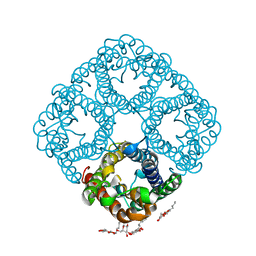
3NK5
 | | Crystal structure of AqpZ mutant F43W | | 分子名称: | Aquaporin Z, octyl beta-D-glucopyranoside | | 著者 | Savage, D.F, O'Connell, J.D, Stroud, R.M, Finer-Moore, J.S. | | 登録日 | 2010-06-18 | | 公開日 | 2010-08-11 | | 最終更新日 | 2024-04-03 | | 実験手法 | X-RAY DIFFRACTION (2.4 Å) | | 主引用文献 | Structural context shapes the aquaporin selectivity filter.
Proc.Natl.Acad.Sci.USA, 107, 2010
|
|
4JSS
 | | Human carbonic anhydrase II H94D bound to a bidentate inhibitor | | 分子名称: | 1-hydroxy-2-sulfanylpyridinium, Carbonic anhydrase 2, MERCURIBENZOIC ACID, ... | | 著者 | Martin, D.P, Hann, Z.S, Cohen, S.M. | | 登録日 | 2013-03-22 | | 公開日 | 2013-06-19 | | 最終更新日 | 2024-02-28 | | 実験手法 | X-RAY DIFFRACTION (1.5 Å) | | 主引用文献 | Metalloprotein-Inhibitor Binding: Human Carbonic Anhydrase II as a Model for Probing Metal-Ligand Interactions in a Metalloprotein Active Site.
Inorg.Chem., 52, 2013
|
|
5CQZ
 | |
3UKF
 | |
1RAY
 | |
1RC2
 | | 2.5 Angstrom Resolution X-ray Structure of Aquaporin Z | | 分子名称: | 2-O-octyl-beta-D-glucopyranose, Aquaporin Z | | 著者 | Savage, D.F, Egea, P.F, Robles, Y.C, O'Connell III, J.D, Stroud, R.M. | | 登録日 | 2003-11-03 | | 公開日 | 2003-11-25 | | 最終更新日 | 2023-08-23 | | 実験手法 | X-RAY DIFFRACTION (2.5 Å) | | 主引用文献 | Architecture and selectivity in aquaporins: 2.5 a X-ray structure of aquaporin Z
Plos Biol., 1, 2003
|
|
6KRP
 | |
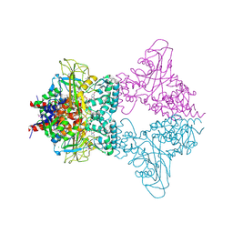
3GZN
 | | Structure of NEDD8-activating enzyme in complex with NEDD8 and MLN4924 | | 分子名称: | NEDD8, NEDD8-activating enzyme E1 catalytic subunit, NEDD8-activating enzyme E1 regulatory subunit, ... | | 著者 | Sintchak, M.D. | | 登録日 | 2009-04-07 | | 公開日 | 2010-02-02 | | 最終更新日 | 2024-11-20 | | 実験手法 | X-RAY DIFFRACTION (3 Å) | | 主引用文献 | Substrate-assisted inhibition of ubiquitin-like protein-activating enzymes: the NEDD8 E1 inhibitor MLN4924 forms a NEDD8-AMP mimetic in situ.
Mol.Cell, 37, 2010
|
|


