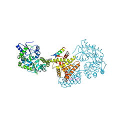
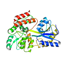
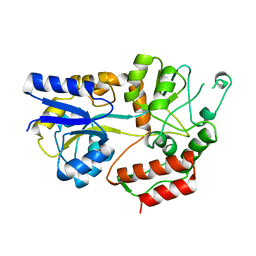
3SLO
 
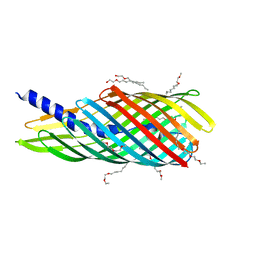 | | Pre-cleavage Structure of the Autotransporter EspP - N1023D mutant | | 分子名称: | (HYDROXYETHYLOXY)TRI(ETHYLOXY)OCTANE, Serine protease espP | | 著者 | Barnard, T.B, Noinaj, N, Easley, N.C, Kuszak, A.J, Buchanan, S.K. | | 登録日 | 2011-06-24 | | 公開日 | 2011-11-16 | | 最終更新日 | 2024-02-28 | | 実験手法 | X-RAY DIFFRACTION (2.52 Å) | | 主引用文献 | Molecular basis for the activation of a catalytic asparagine residue in a self-cleaving bacterial autotransporter.
J.Mol.Biol., 415, 2012
|
|
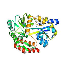
4GIZ
 | | Crystal structure of full-length human papillomavirus oncoprotein E6 in complex with LXXLL peptide of ubiquitin ligase E6AP at 2.55 A resolution | | 分子名称: | Maltose-binding periplasmic protein, UBIQUITIN LIGASE EA6P: chimeric protein, Protein E6, ... | | 著者 | McEwen, A.G, Zanier, K, Charbonnier, S, Poussin, P, Cura, V, Vande Pol, S, Trave, G, Cavarelli, J. | | 登録日 | 2012-08-09 | | 公開日 | 2013-01-23 | | 最終更新日 | 2024-02-28 | | 実験手法 | X-RAY DIFFRACTION (2.55 Å) | | 主引用文献 | Structural basis for hijacking of cellular LxxLL motifs by papillomavirus E6 oncoproteins.
Science, 339, 2013
|
|
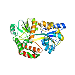
5ZNZ
 | | Structure of mDR3 DD with MBP tag mutant-I387V | | 分子名称: | Maltose-binding periplasmic protein,Tumor necrosis factor receptor superfamily, member 25, SULFATE ION | | 著者 | Jin, T, Yin, X. | | 登録日 | 2018-04-12 | | 公開日 | 2019-04-17 | | 最終更新日 | 2023-11-22 | | 実験手法 | X-RAY DIFFRACTION (2.55 Å) | | 主引用文献 | Crystal structure and activation mechanism of DR3 death domain.
Febs J., 286, 2019
|
|
7MHW
 | | Crystal structure of the protease inhibitor U-Omp19 from Brucella abortus fused to Maltose-binding protein | | 分子名称: | Maltose/maltodextrin-binding periplasmic protein,Outer membrane lipoprotein omp19, SULFATE ION | | 著者 | Darriba, M.L, Klinke, S, Otero, L.H, Cerutti, M.L, Cassataro, J, Pasquevich, K.A. | | 登録日 | 2021-04-15 | | 公開日 | 2022-04-20 | | 最終更新日 | 2023-10-18 | | 実験手法 | X-RAY DIFFRACTION (2.55 Å) | | 主引用文献 | A disordered region retains the full protease inhibitor activity and the capacity to induce CD8 + T cells in vivo of the oral vaccine adjuvant U-Omp19.
Comput Struct Biotechnol J, 20, 2022
|
|
6D67
 | | Crystal structure of the human dual specificity phosphatase 1 catalytic domain (C258S) as a maltose binding protein fusion (maltose bound form) in complex with the designed AR protein mbp3_16 | | 分子名称: | 1,2-ETHANEDIOL, DI(HYDROXYETHYL)ETHER, Designed AR protein mbp3_16, ... | | 著者 | Gumpena, R, Lountos, G.T, Waugh, D.S. | | 登録日 | 2018-04-20 | | 公開日 | 2018-09-19 | | 最終更新日 | 2023-10-04 | | 実験手法 | X-RAY DIFFRACTION (2.55 Å) | | 主引用文献 | MBP-binding DARPins facilitate the crystallization of an MBP fusion protein.
Acta Crystallogr F Struct Biol Commun, 74, 2018
|
|
7RW6
 | | BORF2-APOBEC3Bctd Complex | | 分子名称: | DNA dC->dU-editing enzyme APOBEC-3B, Maltose/maltodextrin-binding periplasmic protein,Ribonucleoside-diphosphate reductase large subunit, ZINC ION | | 著者 | Shaban, N.M, Yan, R, Shi, K, McLellan, J.S, Yu, Z, Harris, R.S. | | 登録日 | 2021-08-19 | | 公開日 | 2022-04-27 | | 最終更新日 | 2022-05-04 | | 実験手法 | ELECTRON MICROSCOPY (2.55 Å) | | 主引用文献 | Cryo-EM structure of the EBV ribonucleotide reductase BORF2 and mechanism of APOBEC3B inhibition.
Sci Adv, 8, 2022
|
|
5AZ6
 | |
8J25
 | | Crystal structure of PML B-box2 mutant | | 分子名称: | Maltose/maltodextrin-binding periplasmic protein,Protein PML, ZINC ION, alpha-D-glucopyranose-(1-4)-alpha-D-glucopyranose | | 著者 | Zhou, C, Zang, N, Zhang, J. | | 登録日 | 2023-04-14 | | 公開日 | 2023-09-20 | | 最終更新日 | 2023-12-20 | | 実験手法 | X-RAY DIFFRACTION (2.6 Å) | | 主引用文献 | Structural Basis of PML-RARA Oncoprotein Targeting by Arsenic Unravels a Cysteine Rheostat Controlling PML Body Assembly and Function.
Cancer Discov, 13, 2023
|
|
1PEB
 | |
3MP1
 | | Complex structure of Sgf29 and trimethylated H3K4 | | 分子名称: | ACETATE ION, H3K4me3 peptide, Maltose-binding periplasmic protein,LINKER,SAGA-associated factor 29, ... | | 著者 | Li, J, Ruan, J, Wu, M, Xue, X, Zang, J. | | 登録日 | 2010-04-24 | | 公開日 | 2011-05-04 | | 最終更新日 | 2023-11-01 | | 実験手法 | X-RAY DIFFRACTION (2.6 Å) | | 主引用文献 | Sgf29 binds histone H3K4me2/3 and is required for SAGA complex recruitment and histone H3 acetylation
Embo J., 30, 2011
|
|
8C5L
 | |
7U0G
 | | structure of LIN28b nucleosome bound 3 OCT4 | | 分子名称: | DNA (162-MER), Histone H2A type 2-C, Histone H2B type 2-E, ... | | 著者 | Lian, T, Guan, R, Bai, Y. | | 登録日 | 2022-02-18 | | 公開日 | 2023-06-28 | | 実験手法 | ELECTRON MICROSCOPY (2.6 Å) | | 主引用文献 | Structural mechanism of LIN28B nucleosome targeting by OCT4.
Mol.Cell, 83, 2023
|
|
7U0I
 | | Structure of LIN28b nucleosome bound 2 OCT4 | | 分子名称: | DNA (162-MER), Histone H2A type 2-C, Histone H2B type 2-E, ... | | 著者 | Tengfei, L, Guan, R, Bai, Y. | | 登録日 | 2022-02-18 | | 公開日 | 2023-06-28 | | 実験手法 | ELECTRON MICROSCOPY (2.6 Å) | | 主引用文献 | Structural mechanism of LIN28B nucleosome targeting by OCT4.
Mol.Cell, 83, 2023
|
|
5BK2
 | |
1N3W
 | | Engineered High-Affinity Maltose-Binding Protein | | 分子名称: | Maltose-binding periplasmic protein, alpha-D-glucopyranose-(1-4)-alpha-D-glucopyranose | | 著者 | Telmer, P.G, Shilton, B.H. | | 登録日 | 2002-10-29 | | 公開日 | 2003-08-12 | | 最終更新日 | 2024-02-14 | | 実験手法 | X-RAY DIFFRACTION (2.6 Å) | | 主引用文献 | Insights into the Conformational Equilibria of Maltose-binding Protein by Analysis of High Affinity Mutants.
J.Biol.Chem., 278, 2003
|
|
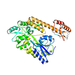
4KYE
 | | Partial Structure of the C-terminal domain of the HPIV4B phosphoprotein, fused to MBP. | | 分子名称: | Maltose-binding periplasmic protein, Phosphoprotein, chimeric construct, ... | | 著者 | Yegambaram, K, Bulloch, E.M.M, Kingston, R.L. | | 登録日 | 2013-05-28 | | 公開日 | 2013-09-25 | | 最終更新日 | 2024-02-28 | | 実験手法 | X-RAY DIFFRACTION (2.6 Å) | | 主引用文献 | Protein domain definition should allow for conditional disorder.
Protein Sci., 22, 2013
|
|
3F5F
 | | Crystal structure of heparan sulfate 2-O-sulfotransferase from gallus gallus as a maltose binding protein fusion. | | 分子名称: | ADENOSINE-3'-5'-DIPHOSPHATE, Maltose-binding periplasmic protein, Heparan sulfate 2-O-sulfotransferase 1, ... | | 著者 | Bethea, H.N, Xu, D, Liu, J, Pedersen, L.C. | | 登録日 | 2008-11-03 | | 公開日 | 2008-12-16 | | 最終更新日 | 2023-09-06 | | 実験手法 | X-RAY DIFFRACTION (2.65 Å) | | 主引用文献 | Redirecting the substrate specificity of heparan sulfate 2-O-sulfotransferase by structurally guided mutagenesis.
Proc.Natl.Acad.Sci.USA, 105, 2008
|
|
8TLV
 | |
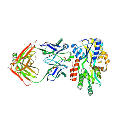
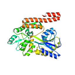
2QOM
 | | The crystal structure of the E.coli EspP autotransporter Beta-domain. | | 分子名称: | Serine protease espP | | 著者 | Barnard, T.J, Dautin, N, Lukacik, P, Bernstein, H.D, Buchanan, S.K. | | 登録日 | 2007-07-20 | | 公開日 | 2007-11-13 | | 最終更新日 | 2024-04-03 | | 実験手法 | X-RAY DIFFRACTION (2.66 Å) | | 主引用文献 | Autotransporter structure reveals intra-barrel cleavage followed by conformational changes.
Nat.Struct.Mol.Biol., 14, 2007
|
|
5YGS
 | | Human TNFRSF25 death domain | | 分子名称: | Human TNRSF25 death domain, SULFATE ION, alpha-D-glucopyranose-(1-4)-alpha-D-glucopyranose-(1-4)-alpha-D-glucopyranose | | 著者 | Yin, X, Jin, T. | | 登録日 | 2017-09-26 | | 公開日 | 2018-10-03 | | 最終更新日 | 2023-11-22 | | 実験手法 | X-RAY DIFFRACTION (2.691 Å) | | 主引用文献 | Crystal structure and activation mechanism of DR3 death domain.
Febs J., 286, 2019
|
|
4LOG
 | | The crystal structure of the orphan nuclear receptor PNR ligand binding domain fused with MBP | | 分子名称: | Maltose ABC transporter periplasmic protein and NR2E3 protein chimeric construct | | 著者 | Tan, M.E, Zhou, X.E, Soon, F.-F, Li, X, Li, J, Yong, E.-L, Melcher, K, Xu, H.E. | | 登録日 | 2013-07-12 | | 公開日 | 2013-10-09 | | 最終更新日 | 2023-09-20 | | 実験手法 | X-RAY DIFFRACTION (2.7 Å) | | 主引用文献 | The Crystal Structure of the Orphan Nuclear Receptor NR2E3/PNR Ligand Binding Domain Reveals a Dimeric Auto-Repressed Conformation.
Plos One, 8, 2013
|
|
1IUD
 | | MALTODEXTRIN-BINDING PROTEIN INSERTION/DELETION MUTANT WITH AN INSERTED B-CELL EPITOPE FROM THE PRES2 REGION OF HEPATITIS B VIRUS | | 分子名称: | MALTODEXTRIN-BINDING PROTEIN MALE-B133, alpha-D-glucopyranose-(1-4)-alpha-D-glucopyranose | | 著者 | Saul, F.A, Vulliez-Le Normand, B, Lema, F, Bentley, G.A. | | 登録日 | 1996-05-29 | | 公開日 | 1997-06-05 | | 最終更新日 | 2024-04-03 | | 実験手法 | X-RAY DIFFRACTION (2.7 Å) | | 主引用文献 | Crystal structure of a recombinant form of the maltodextrin-binding protein carrying an inserted sequence of a B-cell epitope from the preS2 region of hepatitis B virus.
Proteins, 27, 1997
|
|
5I69
 | | MBP-MamC magnetite-interaction component mutant-D70A | | 分子名称: | Maltose-binding periplasmic protein,Tightly bound bacterial magnetic particle protein,Maltose-binding periplasmic protein, SULFATE ION, alpha-D-glucopyranose-(1-4)-alpha-D-glucopyranose | | 著者 | Nudelman, H, Tercedor, C.V, Kolusheva, S, Gonzalez, T.P, Widdrat, M, Grimberg, N, Levi, H, Nelkenbaum, O, Davidove, G, Faivre, D, Jimenez-Lopez, C, Zarivach, R. | | 登録日 | 2016-02-16 | | 公開日 | 2016-03-23 | | 最終更新日 | 2024-01-10 | | 実験手法 | X-RAY DIFFRACTION (2.7 Å) | | 主引用文献 | Structure-function studies of the magnetite-biomineralizing magnetosome-associated protein MamC.
J.Struct.Biol., 194, 2016
|
|
6QUG
 | | GHK tagged MBP-Nup98(1-29) | | 分子名称: | COPPER (II) ION, Maltodextrin-binding protein,Nucleoporin, putative, ... | | 著者 | Huyton, T, Gorlich, D. | | 登録日 | 2019-02-27 | | 公開日 | 2020-05-27 | | 最終更新日 | 2024-05-15 | | 実験手法 | X-RAY DIFFRACTION (2.7 Å) | | 主引用文献 | The copper(II)-binding tripeptide GHK, a valuable crystallization and phasing tag for macromolecular crystallography.
Acta Crystallogr D Struct Biol, 76, 2020
|
|
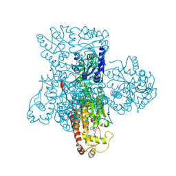
8GKH
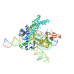 | | Structure of the Spizellomyces punctatus Fanzor (SpuFz) in complex with omega RNA and target DNA | | 分子名称: | DNA (35-MER), DNA (5'-D(P*CP*GP*GP*TP*AP*CP*CP*CP*GP*GP*GP*CP*AP*TP*A)-3'), MAGNESIUM ION, ... | | 著者 | Xu, P, Saito, M, Zhang, F. | | 登録日 | 2023-03-19 | | 公開日 | 2023-07-05 | | 最終更新日 | 2023-08-30 | | 実験手法 | ELECTRON MICROSCOPY (2.7 Å) | | 主引用文献 | Fanzor is a eukaryotic programmable RNA-guided endonuclease.
Nature, 620, 2023
|
|


