1XEJ
 
 | |
1XEK
 | |
1XEL
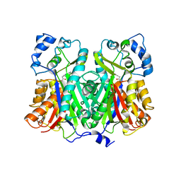
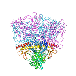
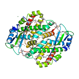
 | | UDP-GALACTOSE 4-EPIMERASE FROM ESCHERICHIA COLI | | 分子名称: | 1,2-ETHANEDIOL, DI(HYDROXYETHYL)ETHER, NICOTINAMIDE-ADENINE-DINUCLEOTIDE, ... | | 著者 | Thoden, J, Holden, H. | | 登録日 | 1996-01-25 | | 公開日 | 1997-02-12 | | 最終更新日 | 2024-02-14 | | 実験手法 | X-RAY DIFFRACTION (1.8 Å) | | 主引用文献 | Molecular structure of the NADH/UDP-glucose abortive complex of UDP-galactose 4-epimerase from Escherichia coli: implications for the catalytic mechanism.
Biochemistry, 35, 1996
|
|
1XEM
 | | High Resolution Crystal Structure of Escherichia coli Zinc- Peptide Deformylase bound to formate | | 分子名称: | FORMIC ACID, Peptide deformylase, ZINC ION | | 著者 | Jain, R, Hao, B, Liu, R.-P, Chan, M.K. | | 登録日 | 2004-09-10 | | 公開日 | 2005-03-29 | | 最終更新日 | 2024-02-14 | | 実験手法 | X-RAY DIFFRACTION (1.76 Å) | | 主引用文献 | Structures of E. coli peptide deformylase bound to formate: insight into the preference for Fe2+ over Zn2+ as the active site metal
J.Am.Chem.Soc., 127, 2005
|
|
1XEN
 | | High Resolution Crystal Structure of Escherichia coli Iron- Peptide Deformylase Bound To Formate | | 分子名称: | FE (III) ION, FORMIC ACID, Peptide deformylase | | 著者 | Jain, R, Hao, B, Liu, R.-P, Chan, M.K. | | 登録日 | 2004-09-10 | | 公開日 | 2005-03-29 | | 最終更新日 | 2024-02-14 | | 実験手法 | X-RAY DIFFRACTION (1.85 Å) | | 主引用文献 | Structures of E. coli peptide deformylase bound to formate: insight into the preference for Fe2+ over Zn2+ as the active site metal
J.Am.Chem.Soc., 127, 2005
|
|
1XEO
 | | High Resolution Crystals Structure of Cobalt- Peptide Deformylase Bound To Formate | | 分子名称: | COBALT (II) ION, FORMIC ACID, Peptide deformylase | | 著者 | Jain, R, Hao, B, Liu, R.-P, Chan, M.K. | | 登録日 | 2004-09-10 | | 公開日 | 2005-03-29 | | 最終更新日 | 2024-02-14 | | 実験手法 | X-RAY DIFFRACTION (1.3 Å) | | 主引用文献 | Structures of E. coli peptide deformylase bound to formate: insight into the preference for Fe2+ over Zn2+ as the active site metal
J.Am.Chem.Soc., 127, 2005
|
|
1XEP
 | | Catechol in complex with T4 lysozyme L99A/M102Q | | 分子名称: | BETA-MERCAPTOETHANOL, CATECHOL, Lysozyme, ... | | 著者 | Graves, A.P, Brenk, R, Shoichet, B.K. | | 登録日 | 2004-09-10 | | 公開日 | 2005-05-31 | | 最終更新日 | 2024-02-14 | | 実験手法 | X-RAY DIFFRACTION (1.55 Å) | | 主引用文献 | Decoys for docking.
J.Med.Chem., 48, 2005
|
|
1XEQ
 | | Crystal tructure of RNA binding domain of influenza B virus non-structural protein | | 分子名称: | BROMIDE ION, Nonstructural protein NS1 | | 著者 | Khan, J.A, Yin, C, Krug, R.M, Montelione, G.T, Tong, L, Northeast Structural Genomics Consortium (NESG) | | 登録日 | 2004-09-11 | | 公開日 | 2005-09-20 | | 最終更新日 | 2024-02-14 | | 実験手法 | X-RAY DIFFRACTION (2.1 Å) | | 主引用文献 | Conserved surface features form the double-stranded RNA binding site of non-structural protein 1 (NS1) from influenza A and B viruses.
J.Biol.Chem., 282, 2007
|
|
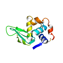
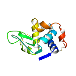
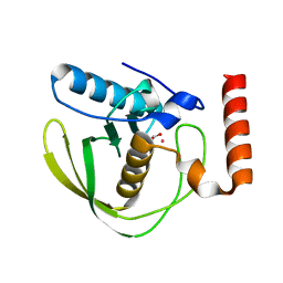
1XER
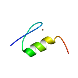
 | | STRUCTURE OF FERREDOXIN | | 分子名称: | FE3-S4 CLUSTER, FERREDOXIN, ZINC ION | | 著者 | Fujii, T, Hata, Y, Moriyama, H, Wakagi, T, Tanaka, N, Oshima, T. | | 登録日 | 1996-08-28 | | 公開日 | 1997-09-04 | | 最終更新日 | 2011-07-13 | | 実験手法 | X-RAY DIFFRACTION (2 Å) | | 主引用文献 | Novel zinc-binding centre in thermoacidophilic archaeal ferredoxins.
Nat.Struct.Biol., 3, 1996
|
|
1XES
 | | Crystal structure of stilbene synthase from Pinus sylvestris | | 分子名称: | 3-(1H-INDOL-3-YL)-2-OXOPROPANOIC ACID, Dihydropinosylvin synthase | | 著者 | Ng, S.H, Chirgadze, D, Spiteller, D, Li, T.L, Spencer, J.B, Blundell, T.L. | | 登録日 | 2004-09-12 | | 公開日 | 2006-04-11 | | 最終更新日 | 2023-08-23 | | 実験手法 | X-RAY DIFFRACTION (1.7 Å) | | 主引用文献 | Crystal structure of stilbene synthase from Pinus sylvestris
To be Published
|
|
1XET
 | | Crystal structure of stilbene synthase from Pinus sylvestris, complexed with methylmalonyl CoA | | 分子名称: | 3-(1H-INDOL-3-YL)-2-OXOPROPANOIC ACID, Dihydropinosylvin synthase, METHYLMALONYL-COENZYME A | | 著者 | Ng, S.H, Chirgadze, D, Spiteller, D, Li, T.L, Spencer, J.B, Blundell, T.L. | | 登録日 | 2004-09-12 | | 公開日 | 2006-04-11 | | 最終更新日 | 2024-02-14 | | 実験手法 | X-RAY DIFFRACTION (2 Å) | | 主引用文献 | Crystal structure of stilbene synthase from Pinus sylvestris, complexed with methylmalonyl CoA
To be Published
|
|
1XEU
 | | Crystal Structure of Internalin C from Listeria monocytogenes | | 分子名称: | internalin C | | 著者 | Ooi, A, Hussain, S, Seyedarabi, A, Pickersgill, R.W. | | 登録日 | 2004-09-13 | | 公開日 | 2005-08-30 | | 最終更新日 | 2024-04-03 | | 実験手法 | X-RAY DIFFRACTION (2.05 Å) | | 主引用文献 | Structure of internalin C from Listeria monocytogenes.
Acta Crystallogr.,Sect.D, 62, 2006
|
|
1XEV
 | |
1XEW
 | |
1XEX
 | |
1XEY
 | | Crystal structure of the complex of Escherichia coli GADA with glutarate at 2.05 A resolution | | 分子名称: | ACETATE ION, GLUTARIC ACID, Glutamate decarboxylase alpha, ... | | 著者 | Dutyshev, D.I, Darii, E.L, Fomenkova, N.P, Pechik, I.V, Polyakov, K.M, Nikonov, S.V, Andreeva, N.S, Sukhareva, B.S. | | 登録日 | 2004-09-13 | | 公開日 | 2004-10-05 | | 最終更新日 | 2023-08-23 | | 実験手法 | X-RAY DIFFRACTION (2.05 Å) | | 主引用文献 | Structure of Escherichia coli glutamate decarboxylase (GADalpha) in complex with glutarate at 2.05 angstroms resolution.
Acta Crystallogr.,Sect.D, 61, 2005
|
|
1XEZ
 | |
1XF0
 | | Crystal structure of human 17beta-hydroxysteroid dehydrogenase type 5 (AKR1C3) complexed with delta4-androstene-3,17-dione and NADP | | 分子名称: | 4-ANDROSTENE-3-17-DIONE, ACETATE ION, Aldo-keto reductase family 1 member C3, ... | | 著者 | Qiu, W, Zhou, M, Labrie, F, Lin, S.-X. | | 登録日 | 2004-09-13 | | 公開日 | 2004-10-26 | | 最終更新日 | 2023-08-23 | | 実験手法 | X-RAY DIFFRACTION (2 Å) | | 主引用文献 | Crystal structures of the multispecific 17beta-hydroxysteroid dehydrogenase type 5: critical androgen regulation in human peripheral tissues
Mol.Endocrinol., 18, 2004
|
|
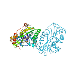
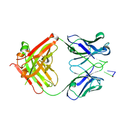
1XF1
 | | Structure of C5a peptidase- a key virulence factor from Streptococcus | | 分子名称: | ACETATE ION, C5a peptidase, CALCIUM ION, ... | | 著者 | Brown, C.K, Gu, Z.Y, Cleary, P.P, Matsuka, Y, Olmstead, S, Ohlendorf, D.H, Earhart, C.A. | | 登録日 | 2004-09-13 | | 公開日 | 2005-11-22 | | 最終更新日 | 2021-10-20 | | 実験手法 | X-RAY DIFFRACTION (1.9 Å) | | 主引用文献 | Structure of the streptococcal cell wall C5a peptidase
Proc.Natl.Acad.Sci.Usa, 102, 2005
|
|
1XF2
 | | Structure of Fab DNA-1 complexed with dT3 | | 分子名称: | 5'-D(*TP*TP*T)-3', SULFATE ION, antibody heavy chain Fab, ... | | 著者 | Schuermann, J.P, Prewitt, S.P, Deutscher, S.L, Tanner, J.J. | | 登録日 | 2004-09-13 | | 公開日 | 2005-04-12 | | 最終更新日 | 2023-08-23 | | 実験手法 | X-RAY DIFFRACTION (2.3 Å) | | 主引用文献 | Evidence for Structural Plasticity of Heavy Chain Complementarity-determining Region 3 in Antibody-ssDNA Recognition
J.Mol.Biol., 347, 2005
|
|
1XF3
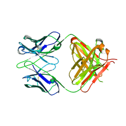 | | Structure of ligand-free Fab DNA-1 in space group P65 | | 分子名称: | Fab Light chain, Fab heavy chain | | 著者 | Schuermann, J.P, Prewitt, S.P, Deutscher, S.L, Tanner, J.J. | | 登録日 | 2004-09-13 | | 公開日 | 2005-04-12 | | 最終更新日 | 2024-10-16 | | 実験手法 | X-RAY DIFFRACTION (2.3 Å) | | 主引用文献 | Evidence for Structural Plasticity of Heavy Chain Complementarity-determining Region 3 in Antibody-ssDNA Recognition
J.Mol.Biol., 347, 2005
|
|
1XF4
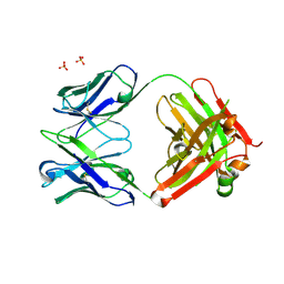 | | Structure of ligand-free Fab DNA-1 in space group P321 solved from crystals with perfect hemihedral twinning | | 分子名称: | Fab heavy chain, Fab light chain, SULFATE ION | | 著者 | Schuermann, J.P, Prewitt, S.P, Deutscher, S.L, Tanner, J.J. | | 登録日 | 2004-09-13 | | 公開日 | 2005-04-12 | | 最終更新日 | 2023-08-23 | | 実験手法 | X-RAY DIFFRACTION (2.5 Å) | | 主引用文献 | Evidence for Structural Plasticity of Heavy Chain Complementarity-determining Region 3 in Antibody-ssDNA Recognition
J.Mol.Biol., 347, 2005
|
|
1XF5
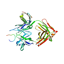 | | Complex HCV core-Fab 19D9D6-Protein L mutant (H74C, Y64W)in space group P21212 | | 分子名称: | Capsid protein C, Monoclonal antibody 19D9D6 Heavy chain, Monoclonal antibody 19D9D6 Light chain, ... | | 著者 | Menez, R, Housden, N.G, Harrison, S, Jolivet-Reynaud, C, Gore, M.G, Stura, E.A. | | 登録日 | 2004-09-14 | | 公開日 | 2005-05-31 | | 最終更新日 | 2011-07-13 | | 実験手法 | X-RAY DIFFRACTION (2.6 Å) | | 主引用文献 | Different crystal packing in Fab-protein L semi-disordered peptide complex.
Acta Crystallogr.,Sect.D, 61, 2005
|
|
1XF6
 | | High resolution crystal structure of phycoerythrin 545 from the marine cryptophyte rhodomonas CS24 | | 分子名称: | 15,16-DIHYDROBILIVERDIN, B-phycoerythrin beta chain, CHLORIDE ION, ... | | 著者 | Doust, A.B, Marai, C.N.J, Harrop, S.J, Wilk, K.E, Curmi, P.M.G, Scholes, G.D. | | 登録日 | 2004-09-14 | | 公開日 | 2004-11-30 | | 最終更新日 | 2023-10-25 | | 実験手法 | X-RAY DIFFRACTION (1.1 Å) | | 主引用文献 | Developing a structure-function model for the cryptophyte phycoerythrin 545 using ultrahigh resolution crystallography and ultrafast laser spectroscopy
J.Mol.Biol., 344, 2004
|
|
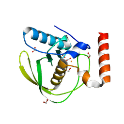
1XF7
 | | High Resolution NMR Structure of the Wilms' Tumor Suppressor Protein (WT1) Finger 3 | | 分子名称: | Wilms' Tumor Protein, ZINC ION | | 著者 | Lachenmann, M.J, Ladbury, J.E, Dong, J, Huang, K, Carey, P, Weiss, M.A. | | 登録日 | 2004-09-14 | | 公開日 | 2004-12-14 | | 最終更新日 | 2024-05-22 | | 実験手法 | SOLUTION NMR | | 主引用文献 | Why zinc fingers prefer zinc: ligand-field symmetry and the hidden thermodynamics of metal ion selectivity
Biochemistry, 43, 2004
|
|


