Loading
PDBj
MenuPDBj@FacebookPDBj@TwitterPDBj@YouTubewwPDB FoundationwwPDB
RCSB PDBPDBeBMRBAdv. SearchSearch help

3O0H
DownloadVisualize
BU of 3o0h by Molmil
Crystal structure of glutathione reductase from Bartonella henselae
Descriptor: CHLORIDE ION, FLAVIN-ADENINE DINUCLEOTIDE, Glutathione reductase
Authors:Seattle Structural Genomics Center for Infectious Disease (SSGCID)
Deposit date:2010-07-19
Release date:2010-08-18
Last modified:2023-09-06
Method:X-RAY DIFFRACTION (1.9 Å)
Cite:Crystal structure of glutathione reductase from Bartonella henselae
To be Published
4QFF
DownloadVisualize
BU of 4qff by Molmil
Structure of a 16 nm protein cage designed by fusing symmetric oligomeric domains, quadruple mutant, P212121 form
Descriptor: Non-haem bromoperoxidase BPO-A2, Matrix protein 1 chimera
Authors:Lai, Y.-T, Yeates, T.O.
Deposit date:2014-05-20
Release date:2015-05-20
Last modified:2024-02-28
Method:X-RAY DIFFRACTION (7.811 Å)
Cite:Structural transition of a protein nanocage as a function of pH and salt concentration
To be Published
4MPQ
DownloadVisualize
BU of 4mpq by Molmil
Crystal structure of1-pyrroline-4-hydroxy-2-carboxylate deaminase from Brucella melitensis ATCC 23457
Descriptor: 1,2-ETHANEDIOL, Dihydrodipicolinate synthetase
Authors:Seattle Structural Genomics Center for Infectious Disease (SSGCID)
Deposit date:2013-09-13
Release date:2013-09-25
Last modified:2023-09-20
Method:X-RAY DIFFRACTION (1.75 Å)
Cite:Crystal structure of1-pyrroline-4-hydroxy-2-carboxylate deaminase from Brucella melitensis ATCC 23457
To be Published
8J0P
DownloadVisualize
BU of 8j0p by Molmil
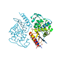
Chitin binding SusD-like protein AqSusD from a marine Bacteroidetes
Descriptor: Chitin binding SusD-like protein
Authors:Yang, J.
Deposit date:2023-04-11
Release date:2023-11-01
Last modified:2024-02-28
Method:X-RAY DIFFRACTION (2.4 Å)
Cite:Structural insights of a SusD-like protein in marine Bacteroidetes bacteria reveal the molecular basis for chitin recognition and acquisition.
Febs J., 291, 2024
4N0Q
DownloadVisualize
BU of 4n0q by Molmil
Crystal Structure of an ABC transporter, substrate-binding protein from Brucella melitensis 16M in complex with L-Leucine using a crystal grown in a Crystal Former (Microlytic)
Descriptor: LEUCINE, Leu/Ile/Val-binding protein homolog 3
Authors:Seattle Structural Genomics Center for Infectious Disease (SSGCID)
Deposit date:2013-10-02
Release date:2013-11-06
Last modified:2023-09-20
Method:X-RAY DIFFRACTION (2.3 Å)
Cite:Crystal Structure of an ABC transporter, substrate-binding protein from Brucella melitensis 16M in complex with L-Leucine using a crystal grown in a Crystal Former (Microlytic)
To be Published
3OA1
DownloadVisualize
BU of 3oa1 by Molmil
Crystal structure of phosphoprotein/Protein P/Protein M1 residues 69-297 from Rabies virus reveals degradation to C-terminal domain only
Descriptor: Phosphoprotein, SULFATE ION
Authors:Seattle Structural Genomics Center for Infectious Disease (SSGCID)
Deposit date:2010-08-04
Release date:2010-08-18
Last modified:2024-02-21
Method:X-RAY DIFFRACTION (2.2 Å)
Cite:Crystal structure of phosphoprotein/Protein P/Protein M1 residues 69-297 from Rabies virus reveals degradation to C-terminal domain only
To be Published
8IWV
DownloadVisualize
BU of 8iwv by Molmil
Crystal structure of BlaA-1 APO
Descriptor: Beta-lactamase, PHOSPHATE ION
Authors:Bhattacharya, S, Hazra, S.
Deposit date:2023-03-31
Release date:2024-04-03
Method:X-RAY DIFFRACTION (2.4 Å)
Cite:Crystal structure of BlaA-1 APO
To be published
4OGX
DownloadVisualize
BU of 4ogx by Molmil
Crystal structure of Fab DX-2930 in complex with human plasma kallikrein at 2.4 Angstrom resolution
Descriptor: DX-2930 HEAVY CHAIN, DX-2930 LIGHT CHAIN, Plasma kallikrein, ...
Authors:Edwards, T.E, Clifton, M.C, Abendroth, J, Nixon, A, Ladner, R.
Deposit date:2014-01-16
Release date:2014-07-09
Last modified:2023-09-20
Method:X-RAY DIFFRACTION (2.4 Å)
Cite:Inhibition of plasma kallikrein by a highly specific active site blocking antibody.
J.Biol.Chem., 289, 2014
8IY4
DownloadVisualize
BU of 8iy4 by Molmil
Crystal structure of Class A beta-lactamase BlaA WT - complex with Meropenem
Descriptor: (4R,5S)-3-{[(3S,5S)-5-(dimethylcarbamoyl)pyrrolidin-3-yl]sulfanyl}-5-[(2S,3R)-3-hydroxy-1-oxobutan-2-yl]-4-methyl-4,5-d ihydro-1H-pyrrole-2-carboxylic acid, Beta-lactamase, PHOSPHATE ION
Authors:Bhattacharya, S, Hazra, S.
Deposit date:2023-04-03
Release date:2024-04-03
Method:X-RAY DIFFRACTION (2 Å)
Cite:Crystal structure of Class A beta-lactamase BlaA WT - complex with Meropenem
To be published
8IX8
DownloadVisualize
BU of 8ix8 by Molmil
Crystal structure of Class A beta-lactamase BlaA WT - complex with Tebipenem
Descriptor: (4R,5S)-3-(1-(4,5-dihydrothiazol-2-yl)azetidin-3-ylthio)-5-((2S,3R)-3-hydroxy-1-oxobutan-2-yl)-4-methyl-4,5- dihydro-1H-pyrrole-2-carboxylic acid, Beta-lactamase, PHOSPHATE ION
Authors:Bhattacharya, S, Hazra, S.
Deposit date:2023-03-31
Release date:2024-04-03
Method:X-RAY DIFFRACTION (1.5 Å)
Cite:Crystal structure of Class A beta-lactamase BlaA WT - complex with Tebipenem
To be published
8IXX
DownloadVisualize
BU of 8ixx by Molmil
Crystal structure of Class A beta-lactamase BlaA WT - complex with Ertapenem
Descriptor: (4R,5S)-3-({(3S,5S)-5-[(3-carboxyphenyl)carbamoyl]pyrrolidin-3-yl}sulfanyl)-5-[(1S,2R)-1-formyl-2-hydroxypropyl]-4-methyl-4,5-dihydro-1H-pyrrole-2-carboxylic acid, Beta-lactamase, PHOSPHATE ION
Authors:Bhattacharya, S, Hazra, S.
Deposit date:2023-04-03
Release date:2024-04-03
Method:X-RAY DIFFRACTION (1.7 Å)
Cite:Crystal structure of Class A beta-lactamase BlaA WT - complex with Ertapenem
To be published
7JQW
DownloadVisualize
BU of 7jqw by Molmil
The external aldimine crystal structure of Salmonella typhimurium Tryptophan Synthase mutant beta-S377A in complex with cesium ion at the metal coordination site. The single beta-Q114 rotamer conformation allows a hydrogen bond to form with the PLP oxygen at the position 3 in the ring
Descriptor: (E)-N-({3-hydroxy-2-methyl-5-[(phosphonooxy)methyl]pyridin-4-yl}methylidene)-L-serine, 1,2-ETHANEDIOL, 2-AMINOPHENOL, ...
Authors:Hilario, E, Dunn, M.F, Mueller, L.J.
Deposit date:2020-08-11
Release date:2021-08-25
Last modified:2023-10-18
Method:X-RAY DIFFRACTION (1.7 Å)
Cite:The external aldimine crystal structure of Salmonella typhimurium Tryptophan Synthase mutant beta-S377A in complex with cesium ion at the metal coordination site. The single beta-Q114 rotamer conformation allows a hydrogen bond to form with the PLP oxygen at the position 3 in the ring.
To be Published
8J6Y
DownloadVisualize
BU of 8j6y by Molmil
Crystal structure of class A beta-lactamase PSE-4 WT- Apo state
Descriptor: Beta-lactamase PSE-4, SULFATE ION
Authors:Sarkar, M, Bhattacharya, S, Hazra, S.
Deposit date:2023-04-26
Release date:2024-05-01
Method:X-RAY DIFFRACTION (2.4 Å)
Cite:Crystal structure of class A beta-lactamase PSE-4 WT- Apo state
To be published
3OEC
DownloadVisualize
BU of 3oec by Molmil
Crystal structure of carveol dehydrogenase from Mycobacterium thermoresistibile
Descriptor: Carveol dehydrogenase (MythA.01326.c, A0R518 homolog), SODIUM ION
Authors:Seattle Structural Genomics Center for Infectious Disease (SSGCID)
Deposit date:2010-08-12
Release date:2010-09-01
Last modified:2023-09-06
Method:X-RAY DIFFRACTION (1.95 Å)
Cite:Mycofactocin-associated mycobacterial dehydrogenases with non-exchangeable NAD cofactors.
Sci Rep, 7, 2017
8JHW
DownloadVisualize
BU of 8jhw by Molmil
The first crystal structure of a H-2Kb-restricted decapeptide from Cryptosporidium parvum
Descriptor: 1,2-ETHANEDIOL, Beta-2-microglobulin, H-2 class I histocompatibility antigen, ...
Authors:Fan, S, Wang, Y.
Deposit date:2023-05-25
Release date:2024-05-29
Method:X-RAY DIFFRACTION (3.12 Å)
Cite:The first crystal structure of a H-2Kb-restricted decapeptide from Cryptosporidium parvum
To Be Published
8OEM
DownloadVisualize
BU of 8oem by Molmil
Crystal structure of FutA bound to Fe(II)
Descriptor: FE (II) ION, Putative iron ABC transporter, substrate binding protein
Authors:Bolton, R, Tews, I.
Deposit date:2023-03-10
Release date:2023-08-30
Last modified:2024-03-27
Method:X-RAY DIFFRACTION (1.7 Å)
Cite:A redox switch allows binding of Fe(II) and Fe(III) ions in the cyanobacterial iron-binding protein FutA from Prochlorococcus.
Proc.Natl.Acad.Sci.USA, 121, 2024
3OFV
DownloadVisualize
BU of 3ofv by Molmil
Crystal structure of peptidyl-tRNA hydrolase from Escherichia Coli, I222 crystal form
Descriptor: Peptidyl-tRNA hydrolase
Authors:Lam, R, McGrath, T.E, Romanov, V, Gothe, S.A, Peddi, S.R, Razumova, E, Lipman, R.S, Branstrom, A.A, Chirgadze, N.Y.
Deposit date:2010-08-16
Release date:2011-08-17
Last modified:2023-09-06
Method:X-RAY DIFFRACTION (3.2 Å)
Cite:Crystal structure of peptidyl-tRNA hydrolase from Escherichia Coli, I222 crystal form
To be Published
4MVH
DownloadVisualize
BU of 4mvh by Molmil
Crystal Structure of PDE10A with Novel Keto-Benzimidazole Inhibitor
Descriptor: 1H-benzimidazol-2-yl(4-{[3-(morpholin-4-yl)pyrazin-2-yl]oxy}phenyl)methanone, SULFATE ION, ZINC ION, ...
Authors:Chmait, S, Jordan, S.
Deposit date:2013-09-24
Release date:2013-10-23
Last modified:2014-01-15
Method:X-RAY DIFFRACTION (2.496 Å)
Cite:Design, Optimization, and Biological Evaluation of Novel Keto-Benzimidazoles as Potent and Selective Inhibitors of Phosphodiesterase 10A (PDE10A).
J.Med.Chem., 56, 2013
3OMC
DownloadVisualize
BU of 3omc by Molmil
Structure of human SND1 extended tudor domain in complex with the symmetrically dimethylated arginine PIWIL1 peptide R4me2s
Descriptor: CHLORIDE ION, SYNTHETIC PEPTIDE, Staphylococcal nuclease domain-containing protein 1
Authors:Lam, R, Liu, K, Guo, Y.H, Bian, C.B, Xu, C, MacKenzie, F, Bountra, C, Weigelt, J, Arrowsmith, C.H, Edwards, A.M, Bochkarev, A, Min, J, Structural Genomics Consortium (SGC)
Deposit date:2010-08-26
Release date:2010-09-15
Last modified:2023-09-06
Method:X-RAY DIFFRACTION (1.77 Å)
Cite:Structural basis for recognition of arginine methylated Piwi proteins by the extended Tudor domain.
Proc.Natl.Acad.Sci.USA, 107, 2010
3OL4
DownloadVisualize
BU of 3ol4 by Molmil
Crystal structure of a putative uncharacterized protein from Mycobacterium smegmatis, an ortholog of Rv0543c
Descriptor: Putative uncharacterized protein
Authors:Seattle Structural Genomics Center for Infectious Disease (SSGCID)
Deposit date:2010-08-25
Release date:2010-09-08
Last modified:2023-09-06
Method:X-RAY DIFFRACTION (2 Å)
Cite:Structural diversity in the Mycobacteria DUF3349 superfamily.
Protein Sci., 29, 2020
4QT6
DownloadVisualize
BU of 4qt6 by Molmil
Crystal structure of the SPRY domain of human HERC1
Descriptor: FORMAMIDE, Probable E3 ubiquitin-protein ligase HERC1, UNKNOWN ATOM OR ION
Authors:Dong, A, Hu, J, Guan, X, Wernimont, A, Li, Y, Bountra, C, Arrowsmith, C.H, Edwards, A.M, Tong, Y, Structural Genomics Consortium (SGC)
Deposit date:2014-07-07
Release date:2015-01-07
Last modified:2017-11-22
Method:X-RAY DIFFRACTION (1.64 Å)
Cite:Crystal structure of the SPRY domain of human HERC1
To be Published
3OI7
DownloadVisualize
BU of 3oi7 by Molmil
Structure of the structure of the H13A mutant of Ykr043C in complex with sedoheptulose-1,7-bisphosphate
Descriptor: 1,2-ETHANEDIOL, 1,7-di-O-phosphono-beta-D-altro-hept-2-ulofuranose, GLYCEROL, ...
Authors:Singer, A.U, Xu, X, Dong, A, Cui, H, Clasquin, M.F, Caudy, A.A, Edwards, A.M, Savchenko, A, Joachimiak, A, Yakunin, A.F, Midwest Center for Structural Genomics (MCSG)
Deposit date:2010-08-18
Release date:2010-11-17
Last modified:2023-12-06
Method:X-RAY DIFFRACTION (2.4 Å)
Cite:Riboneogenesis in yeast.
Cell(Cambridge,Mass.), 145, 2011
3OJ6
DownloadVisualize
BU of 3oj6 by Molmil
Crystal structure of Blasticidin S Deaminase from Coccidioides Immitis
Descriptor: 1,2-ETHANEDIOL, Blasticidin-S deaminase, CHLORIDE ION, ...
Authors:Seattle Structural Genomics Center for Infectious Disease (SSGCID)
Deposit date:2010-08-20
Release date:2010-09-15
Last modified:2023-09-06
Method:X-RAY DIFFRACTION (1.7 Å)
Cite:Crystal structure of Blasticidin S Deaminase from Coccidioides Immitis
To be Published
2ZO7
DownloadVisualize
BU of 2zo7 by Molmil
Crystal Structure of a Kusabira-Cyan Mutant (KCY-R1), a Cyan/Green-Emitting GFP-Like Protein
Descriptor: CYAN/GREEN-EMITTING GFP-LIKE PROTEIN, KUSABIRA-CYAN MUTANT (KCY-R1)
Authors:Kikuchi, A, Fukumura, E, Karasawa, S, Miyawaki, A, Shiro, Y, RIKEN Structural Genomics/Proteomics Initiative (RSGI)
Deposit date:2008-05-06
Release date:2009-05-12
Last modified:2023-11-15
Method:X-RAY DIFFRACTION (1.58 Å)
Cite:Crystal structure of a new cyan fluorescent protein and its hue-shifted variants
Biochemistry, 48, 2009
4QB4
DownloadVisualize
BU of 4qb4 by Molmil
Crystal Structure of Staphylococcal Nuclease mutant V23L/L25V/V66L
Descriptor: Thermonuclease
Authors:Lor, P, Janowska, K, Sakon, J, Stites, W.E.
Deposit date:2014-05-06
Release date:2014-06-11
Last modified:2024-02-28
Method:X-RAY DIFFRACTION (1.6 Å)
Cite:Crystal Structure of Staphylococcal Nuclease mutant V23L/L25V/V66L
To be Published

223790

PDB entries from 2024-08-14

PDB statisticsPDBj update infoContact PDBjnumon