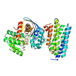
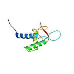
1C1J
 
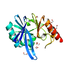
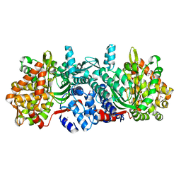
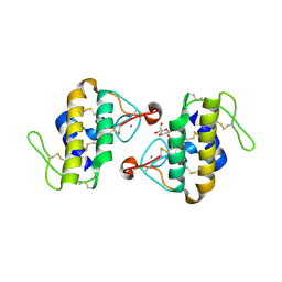 | | STRUCTURE OF CADMIUM-SUBSTITUTED PHOSPHOLIPASE A2 FROM AGKISTRONDON HALYS PALLAS AT 2.8 ANGSTROMS RESOLUTION | | 分子名称: | BASIC PHOSPHOLIPASE A2, CADMIUM ION, octyl beta-D-glucopyranoside | | 著者 | Zhang, H.-l, Zhang, Y.-q, Song, S.-y, Zhou, Y, Lin, Z.-j. | | 登録日 | 1999-07-22 | | 公開日 | 2002-07-05 | | 最終更新日 | 2020-07-29 | | 実験手法 | X-RAY DIFFRACTION (2.8 Å) | | 主引用文献 | Structure of Cadmium-substituted Phospholipase A2 from Agkistrodon halys
Pallas at 2.8 Angstroms Resolution
Protein Pept.Lett., 6, 1999
|
|
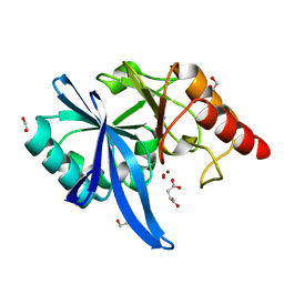
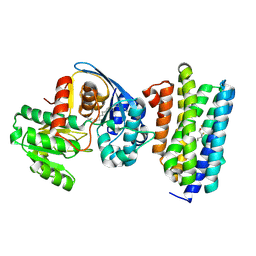
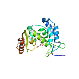
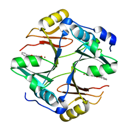
6JYJ
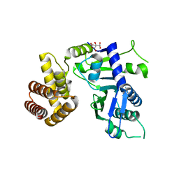 | | Crystal structure of FAM46B (TENT5B) | | 分子名称: | CITRATE ANION, Terminal nucleotidyltransferase 5B | | 著者 | Zhang, H, Hu, J.L, Gao, S. | | 登録日 | 2019-04-26 | | 公開日 | 2020-03-04 | | 最終更新日 | 2024-04-03 | | 実験手法 | X-RAY DIFFRACTION (2.693199 Å) | | 主引用文献 | FAM46B is a prokaryotic-like cytoplasmic poly(A) polymerase essential in human embryonic stem cells.
Nucleic Acids Res., 48, 2020
|
|
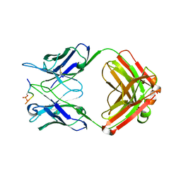
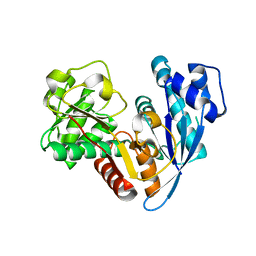
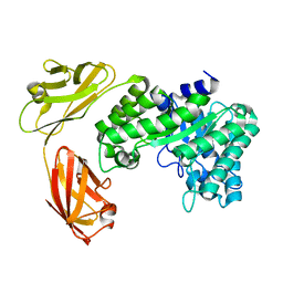
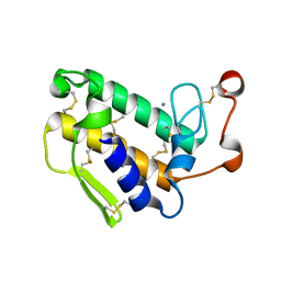
5HEC
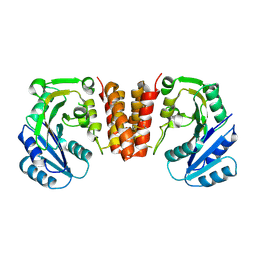 | | CgT structure in dimer | | 分子名称: | Putative glycosyltransferase (GalT1) | | 著者 | Zhang, H, Wu, H. | | 登録日 | 2016-01-05 | | 公開日 | 2016-08-31 | | 最終更新日 | 2024-10-09 | | 実験手法 | X-RAY DIFFRACTION (2.395 Å) | | 主引用文献 | New Helical Binding Domain Mediates a Glycosyltransferase Activity of a Bifunctional Protein.
J.Biol.Chem., 291, 2016
|
|
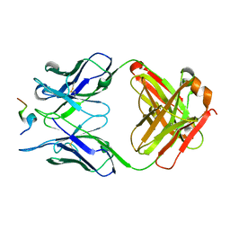
5ZV3
 | |
5ZGX
 | |
5ZGI
 | |
5ZIA
 | |
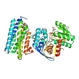
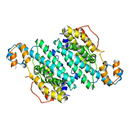
5ZGF
 | | Crystal structure of NDM-1 Q123G mutant | | 分子名称: | HYDROXIDE ION, Metallo-beta-lactamase type 2, ZINC ION | | 著者 | Zhang, H, Hao, Q. | | 登録日 | 2018-03-08 | | 公開日 | 2018-08-22 | | 最終更新日 | 2023-11-22 | | 実験手法 | X-RAY DIFFRACTION (1.2 Å) | | 主引用文献 | Active-Site Conformational Fluctuations Promote the Enzymatic Activity of NDM-1.
Antimicrob. Agents Chemother., 62, 2018
|
|
6JZZ
 | | The crystal structure of AAR-C294S in complex with ADO. | | 分子名称: | Aldehyde decarbonylase, FE (II) ION, HEXADECAN-1-OL, ... | | 著者 | Zhang, H.M, Li, M, Gao, Y. | | 登録日 | 2019-05-04 | | 公開日 | 2020-04-01 | | 最終更新日 | 2023-11-22 | | 実験手法 | X-RAY DIFFRACTION (3.011 Å) | | 主引用文献 | Structural insights into catalytic mechanism and product delivery of cyanobacterial acyl-acyl carrier protein reductase.
Nat Commun, 11, 2020
|
|
6JZY
 | | Crystal structure of AAR with NADPH and stearyl in complex with ADO binding a long chain carbohydrate | | 分子名称: | Aldehyde decarbonylase, FE (II) ION, HEXADECAN-1-OL, ... | | 著者 | Zhang, H.M, Li, M, Gao, Y. | | 登録日 | 2019-05-04 | | 公開日 | 2020-04-01 | | 最終更新日 | 2024-10-09 | | 実験手法 | X-RAY DIFFRACTION (2.1 Å) | | 主引用文献 | Structural insights into catalytic mechanism and product delivery of cyanobacterial acyl-acyl carrier protein reductase.
Nat Commun, 11, 2020
|
|
6JZU
 | | The crystal structure of acyl-acyl carrier protein (acyl-ACP) reductase (AAR) in complex with aldehyde deformylating oxygenase (ADO) | | 分子名称: | Aldehyde decarbonylase, FE (II) ION, HEXADECAN-1-OL, ... | | 著者 | Zhang, H.M, Li, M, Gao, Y. | | 登録日 | 2019-05-03 | | 公開日 | 2020-04-01 | | 最終更新日 | 2024-10-09 | | 実験手法 | X-RAY DIFFRACTION (2.181 Å) | | 主引用文献 | Structural insights into catalytic mechanism and product delivery of cyanobacterial acyl-acyl carrier protein reductase.
Nat Commun, 11, 2020
|
|
6JZQ
 | |
3CHT
 | |
5ZUM
 | | Structure of dipeptidyl-peptidase III from Corallococcus sp. strain EGB | | 分子名称: | ZINC ION, dipeptidyl-peptidase III | | 著者 | Zhang, H, Duan, Y.J, Li, Z.K, Liu, W.D, Huang, Y, Cui, Z.L. | | 登録日 | 2018-05-08 | | 公開日 | 2019-06-12 | | 実験手法 | X-RAY DIFFRACTION (1.9 Å) | | 主引用文献 | Structure of dipeptidyl peptidase III from Corallococcus sp. strain EGB
To Be Published
|
|
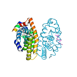
3OFS
 | | Dynamic conformations of the CD38-mediated NAD cyclization captured using multi-copy crystallography | | 分子名称: | ADP-ribosyl cyclase 1, [(2R,3S,4R,5R)-5-(6-amino-9H-purin-9-yl)-3,4-dihydroxytetrahydrofuran-2-yl]methyl [(2R,3R,4R)-4-fluoro-3-hydroxytetrahydrofuran-2-yl]methyl dihydrogen diphosphate | | 著者 | Zhang, H, Lee, H.C, Hao, Q. | | 登録日 | 2010-08-16 | | 公開日 | 2010-12-15 | | 最終更新日 | 2023-11-01 | | 実験手法 | X-RAY DIFFRACTION (2.2 Å) | | 主引用文献 | Dynamic Conformations of the CD38-Mediated NAD Cyclization Captured in a Single Crystal
J.Mol.Biol., 405, 2011
|
|
5E0C
 | |
1VQF
 | | GENE V PROTEIN MUTANT WITH VAL 35 REPLACED BY ILE 35 AND ILE 47 REPLACED BY VAL 47 (V35I, I47V) | | 分子名称: | GENE V PROTEIN | | 著者 | Zhang, H, Skinner, M.M, Sandberg, W.S, Wang, A.H.-J, Terwilliger, T.C. | | 登録日 | 1996-08-14 | | 公開日 | 1997-02-12 | | 最終更新日 | 2024-02-14 | | 実験手法 | X-RAY DIFFRACTION (1.8 Å) | | 主引用文献 | Context dependence of mutational effects in a protein: the crystal structures of the V35I, I47V and V35I/I47V gene V protein core mutants.
J.Mol.Biol., 259, 1996
|
|
1VQI
 | | GENE V PROTEIN MUTANT WITH ILE 47 REPLACED BY VAL 47 (I47V) | | 分子名称: | GENE V PROTEIN | | 著者 | Zhang, H, Skinner, M.M, Sandberg, W.S, Wang, A.H.-J, Terwilliger, T.C. | | 登録日 | 1996-08-14 | | 公開日 | 1997-02-12 | | 最終更新日 | 2024-02-14 | | 実験手法 | X-RAY DIFFRACTION (1.8 Å) | | 主引用文献 | Context dependence of mutational effects in a protein: the crystal structures of the V35I, I47V and V35I/I47V gene V protein core mutants.
J.Mol.Biol., 259, 1996
|
|
1VQJ
 | | GENE V PROTEIN MUTANT WITH VAL 35 REPLACED BY ILE 35 (V35I) | | 分子名称: | GENE V PROTEIN | | 著者 | Zhang, H, Skinner, M.M, Sandberg, W.S, Wang, A.H.-J, Terwilliger, T.C. | | 登録日 | 1996-08-14 | | 公開日 | 1997-02-12 | | 最終更新日 | 2024-02-14 | | 実験手法 | X-RAY DIFFRACTION (1.8 Å) | | 主引用文献 | Context dependence of mutational effects in a protein: the crystal structures of the V35I, I47V and V35I/I47V gene V protein core mutants.
J.Mol.Biol., 259, 1996
|
|
3PCU
 | | Crystal structure of human retinoic X receptor alpha ligand-binding domain complexed with LX0278 and SRC1 peptide | | 分子名称: | 2-[(2S)-6-(2-methylbut-3-en-2-yl)-7-oxo-2,3-dihydro-7H-furo[3,2-g]chromen-2-yl]propan-2-yl acetate, Nuclear receptor coactivator 2, Retinoic acid receptor RXR-alpha | | 著者 | Zhang, H, Zhang, Y, Shen, H, Chen, J, Li, C, Chen, L, Hu, L, Jiang, H, Shen, X. | | 登録日 | 2010-10-22 | | 公開日 | 2011-11-16 | | 最終更新日 | 2023-11-01 | | 実験手法 | X-RAY DIFFRACTION (2 Å) | | 主引用文献 | (+)-Rutamarin as a Dual Inducer of Both GLUT4 Translocation and Expression Efficiently Ameliorates Glucose Homeostasis in Insulin-Resistant Mice.
Plos One, 7, 2012
|
|
2YSR
 | |
4X2A
 | | Crystal structure of mouse glyoxalase I complexed with baicalein | | 分子名称: | 5,6,7-trihydroxy-2-phenyl-4H-chromen-4-one, Lactoylglutathione lyase, ZINC ION | | 著者 | Zhang, H, Zhai, J, Zhang, L, Li, C, Zhao, Y, Hu, X. | | 登録日 | 2014-11-26 | | 公開日 | 2015-09-16 | | 最終更新日 | 2023-11-29 | | 実験手法 | X-RAY DIFFRACTION (2 Å) | | 主引用文献 | In Vitro Inhibition of Glyoxalase І by Flavonoids: New Insights from Crystallographic Analysis.
Curr Top Med Chem, 16, 2016
|
|
6KX2
 | | Crystal structure of GDP bound RhoA protein | | 分子名称: | GUANOSINE-5'-DIPHOSPHATE, Transforming protein RhoA | | 著者 | Zhang, H, Luo, C. | | 登録日 | 2019-09-09 | | 公開日 | 2020-08-19 | | 最終更新日 | 2023-11-22 | | 実験手法 | X-RAY DIFFRACTION (1.454 Å) | | 主引用文献 | Covalent Inhibitors Allosterically Block the Activation of Rho Family Proteins and Suppress Cancer Cell Invasion.
Adv Sci, 7, 2020
|
|
6KX3
 | | Crystal structure of RhoA protein with covalent inhibitor DC-Rhoin | | 分子名称: | GUANOSINE-5'-DIPHOSPHATE, Transforming protein RhoA, prop-2-enyl (3R)-1,1-bis(oxidanylidene)-2,3-dihydro-1-benzothiophene-3-carboxylate | | 著者 | Zhang, H, Luo, C. | | 登録日 | 2019-09-09 | | 公開日 | 2020-08-19 | | 最終更新日 | 2023-11-22 | | 実験手法 | X-RAY DIFFRACTION (1.981 Å) | | 主引用文献 | Covalent Inhibitors Allosterically Block the Activation of Rho Family Proteins and Suppress Cancer Cell Invasion.
Adv Sci, 7, 2020
|
|
1GP7
 | |


