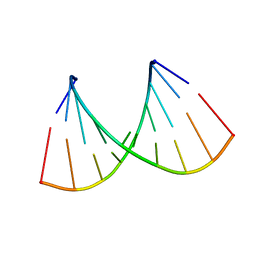
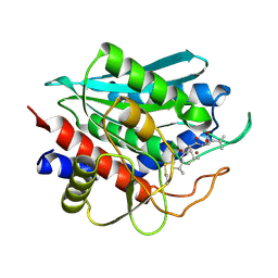
2NCQ
 
 | |
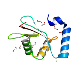
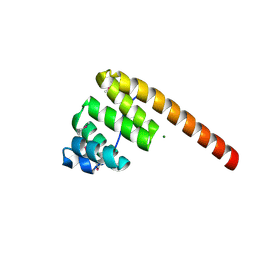
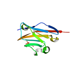
7GAA
 | | PanDDA analysis group deposition -- Crystal Structure of MAP1LC3B in complex with Z1198233191 | | 分子名称: | (4S)-imidazo[1,2-a]pyrimidine-5,7-diol, 1,2-ETHANEDIOL, CHLORIDE ION, ... | | 著者 | Kumar, A, Marples, P.G, Tomlinson, C.W.E, Fearon, D, von-Delft, F, Knapp, S, Structural Genomics Consortium (SGC) | | 登録日 | 2023-08-10 | | 公開日 | 2024-09-04 | | 実験手法 | X-RAY DIFFRACTION (2.03 Å) | | 主引用文献 | PanDDA analysis group deposition
To Be Published
|
|
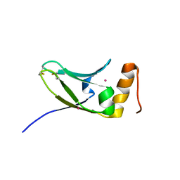
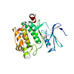
5AX2
 | | Crystal structure of S.cerevisiae Kti11p | | 分子名称: | CADMIUM ION, Diphthamide biosynthesis protein 3 | | 著者 | Kumar, A, Nagarathinam, K, Tanabe, M, Balbach, J. | | 登録日 | 2015-07-13 | | 公開日 | 2016-07-20 | | 最終更新日 | 2024-10-30 | | 実験手法 | X-RAY DIFFRACTION (2.4 Å) | | 主引用文献 | Hyperbolic Pressure-Temperature Phase Diagram of the Zinc-Finger Protein apoKti11 Detected by NMR Spectroscopy.
J Phys Chem B, 123, 2019
|
|
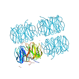
5C9L
 | | Crystal structure of native PLL lectin from Photorhabdus luminescens at 1.65 A resolution | | 分子名称: | 1,2-ETHANEDIOL, CALCIUM ION, CHLORIDE ION, ... | | 著者 | Kumar, A, Sykorova, P, Demo, G, Dobes, P, Hyrsl, P, Wimmerova, M. | | 登録日 | 2015-06-27 | | 公開日 | 2016-10-19 | | 最終更新日 | 2024-11-13 | | 実験手法 | X-RAY DIFFRACTION (1.65 Å) | | 主引用文献 | A Novel Fucose-binding Lectin from Photorhabdus luminescens (PLL) with an Unusual Heptabladed beta-Propeller Tetrameric Structure.
J.Biol.Chem., 291, 2016
|
|
3KK0
 | | Crystal structure of partially folded intermediate state of peptidyl-tRNA hydrolase from Mycobacterium smegmatis | | 分子名称: | 1,2-ETHANEDIOL, BETA-MERCAPTOETHANOL, DI(HYDROXYETHYL)ETHER, ... | | 著者 | Kumar, A, Singh, N, Yadav, R, Sharma, S, Arora, A, Singh, T.P. | | 登録日 | 2009-11-04 | | 公開日 | 2010-11-10 | | 最終更新日 | 2023-11-01 | | 実験手法 | X-RAY DIFFRACTION (2.65 Å) | | 主引用文献 | Structures of Fully-Folded Native and Partially-Folded Intermediate States of Peptidyl-tRNA Hydrolase from Mycobacterium smegmatis
To be Published
|
|
1HXI
 | | AN UNEXPECTED EXTENDED CONFORMATION FOR THE THIRD TPR MOTIF OF THE PEROXIN PEX5 FROM TRYPANOSOMA BRUCEI | | 分子名称: | MAGNESIUM ION, PEROXISOME TARGETING SIGNAL 1 RECEPTOR PEX5 | | 著者 | Kumar, A, Roach, C, Hirsh, I.S, Turley, S, deWalque, S, Michels, P.A.M, Hol, W.G.J. | | 登録日 | 2001-01-15 | | 公開日 | 2001-03-21 | | 最終更新日 | 2024-10-16 | | 実験手法 | X-RAY DIFFRACTION (1.6 Å) | | 主引用文献 | An unexpected extended conformation for the third TPR motif of the peroxin PEX5 from Trypanosoma brucei.
J.Mol.Biol., 307, 2001
|
|
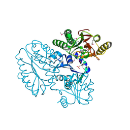
5CDE
 | | R372A mutant of Xaa-Pro dipeptidase from Xanthomonas campestris | | 分子名称: | Proline dipeptidase, SULFATE ION, ZINC ION | | 著者 | Kumar, A, Are, V, Ghosh, B, Jamdar, S, Makde, R. | | 登録日 | 2015-07-03 | | 公開日 | 2016-09-14 | | 最終更新日 | 2024-03-20 | | 実験手法 | X-RAY DIFFRACTION (1.85 Å) | | 主引用文献 | R372A mutant of Xaa-Pro dipeptidase from Xanthomonas campestris at 1.85 Angstrom resolution
To Be Published
|
|
3P9N
 | | Rv2966c of M. tuberculosis is a RsmD-like methyltransferase | | 分子名称: | ACETATE ION, POSSIBLE METHYLTRANSFERASE (METHYLASE) | | 著者 | Kumar, A, Malhotra, K, Saigal, K, Sinha, K.M, Taneja, B. | | 登録日 | 2010-10-18 | | 公開日 | 2011-04-06 | | 最終更新日 | 2023-11-01 | | 実験手法 | X-RAY DIFFRACTION (1.9 Å) | | 主引用文献 | Structural and functional characterization of Rv2966c protein reveals an RsmD-like methyltransferase from Mycobacterium tuberculosis and the role of its N-terminal domain in target recognition
J.Biol.Chem., 286, 2011
|
|
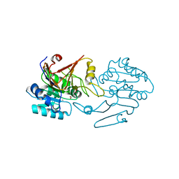
5CNX
 | | Crystal structure of Xaa-Pro aminopeptidase from Escherichia coli K12 | | 分子名称: | Aminopeptidase YpdF, CACODYLATE ION, GLYCEROL, ... | | 著者 | Kumar, A, Are, V, Ghosh, B, Jamdar, S, Makde, R.D. | | 登録日 | 2015-07-18 | | 公開日 | 2016-07-20 | | 最終更新日 | 2023-11-08 | | 実験手法 | X-RAY DIFFRACTION (2.6 Å) | | 主引用文献 | Structures and activities of widely conserved small prokaryotic aminopeptidases-P clarify classification of M24B peptidases
Proteins, 2018
|
|
3DSR
 | | ADP in transition binding site in the subunit B of the energy converter A1Ao ATP synthase | | 分子名称: | ADENOSINE-5'-DIPHOSPHATE, V-type ATP synthase beta chain | | 著者 | Kumar, A, Manimekalai, S.M.S, Balakrishna, A.M, Gruber, G. | | 登録日 | 2008-07-14 | | 公開日 | 2009-06-16 | | 最終更新日 | 2023-11-01 | | 実験手法 | X-RAY DIFFRACTION (2.7 Å) | | 主引用文献 | Structure of the nucleotide-binding subunit B of the energy producer A1A0 ATP synthase in complex with adenosine diphosphate
Acta Crystallogr.,Sect.D, 64, 2008
|
|
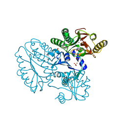
5CDL
 | | Proline dipeptidase from Deinococcus radiodurans (selenomethionine derivative) | | 分子名称: | MANGANESE (II) ION, PHOSPHATE ION, Proline dipeptidase | | 著者 | Kumar, A, Are, V, Ghosh, B, Jamdar, S, Makde, R. | | 登録日 | 2015-07-04 | | 公開日 | 2016-08-10 | | 最終更新日 | 2024-10-30 | | 実験手法 | X-RAY DIFFRACTION (1.8 Å) | | 主引用文献 | Proline dipeptidase from Deinococcus radiodurans (selenomethionine derivative)
To Be Published
|
|
2MS5
 | |
7FFP
 | |
4ERX
 | | Crystal structure of the complex of peptidyl-tRNA hydrolase from Pseudomonas aeruginosa with diethylene glycol at 2.5 Angstrom resolution | | 分子名称: | DI(HYDROXYETHYL)ETHER, Peptidyl-tRNA hydrolase | | 著者 | Kumar, A, Singh, A, Singh, N, Sinha, M, Sharma, S, Arora, A, Singh, T.P. | | 登録日 | 2012-04-21 | | 公開日 | 2012-05-09 | | 最終更新日 | 2023-11-08 | | 実験手法 | X-RAY DIFFRACTION (2.5 Å) | | 主引用文献 | Crystal structure of the complex of peptidyl-tRNA hydrolase from Pseudomonas aeruginosa with diethylene glycol at 2.5 Angstrom resolution
To be Published
|
|
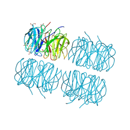
5CDV
 | | Proline dipeptidase from Deinococcus radiodurans R1 | | 分子名称: | MANGANESE (II) ION, PHOSPHATE ION, Proline dipeptidase, ... | | 著者 | Kumar, A, Are, V, Ghosh, B, Jamdar, S, Makde, R. | | 登録日 | 2015-07-05 | | 公開日 | 2016-08-10 | | 最終更新日 | 2023-11-08 | | 実験手法 | X-RAY DIFFRACTION (1.45 Å) | | 主引用文献 | Proline dipeptidase from Deinococcus radiodurans R1 at 1.45 Angstrom resolution
To Be Published
|
|
5C9P
 | | Crystal structure of recombinant PLL lectin complexed with L-fucose from Photorhabdus luminescens at 1.75 A resolution | | 分子名称: | GLYCEROL, PLL lectin, alpha-L-fucopyranose | | 著者 | Kumar, A, Sykorova, P, Demo, G, Dobes, P, Hyrsl, P, Wimmerova, M. | | 登録日 | 2015-06-28 | | 公開日 | 2016-10-19 | | 最終更新日 | 2024-10-23 | | 実験手法 | X-RAY DIFFRACTION (1.75 Å) | | 主引用文献 | A Novel Fucose-binding Lectin from Photorhabdus luminescens (PLL) with an Unusual Heptabladed beta-Propeller Tetrameric Structure.
J.Biol.Chem., 291, 2016
|
|
2IQ6
 | | Crystal Structure of the Aminopeptidase from Vibrio proteolyticus in Complexation with Leucyl-leucyl-leucine. | | 分子名称: | Bacterial leucyl aminopeptidase, Peptide, (Leucyl-leucyl-leucine), ... | | 著者 | Kumar, A, Narayanan, B, Kim, J.-J.P, Bennett, B. | | 登録日 | 2006-10-13 | | 公開日 | 2007-08-28 | | 最終更新日 | 2024-11-20 | | 実験手法 | X-RAY DIFFRACTION (2 Å) | | 主引用文献 | Experimental evidence for a metallohydrolase mechanism in which the nucleophile is not delivered by a metal ion: EPR spectrokinetic and structural studies of aminopeptidase from Vibrio proteolyticus
Biochem.J., 403, 2007
|
|
3P2J
 | | Crystal structure of peptidyl-tRNA hydrolase from Mycobacterium smegmatis at 2.2 A resolution | | 分子名称: | Peptidyl-tRNA hydrolase | | 著者 | Kumar, A, Singh, A, Yadav, R, Sinha, M, Arora, A, Sharma, S, Singh, T.P. | | 登録日 | 2010-10-02 | | 公開日 | 2010-11-17 | | 最終更新日 | 2023-11-01 | | 実験手法 | X-RAY DIFFRACTION (2.22 Å) | | 主引用文献 | Crystal Structure of peptidyl-tRNA hydrolase from Mycobacterium smegmatis at 2.2 A resolution
To be Published
|
|
5C9O
 | | Crystal structure of recombinant PLL lectin from Photorhabdus luminescens at 1.5 A resolution | | 分子名称: | GLYCEROL, PLL lectin | | 著者 | Kumar, A, Sykorova, P, Demo, G, Dobes, P, Hyrsl, P, Wimmerova, M. | | 登録日 | 2015-06-28 | | 公開日 | 2016-10-19 | | 最終更新日 | 2024-10-16 | | 実験手法 | X-RAY DIFFRACTION (1.5 Å) | | 主引用文献 | A Novel Fucose-binding Lectin from Photorhabdus luminescens (PLL) with an Unusual Heptabladed beta-Propeller Tetrameric Structure.
J.Biol.Chem., 291, 2016
|
|
5C23
 | | Parkin (S65DUblR0RBR) | | 分子名称: | CHLORIDE ION, E3 ubiquitin-protein ligase parkin, GLYCEROL, ... | | 著者 | Kumar, A, Aguirre, J.D, Condos, T.E.C, Martinez-Torres, R.J, Chaugule, V.K, Toth, R, Sundaramoorthy, R, Mercier, P, Knebel, A, Spratt, D.E, Barber, K.R, Shaw, G.S, Walden, H. | | 登録日 | 2015-06-15 | | 公開日 | 2015-07-29 | | 最終更新日 | 2024-01-10 | | 実験手法 | X-RAY DIFFRACTION (2.37 Å) | | 主引用文献 | Disruption of the autoinhibited state primes the E3 ligase parkin for activation and catalysis.
Embo J., 34, 2015
|
|
5C1Z
 | | Parkin (UblR0RBR) | | 分子名称: | CHLORIDE ION, E3 ubiquitin-protein ligase parkin, GLYCEROL, ... | | 著者 | kumar, A, Aguirre, J.D, Condos, T.E.C, Martinez-Torres, R.J, Chaugule, V.K, Toth, R, Sundaramoorthy, R, Mercier, P, Knebel, A, Spratt, D.E, Barber, K.R, Shaw, G.S, Walden, H. | | 登録日 | 2015-06-15 | | 公開日 | 2015-07-29 | | 最終更新日 | 2024-01-10 | | 実験手法 | X-RAY DIFFRACTION (1.79 Å) | | 主引用文献 | Disruption of the autoinhibited state primes the E3 ligase parkin for activation and catalysis.
Embo J., 34, 2015
|
|
4K55
 | |
4JKW
 | |
1YXU
 | | Crystal Structure of Kinase Pim1 in Complex with AMP | | 分子名称: | ADENOSINE MONOPHOSPHATE, IMIDAZOLE, Proto-oncogene serine/threonine-protein kinase Pim-1 | | 著者 | Kumar, A, Mandiyan, V, Suzuki, Y, Zhang, C, Rice, J, Tsai, J, Artis, D.R, Ibrahim, P, Bremer, R. | | 登録日 | 2005-02-22 | | 公開日 | 2005-04-26 | | 最終更新日 | 2024-02-14 | | 実験手法 | X-RAY DIFFRACTION (2.24 Å) | | 主引用文献 | Crystal Structures of Proto-oncogene Kinase Pim1: A Target of Aberrant Somatic Hypermutations in Diffuse Large Cell Lymphoma.
J.Mol.Biol., 348, 2005
|
|
1YXX
 | | Crystal Structure of Kinase Pim1 in complex with (3E)-3-[(4-HYDROXYPHENYL)IMINO]-1H-INDOL-2(3H)-ONE | | 分子名称: | (3E)-3-[(4-HYDROXYPHENYL)IMINO]-1H-INDOL-2(3H)-ONE, IMIDAZOLE, Proto-oncogene serine/threonine-protein kinase Pim-1 | | 著者 | Kumar, A, Mandiyan, V, Suzuki, Y, Zhang, C, Rice, J, Tsai, J, Artis, D.R, Ibrahim, P, Bremer, R. | | 登録日 | 2005-02-22 | | 公開日 | 2005-04-26 | | 最終更新日 | 2024-02-14 | | 実験手法 | X-RAY DIFFRACTION (2 Å) | | 主引用文献 | Crystal structures of proto-oncogene kinase Pim1: a target of aberrant somatic hypermutations in diffuse large cell lymphoma.
J.Mol.Biol., 348, 2005
|
|


