Loading
PDBj
MenuPDBj@FacebookPDBj@X(formerly Twitter)PDBj@BlueSkyPDBj@YouTubewwPDB FoundationwwPDB
RCSB PDBPDBeBMRBAdv. SearchSearch help
PDB: 334 results

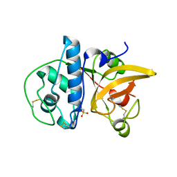
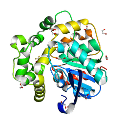
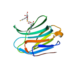
7GAK
DownloadVisualize
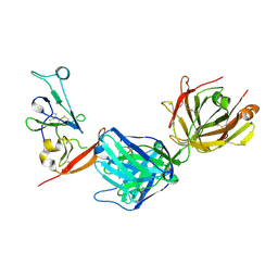
BU of 7gak by Molmil
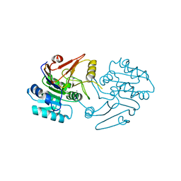
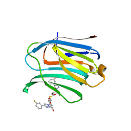
PanDDA analysis group deposition -- Crystal Structure of MAP1LC3B in complex with Z287121492
Descriptor: 1,2-ETHANEDIOL, CHLORIDE ION, Microtubule-associated proteins 1A/1B light chain 3B, ...
Authors:Kumar, A, Marples, P.G, Tomlinson, C.W.E, Fearon, D, von-Delft, F, Knapp, S, Structural Genomics Consortium (SGC)
Deposit date:2023-08-10
Release date:2024-09-04
Method:X-RAY DIFFRACTION (1.77 Å)
Cite:PanDDA analysis group deposition
To Be Published
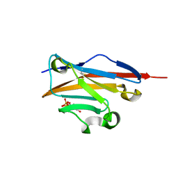
5Y05
DownloadVisualize
BU of 5y05 by Molmil
Structural characterization of msmeg_4306 from Mycobacterium smegmatis
Descriptor: ZINC ION, msmeg_4306
Authors:Kumar, A, Karthikeyan, S.
Deposit date:2017-07-14
Release date:2018-05-02
Last modified:2024-03-27
Method:X-RAY DIFFRACTION (2.799 Å)
Cite:Crystal structure of the MSMEG_4306 gene product from Mycobacterium smegmatis
Acta Crystallogr F Struct Biol Commun, 74, 2018
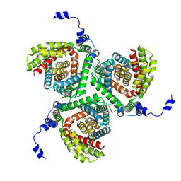
7VRR
DownloadVisualize
BU of 7vrr by Molmil
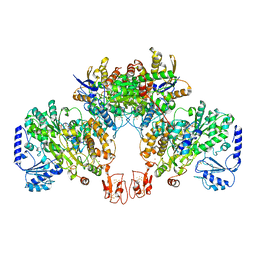
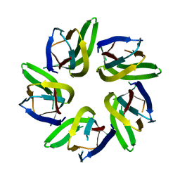
Crystal structure of Arabidopsis thaliana HDT1
Descriptor: Histone deacetylase HDT1
Authors:Kumar, A, Bobde, R.C, Vasudevan, D.
Deposit date:2021-10-23
Release date:2022-09-21
Last modified:2023-11-29
Method:X-RAY DIFFRACTION (2.95 Å)
Cite:Plant-specific HDT family histone deacetylases are nucleoplasmins.
Plant Cell, 34, 2022
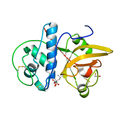
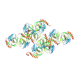
8JCR
DownloadVisualize
BU of 8jcr by Molmil
Crystal structure of Calotropain FI from Calotropis gigantea (pH 6.0)
Descriptor: GLYCEROL, N-[N-[1-HYDROXYCARBOXYETHYL-CARBONYL]LEUCYLAMINO-BUTYL]-GUANIDINE, Procerain, ...
Authors:Kumar, A, Jamdar, S.N, Srivastava, G, Makde, R.D.
Deposit date:2023-05-11
Release date:2024-05-22
Last modified:2024-11-20
Method:X-RAY DIFFRACTION (1.4 Å)
Cite:Crystal structure of Calotropain FI from Calotropis gigantea
To Be Published
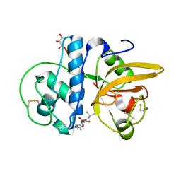
8JCS
DownloadVisualize
BU of 8jcs by Molmil
Crystal structure of Procerain-B from Calotropis gigantea
Descriptor: Procerain B
Authors:Kumar, A, Jamdar, S.N, Srivastava, G, Makde, R.D.
Deposit date:2023-05-11
Release date:2024-05-22
Method:X-RAY DIFFRACTION (1.25 Å)
Cite:Crystal structure of Procerain-B from Calotropis gigantea
To Be Published
8JCQ
DownloadVisualize
BU of 8jcq by Molmil
Crystal structure of calotropain FI from Calotropis gigantea
Descriptor: N-[N-[1-HYDROXYCARBOXYETHYL-CARBONYL]LEUCYLAMINO-BUTYL]-GUANIDINE, Procerain
Authors:Kumar, A, Jamdar, S.N, Srivastava, G, Makde, R.D.
Deposit date:2023-05-11
Release date:2024-05-22
Last modified:2024-11-20
Method:X-RAY DIFFRACTION (1.25 Å)
Cite:Crystal structure of Calotropain FI from Calotropis gigantea
To Be Published
5Y06
DownloadVisualize
BU of 5y06 by Molmil
Structural characterization of msmeg_4306 from Mycobacterium smegmatis
Descriptor: GLYCEROL, ZINC ION, msmeg_4306
Authors:Kumar, A, Karthikeyan, S.
Deposit date:2017-07-14
Release date:2018-05-02
Last modified:2023-11-22
Method:X-RAY DIFFRACTION (2.606 Å)
Cite:Crystal structure of the MSMEG_4306 gene product from Mycobacterium smegmatis
Acta Crystallogr F Struct Biol Commun, 74, 2018
7MWX
DownloadVisualize
BU of 7mwx by Molmil
Structure of the core ectodomain of the hepatitis C virus envelope glycoprotein 2 with tamarin CD81
Descriptor: 2-acetamido-2-deoxy-beta-D-glucopyranose, 2-acetamido-2-deoxy-beta-D-glucopyranose-(1-4)-2-acetamido-2-deoxy-beta-D-glucopyranose, 2A12 Fab Heavy Chain, ...
Authors:Kumar, A, Hossain, R.A, Yost, S.A, Bu, W, Wang, Y, Dearborn, A.D, Grakoui, A, Cohen, J.I, Marcotrigiano, J.
Deposit date:2021-05-17
Release date:2021-09-15
Last modified:2024-10-09
Method:X-RAY DIFFRACTION (3.32 Å)
Cite:Structural insights into hepatitis C virus receptor binding and entry.
Nature, 598, 2021
7MWS
DownloadVisualize
BU of 7mws by Molmil
Crystal structure of tamarin CD81 large extracellular loop
Descriptor: CD81 protein, GLYCEROL, TETRAETHYLENE GLYCOL
Authors:Kumar, A, Hossain, R.A, Yost, S.A, Bu, W, Wang, Y, Dearborn, A.D, Grakoui, A, Cohen, J.I, Marcotrigiano, J.
Deposit date:2021-05-17
Release date:2021-09-15
Last modified:2024-10-16
Method:X-RAY DIFFRACTION (1.8 Å)
Cite:Structural insights into hepatitis C virus receptor binding and entry.
Nature, 598, 2021
7MWW
DownloadVisualize
BU of 7mww by Molmil
Structure of hepatitis C virus envelope full-length glycoprotein 2 (eE2) from J6 genotype
Descriptor: 2-acetamido-2-deoxy-beta-D-glucopyranose, 2-acetamido-2-deoxy-beta-D-glucopyranose-(1-4)-2-acetamido-2-deoxy-beta-D-glucopyranose, 2A12 Fab Heavy chain, ...
Authors:Kumar, A, Hossain, R.A, Yost, S.A, Bu, W, Wang, Y, Dearborn, A.D, Grakoui, A, Cohen, J.I, Marcotrigiano, J.
Deposit date:2021-05-17
Release date:2021-09-15
Last modified:2024-10-30
Method:X-RAY DIFFRACTION (2.71 Å)
Cite:Structural insights into hepatitis C virus receptor binding and entry.
Nature, 598, 2021
6VM0
DownloadVisualize
BU of 6vm0 by Molmil
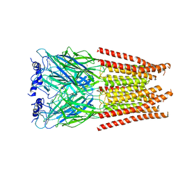
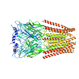
Full length Glycine receptor reconstituted in lipid nanodisc in Gly/IVM-conformation (State-1)
Descriptor: (2aE,4E,5'S,6S,6'R,7S,8E,11R,13R,15S,17aR,20R,20aR,20bS)-6'-[(2S)-butan-2-yl]-20,20b-dihydroxy-5',6,8,19-tetramethyl-17 -oxo-3',4',5',6,6',10,11,14,15,17,17a,20,20a,20b-tetradecahydro-2H,7H-spiro[11,15-methanofuro[4,3,2-pq][2,6]benzodioxacy clooctadecine-13,2'-pyran]-7-yl 2,6-dideoxy-4-O-(2,6-dideoxy-3-O-methyl-alpha-L-arabino-hexopyranosyl)-3-O-methyl-alpha-L-arabino-hexopyranoside, 2-acetamido-2-deoxy-beta-D-glucopyranose-(1-4)-2-acetamido-2-deoxy-beta-D-glucopyranose, GLYCINE, ...
Authors:Kumar, A, Basak, S, Chakrapani, S.
Deposit date:2020-01-27
Release date:2020-07-29
Last modified:2024-03-06
Method:ELECTRON MICROSCOPY (3.14 Å)
Cite:Mechanisms of activation and desensitization of full-length glycine receptor in lipid nanodiscs.
Nat Commun, 11, 2020
6VM2
DownloadVisualize
BU of 6vm2 by Molmil
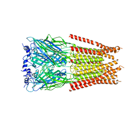
Full length Glycine receptor reconstituted in lipid nanodisc in Gly/IVM-conformation (State-2)
Descriptor: (2aE,4E,5'S,6S,6'R,7S,8E,11R,13R,15S,17aR,20R,20aR,20bS)-6'-[(2S)-butan-2-yl]-20,20b-dihydroxy-5',6,8,19-tetramethyl-17 -oxo-3',4',5',6,6',10,11,14,15,17,17a,20,20a,20b-tetradecahydro-2H,7H-spiro[11,15-methanofuro[4,3,2-pq][2,6]benzodioxacy clooctadecine-13,2'-pyran]-7-yl 2,6-dideoxy-4-O-(2,6-dideoxy-3-O-methyl-alpha-L-arabino-hexopyranosyl)-3-O-methyl-alpha-L-arabino-hexopyranoside, 2-acetamido-2-deoxy-beta-D-glucopyranose-(1-4)-2-acetamido-2-deoxy-beta-D-glucopyranose, GLYCINE, ...
Authors:Kumar, A, Basak, S, Chakrapani, S.
Deposit date:2020-01-27
Release date:2020-07-29
Last modified:2024-03-06
Method:ELECTRON MICROSCOPY (3.34 Å)
Cite:Mechanisms of activation and desensitization of full-length glycine receptor in lipid nanodiscs.
Nat Commun, 11, 2020
6VM3
DownloadVisualize
BU of 6vm3 by Molmil
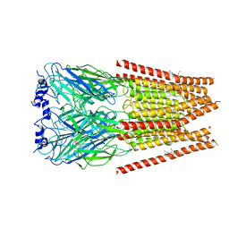
Full length Glycine receptor reconstituted in lipid nanodisc in Gly/IVM-conformation (State-3)
Descriptor: (2aE,4E,5'S,6S,6'R,7S,8E,11R,13R,15S,17aR,20R,20aR,20bS)-6'-[(2S)-butan-2-yl]-20,20b-dihydroxy-5',6,8,19-tetramethyl-17 -oxo-3',4',5',6,6',10,11,14,15,17,17a,20,20a,20b-tetradecahydro-2H,7H-spiro[11,15-methanofuro[4,3,2-pq][2,6]benzodioxacy clooctadecine-13,2'-pyran]-7-yl 2,6-dideoxy-4-O-(2,6-dideoxy-3-O-methyl-alpha-L-arabino-hexopyranosyl)-3-O-methyl-alpha-L-arabino-hexopyranoside, 2-acetamido-2-deoxy-beta-D-glucopyranose-(1-4)-2-acetamido-2-deoxy-beta-D-glucopyranose, GLYCINE, ...
Authors:Kumar, A, Basak, S, Chakrapani, S.
Deposit date:2020-01-27
Release date:2020-07-29
Last modified:2024-03-06
Method:ELECTRON MICROSCOPY (3.07 Å)
Cite:Mechanisms of activation and desensitization of full-length glycine receptor in lipid nanodiscs.
Nat Commun, 11, 2020
8Q53
DownloadVisualize
BU of 8q53 by Molmil
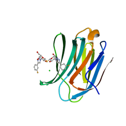
Crystal structure of truncated human Microtubule-associated proteins 1A/1B light chain 3B (MAP1LC3B) in apo form
Descriptor: 1,2-ETHANEDIOL, CHLORIDE ION, Microtubule-associated proteins 1A/1B light chain 3B
Authors:Kumar, A, Knapp, S, Structural Genomics Consortium (SGC)
Deposit date:2023-08-08
Release date:2024-08-21
Method:X-RAY DIFFRACTION (1.36 Å)
Cite:Crystal structure of truncated human Microtubule-associated proteins 1A/1B light chain 3B in apo form
To Be Published
4DJJ
DownloadVisualize
BU of 4djj by Molmil
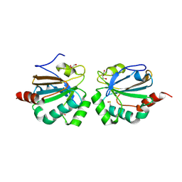
Crystal structure of the complex of Peptidyl-tRNA hydrolase from Pseudomonas aeruginosa with Pimelic acid at 2.9 Angstrom resolution
Descriptor: PIMELIC ACID, Peptidyl-tRNA hydrolase
Authors:Kumar, A, Singh, A, Singh, N, Sinha, M, Sharma, S, Arora, A, Singh, T.P.
Deposit date:2012-02-02
Release date:2012-03-07
Last modified:2023-11-08
Method:X-RAY DIFFRACTION (2.94 Å)
Cite:Crystal structure of the complex of Peptidyl-tRNA hydrolase from Pseudomonas aeruginosa with Pimelic acid at 2.9 Angstrom resolution
To be Published
8A6T
DownloadVisualize
BU of 8a6t by Molmil
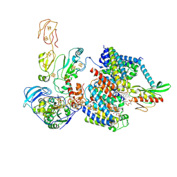
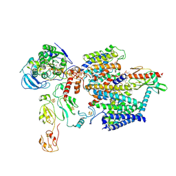
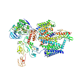
Cryo-EM structure of the electron bifurcating Fe-Fe hydrogenase HydABC complex from Thermoanaerobacter kivui in the reduced state
Descriptor: 2 IRON/2 SULFUR/5 CARBONYL/2 WATER INORGANIC CLUSTER, Electron bifurcating hydrogenase subunit HydA1, Electron bifurcating hydrogenase subunit HydB, ...
Authors:Kumar, A, Saura, P, Gamiz-Hernandez, A.P, Kaila, V.R.I, Mueller, V, Schuller, J.M.
Deposit date:2022-06-19
Release date:2023-02-15
Last modified:2023-03-29
Method:ELECTRON MICROSCOPY (3.1 Å)
Cite:Molecular Basis of the Electron Bifurcation Mechanism in the [FeFe]-Hydrogenase Complex HydABC.
J.Am.Chem.Soc., 145, 2023
8A5E
DownloadVisualize
BU of 8a5e by Molmil
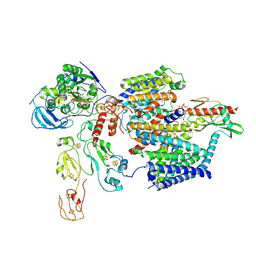
Cryo-EM structure of the electron bifurcating Fe-Fe hydrogenase HydABC complex from Acetobacterium woodii in the reduced state
Descriptor: 1,4-DIHYDRONICOTINAMIDE ADENINE DINUCLEOTIDE, 2 IRON/2 SULFUR/5 CARBONYL/2 WATER INORGANIC CLUSTER, FE2/S2 (INORGANIC) CLUSTER, ...
Authors:Kumar, A, Saura, P, Gamiz-Hernandez, A.P, Kaila, V.R.I, Mueller, V, Schuller, J.M.
Deposit date:2022-06-14
Release date:2023-02-22
Last modified:2023-03-29
Method:ELECTRON MICROSCOPY (3.4 Å)
Cite:Molecular Basis of the Electron Bifurcation Mechanism in the [FeFe]-Hydrogenase Complex HydABC.
J.Am.Chem.Soc., 145, 2023
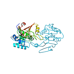
5FCF
DownloadVisualize
BU of 5fcf by Molmil
Crystal Structure of Xaa-Pro dipeptidase from Xanthomonas campestris, phosphate and Mn bound
Descriptor: DI(HYDROXYETHYL)ETHER, GLY-GLY-GLY, GLYCEROL, ...
Authors:Kumar, A, Are, V, Ghosh, B, Jamdar, S, Makde, R.D.
Deposit date:2015-12-15
Release date:2016-12-07
Last modified:2024-03-20
Method:X-RAY DIFFRACTION (1.85 Å)
Cite:Crystal structure and biochemical investigations reveal novel mode of substrate selectivity and illuminate substrate inhibition and allostericity in a subfamily of Xaa-Pro dipeptidases.
Biochim. Biophys. Acta, 1865, 2017
4DHW
DownloadVisualize
BU of 4dhw by Molmil
Crystal structure of Peptidyl-tRNA hydrolase from Pseudomonas aeruginosa with Adipic acid at 2.4 Angstrom resolution
Descriptor: Peptidyl-tRNA hydrolase, hexanedioic acid
Authors:Kumar, A, Singh, A, Singh, N, Sinha, M, Sharma, S, Arora, A, Singh, T.P.
Deposit date:2012-01-30
Release date:2012-02-29
Last modified:2023-11-08
Method:X-RAY DIFFRACTION (2.43 Å)
Cite:Crystal structure of Peptidyl-tRNA hydrolase from Pseudomonas aeruginosa with Adipic acid at 2.4 Angstrom resolution
To be Published
4JKW
DownloadVisualize
BU of 4jkw by Molmil
Structure of the extracellular domain of butyrophilin BTN3A1 in complex with Isopentenyl pyrophosphate (IPP)
Descriptor: 3-METHYLBUT-3-ENYL TRIHYDROGEN DIPHOSPHATE, Butyrophilin subfamily 3 member A1
Authors:Kumar, A, Mori, L, De Libero, G.
Deposit date:2013-03-12
Release date:2013-07-24
Last modified:2024-10-09
Method:X-RAY DIFFRACTION (2.01 Å)
Cite:Butyrophilin 3A1 binds phosphorylated antigens and stimulates human gamma delta T cells.
Nat.Immunol., 14, 2013
4K55
DownloadVisualize
BU of 4k55 by Molmil
Structure of the extracellular domain of butyrophilin BTN3A1 in complex with (E)-4-hydroxy-3-methyl-but-2-enyl pyrophosphate (HMBPP)
Descriptor: (2E)-4-hydroxy-3-methylbut-2-en-1-yl trihydrogen diphosphate, Butyrophilin subfamily 3 member A1
Authors:Kumar, A, Mori, L, De Libero, G.
Deposit date:2013-04-13
Release date:2013-07-24
Last modified:2013-09-25
Method:X-RAY DIFFRACTION (1.91 Å)
Cite:Butyrophilin 3A1 binds phosphorylated antigens and stimulates human gamma delta T cells.
Nat.Immunol., 14, 2013
1CEH
DownloadVisualize
BU of 1ceh by Molmil
STRUCTURE AND FUNCTION OF THE CATALYTIC SITE MUTANT ASP99ASN OF PHOSPHOLIPASE A2: ABSENCE OF CONSERVED STRUCTURAL WATER
Descriptor: CALCIUM ION, PHOSPHOLIPASE A2
Authors:Kumar, A, Sekharudu, C, Ramakrishnan, B, Dupureur, C.M, Zhu, H, Tsai, M.-D, Sundaralingam, M.
Deposit date:1994-11-16
Release date:1995-02-07
Last modified:2024-11-20
Method:X-RAY DIFFRACTION (1.9 Å)
Cite:Structure and function of the catalytic site mutant Asp 99 Asn of phospholipase A2: absence of the conserved structural water.
Protein Sci., 3, 1994
7GAA
DownloadVisualize
BU of 7gaa by Molmil
PanDDA analysis group deposition -- Crystal Structure of MAP1LC3B in complex with Z1198233191
Descriptor: (4S)-imidazo[1,2-a]pyrimidine-5,7-diol, 1,2-ETHANEDIOL, CHLORIDE ION, ...
Authors:Kumar, A, Marples, P.G, Tomlinson, C.W.E, Fearon, D, von-Delft, F, Knapp, S, Structural Genomics Consortium (SGC)
Deposit date:2023-08-10
Release date:2024-09-04
Method:X-RAY DIFFRACTION (2.03 Å)
Cite:PanDDA analysis group deposition
To Be Published
6JMI
DownloadVisualize
BU of 6jmi by Molmil
Crystal structure of M.tuberculosis Rv0081
Descriptor: SULFATE ION, Uncharacterized HTH-type transcriptional regulator Rv0081
Authors:Kumar, A, Phulera, S, Mande, C.S.
Deposit date:2019-03-11
Release date:2019-04-10
Last modified:2024-03-27
Method:X-RAY DIFFRACTION (2.896 Å)
Cite:Structural basis of hypoxic gene regulation by the Rv0081 transcription factor of Mycobacterium tuberculosis.
Febs Lett., 593, 2019
7UCC
DownloadVisualize
BU of 7ucc by Molmil
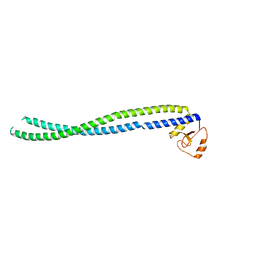
Transcription factor FosB/JunD bZIP domain in the reduced form
Descriptor: CHLORIDE ION, ETHANOL, Protein fosB, ...
Authors:Kumar, A, Machius, M.C, Rudenko, G.
Deposit date:2022-03-16
Release date:2023-01-25
Last modified:2023-10-25
Method:X-RAY DIFFRACTION (1.94 Å)
Cite:Chemically targeting the redox switch in AP1 transcription factor Delta FOSB.
Nucleic Acids Res., 50, 2022

227561

PDB entries from 2024-11-20

PDB statisticsPDBj update infoContact PDBjnumon