7L0Q
 
 | | Structure of NTS-NTSR1-Gi complex in lipid nanodisc, canonical state, with AHD | | 分子名称: | Guanine nucleotide-binding protein G(I)/G(S)/G(T) subunit beta-1, Guanine nucleotide-binding protein G(T) subunit gamma-T1, Guanine nucleotide-binding protein G(i) subunit alpha-1, ... | | 著者 | Zhang, M, Gui, M, Wang, Z, Gorgulla, C, Yu, J.J, Wu, H, Sun, Z, Klenk, C, Merklinger, L, Morstein, L, Hagn, F, Pluckthun, A, Brown, A, Nasr, M.L, Wagner, G. | | 登録日 | 2020-12-12 | | 公開日 | 2021-01-06 | | 最終更新日 | 2021-03-31 | | 実験手法 | ELECTRON MICROSCOPY (4.3 Å) | | 主引用文献 | Cryo-EM structure of an activated GPCR-G protein complex in lipid nanodiscs.
Nat.Struct.Mol.Biol., 28, 2021
|
|
7L0R
 | | Structure of NTS-NTSR1-Gi complex in lipid nanodisc, noncanonical state, without AHD | | 分子名称: | Guanine nucleotide-binding protein G(I)/G(S)/G(T) subunit beta-1, Guanine nucleotide-binding protein G(T) subunit gamma-T1, Guanine nucleotide-binding protein G(i) subunit alpha-1, ... | | 著者 | Zhang, M, Gui, M, Wang, Z, Gorgulla, C, Yu, J.J, Wu, H, Sun, Z, Klenk, C, Merklinger, L, Morstein, L, Hagn, F, Pluckthun, A, Brown, A, Nasr, M.L, Wagner, G. | | 登録日 | 2020-12-12 | | 公開日 | 2021-01-06 | | 最終更新日 | 2021-03-24 | | 実験手法 | ELECTRON MICROSCOPY (4.2 Å) | | 主引用文献 | Cryo-EM structure of an activated GPCR-G protein complex in lipid nanodiscs.
Nat.Struct.Mol.Biol., 28, 2021
|
|
7L0P
 | | Structure of NTS-NTSR1-Gi complex in lipid nanodisc, canonical state, without AHD | | 分子名称: | Guanine nucleotide-binding protein G(I)/G(S)/G(T) subunit beta-1, Guanine nucleotide-binding protein G(T) subunit gamma-T1, Guanine nucleotide-binding protein G(i) subunit alpha-1, ... | | 著者 | Zhang, M, Gui, M, Wang, Z, Gorgulla, C, Yu, J.J, Wu, H, Sun, Z, Klenk, C, Merklinger, L, Morstein, L, Hagn, F, Pluckthun, A, Brown, A, Nasr, M.L, Wagner, G. | | 登録日 | 2020-12-12 | | 公開日 | 2021-01-06 | | 最終更新日 | 2021-03-24 | | 実験手法 | ELECTRON MICROSCOPY (4.1 Å) | | 主引用文献 | Cryo-EM structure of an activated GPCR-G protein complex in lipid nanodiscs.
Nat.Struct.Mol.Biol., 28, 2021
|
|
7L0S
 | | Structure of NTS-NTSR1-Gi complex in lipid nanodisc, noncanonical state, with AHD | | 分子名称: | Guanine nucleotide-binding protein G(I)/G(S)/G(T) subunit beta-1, Guanine nucleotide-binding protein G(T) subunit gamma-T1, Guanine nucleotide-binding protein G(i) subunit alpha-1, ... | | 著者 | Zhang, M, Gui, M, Wang, Z, Gorgulla, C, Yu, J.J, Wu, H, Sun, Z, Klenk, C, Merklinger, L, Morstein, L, Hagn, F, Pluckthun, A, Brown, A, Nasr, M.L, Wagner, G. | | 登録日 | 2020-12-12 | | 公開日 | 2021-01-06 | | 最終更新日 | 2021-03-24 | | 実験手法 | ELECTRON MICROSCOPY (4.5 Å) | | 主引用文献 | Cryo-EM structure of an activated GPCR-G protein complex in lipid nanodiscs.
Nat.Struct.Mol.Biol., 28, 2021
|
|
5XED
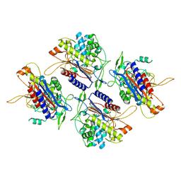
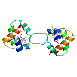 | | Heterodimer constructed from M61A PA cyt c551-HT cyt c552 and HT cyt c552-PA cyt c551 chimeric proteins | | 分子名称: | Cytochrome c-551,Cytochrome c-552, Cytochrome c-552,Cytochrome c-551, HEME C | | 著者 | Zhang, M, Nakanishi, T, Yamanaka, M, Nagao, S, Yanagisawa, S, Shomura, Y, Shibata, N, Ogura, T, Higuchi, Y, Hirota, S. | | 登録日 | 2017-04-04 | | 公開日 | 2017-08-09 | | 最終更新日 | 2023-11-22 | | 実験手法 | X-RAY DIFFRACTION (1.55 Å) | | 主引用文献 | Rational Design of Domain-Swapping-Based c-Type Cytochrome Heterodimers by Using Chimeric Proteins.
Chembiochem, 18, 2017
|
|
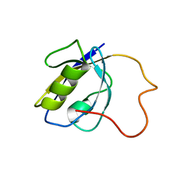
7LH9
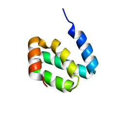
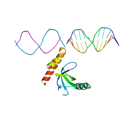 | | Crystal structure of BRPF2 PWWP domain in complex with DNA | | 分子名称: | Bromodomain-containing protein 1, DNA | | 著者 | Zhang, M, Lei, M, Qin, S, Dong, A, Yang, A, Li, Y, Loppnau, P, Hughes, T.R, Arrowsmith, C.H, Edwards, A.M, Min, J, Liu, J, Structural Genomics Consortium (SGC) | | 登録日 | 2021-01-21 | | 公開日 | 2021-02-17 | | 最終更新日 | 2023-10-18 | | 実験手法 | X-RAY DIFFRACTION (2.6 Å) | | 主引用文献 | Crystal structure of the BRPF2 PWWP domain in complex with DNA reveals a different binding mode than the HDGF family of PWWP domains.
Biochim Biophys Acta Gene Regul Mech, 1864, 2021
|
|
1T3B
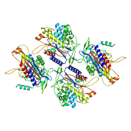
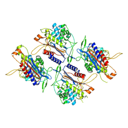
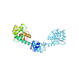 | | X-ray Structure of DsbC from Haemophilus influenzae | | 分子名称: | Thiol:disulfide interchange protein dsbC | | 著者 | Zhang, M, Monzingo, A.F, Segatori, L, Georgiou, G, Robertus, J.D. | | 登録日 | 2004-04-26 | | 公開日 | 2004-09-07 | | 最終更新日 | 2023-08-23 | | 実験手法 | X-RAY DIFFRACTION (2.5 Å) | | 主引用文献 | Structure of DsbC from Haemophilus influenzae.
Acta Crystallogr.,Sect.D, 60, 2004
|
|
7WWV
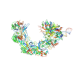 | | DNA bound-ICP1 Csy complex | | 分子名称: | Csy1, Csy2, Csy3, ... | | 著者 | Zhang, M, Peng, R. | | 登録日 | 2022-02-14 | | 公開日 | 2023-04-26 | | 最終更新日 | 2024-06-26 | | 実験手法 | ELECTRON MICROSCOPY (3.2 Å) | | 主引用文献 | Mechanistic insights into DNA binding and cleavage by a compact type I-F CRISPR-Cas system in bacteriophage.
Proc.Natl.Acad.Sci.USA, 120, 2023
|
|
2LA8
 | |
6A0W
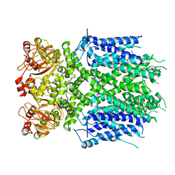
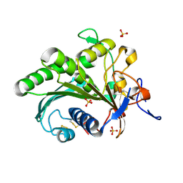 | | Crystal structure of lipase from Rhizopus microsporus var. chinensis | | 分子名称: | Lipase, SULFATE ION | | 著者 | Zhang, M, Yu, X.W, Xu, Y, Huang, C.H, Guo, R.T. | | 登録日 | 2018-06-06 | | 公開日 | 2019-10-09 | | 最終更新日 | 2023-11-22 | | 実験手法 | X-RAY DIFFRACTION (2 Å) | | 主引用文献 | Structural Basis by Which the N-Terminal Polypeptide Segment ofRhizopus chinensisLipase Regulates Its Substrate Binding Affinity.
Biochemistry, 58, 2019
|
|
8IO5
 | | Herg1a-herg1b closed state 2 | | 分子名称: | Potassium voltage-gated channel subfamily H member 2 | | 著者 | Zhang, M.F. | | 登録日 | 2023-03-10 | | 公開日 | 2024-03-13 | | 実験手法 | ELECTRON MICROSCOPY (3.8 Å) | | 主引用文献 | Structure of Herg1a-herg1b open state
To Be Published
|
|
8IOB
 | | Herg1a-herg1b closed state 1 | | 分子名称: | Potassium voltage-gated channel subfamily H member 2 | | 著者 | Zhang, M.F. | | 登録日 | 2023-03-10 | | 公開日 | 2024-03-13 | | 実験手法 | ELECTRON MICROSCOPY (3.9 Å) | | 主引用文献 | Structure of Herg1a-herg1b closed state 1
To Be Published
|
|
8IO4
 | | Herg1a-herg1b open state | | 分子名称: | Potassium voltage-gated channel subfamily H member 2 | | 著者 | Zhang, M.F. | | 登録日 | 2023-03-10 | | 公開日 | 2024-03-13 | | 実験手法 | ELECTRON MICROSCOPY (3.5 Å) | | 主引用文献 | Structure of Herg1a-herg1b open state
To Be Published
|
|
2KBQ
 | |
7VRD
 | |
7V67
 | |
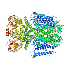
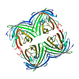
7V1Z
 | | human Serine beta-lactamase-like protein LACTB | | 分子名称: | Serine beta-lactamase-like protein LACTB, mitochondrial | | 著者 | Zhang, M.H, Yang, M.J. | | 登録日 | 2021-08-07 | | 公開日 | 2022-02-16 | | 最終更新日 | 2024-06-12 | | 実験手法 | ELECTRON MICROSCOPY (2.98 Å) | | 主引用文献 | Structural basis for the catalytic activity of filamentous human serine beta-lactamase-like protein LACTB.
Structure, 30, 2022
|
|
7V21
 | |
7V1Y
 | |
7V2J
 | | Crystal Structure of the first bromodomain of human BRD4 in complex with the inhibitor 33 | | 分子名称: | Bromodomain-containing protein 4, ~{N}-(3-ethyl-6-methoxy-1,2-benzoxazol-5-yl)-4-methoxy-benzenesulfonamide | | 著者 | Zhang, M, Wang, C, Zhang, C, Zhang, Y, Xu, Y. | | 登録日 | 2021-08-09 | | 公開日 | 2022-08-17 | | 最終更新日 | 2023-11-29 | | 実験手法 | X-RAY DIFFRACTION (2.24 Å) | | 主引用文献 | Design, synthesis and pharmacological characterization of N-(3-ethylbenzo[d]isoxazol-5-yl) sulfonamide derivatives as BRD4 inhibitors against acute myeloid leukemia.
Acta Pharmacol.Sin., 43, 2022
|
|
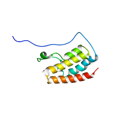
7CSR
 | | Structure of Ephexin4 R676L | | 分子名称: | Rho guanine nucleotide exchange factor 16 | | 著者 | Zhang, M, Lin, L, Wang, C, Zhu, J. | | 登録日 | 2020-08-17 | | 公開日 | 2021-02-24 | | 最終更新日 | 2023-11-29 | | 実験手法 | X-RAY DIFFRACTION (3 Å) | | 主引用文献 | Double inhibition and activation mechanisms of Ephexin family RhoGEFs.
Proc.Natl.Acad.Sci.USA, 118, 2021
|
|
7V1U
 | | Crystal Structure of the first bromodomain of human BRD4 in complex with the inhibitor ZJ12 | | 分子名称: | 1,2-ETHANEDIOL, Bromodomain-containing protein 4, ~{N}-(3-ethyl-6-methoxy-1,2-benzoxazol-5-yl)-2-methoxy-benzenesulfonamide | | 著者 | Zhang, M, Wang, C, Zhang, C, Zhang, Y, Xu, Y. | | 登録日 | 2021-08-06 | | 公開日 | 2022-08-10 | | 最終更新日 | 2023-11-29 | | 実験手法 | X-RAY DIFFRACTION (1.82 Å) | | 主引用文献 | Design, synthesis and pharmacological characterization of N-(3-ethylbenzo[d]isoxazol-5-yl) sulfonamide derivatives as BRD4 inhibitors against acute myeloid leukemia.
Acta Pharmacol.Sin., 43, 2022
|
|
3S05
 | |
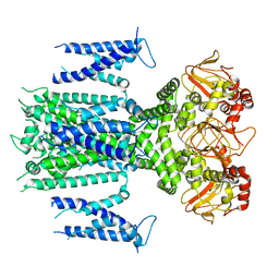
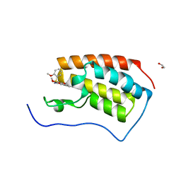
7CSO
 | | Structure of Ephexin4 DH-PH-SH3 | | 分子名称: | Rho guanine nucleotide exchange factor 16, SULFATE ION | | 著者 | Zhang, M, Lin, L, Wang, C, Zhu, J. | | 登録日 | 2020-08-15 | | 公開日 | 2021-02-24 | | 最終更新日 | 2024-03-27 | | 実験手法 | X-RAY DIFFRACTION (2.39 Å) | | 主引用文献 | Double inhibition and activation mechanisms of Ephexin family RhoGEFs.
Proc.Natl.Acad.Sci.USA, 118, 2021
|
|
4KKY
 | |


