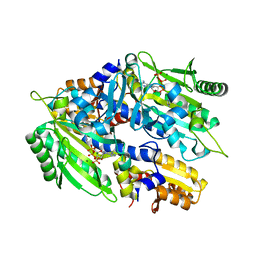
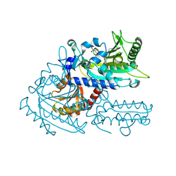
3SPZ
 
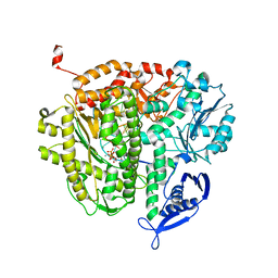 | | DNA Polymerase(L415A/L561A/S565G/Y567A) Ternary Complex with dUpCpp Opposite dA (Ca2+) | | 分子名称: | 2'-deoxy-5'-O-[(R)-hydroxy{[(R)-hydroxy(phosphonooxy)phosphoryl]methyl}phosphoryl]uridine, 5'-D(*GP*CP*GP*GP*AP*CP*TP*GP*CP*TP*TP*AP*C)-3', 5'-D(P*GP*AP*GP*TP*AP*AP*GP*CP*AP*GP*TP*CP*CP*GP*CP*G)-3', ... | | 著者 | Xia, S, Konigsberg, W.H, Wang, J. | | 登録日 | 2011-07-04 | | 公開日 | 2011-10-12 | | 最終更新日 | 2024-02-28 | | 実験手法 | X-RAY DIFFRACTION (2.43 Å) | | 主引用文献 | Structural Insights into Complete Metal Ion Coordination from Ternary Complexes of B Family RB69 DNA Polymerase.
Biochemistry, 50, 2011
|
|
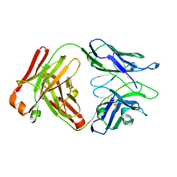
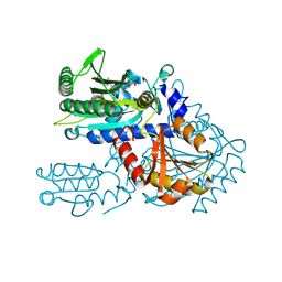
2O9Q
 | | The crystal structure of Bovine Trypsin complexed with a small inhibition peptide ORB2K | | 分子名称: | CALCIUM ION, Cationic trypsin, ORB2K, ... | | 著者 | Li, J, Zhang, C, Xu, X, Wang, J, Gong, W, Lai, R. | | 登録日 | 2006-12-14 | | 公開日 | 2007-12-25 | | 最終更新日 | 2023-10-25 | | 実験手法 | X-RAY DIFFRACTION (1.7 Å) | | 主引用文献 | From protease inhibitor to antibiotics: single point mutation makes tremendous functional shift
To be Published
|
|
4DNV
 | | Crystal structure of the W285F mutant of UVB-resistance protein UVR8 | | 分子名称: | AT5g63860/MGI19_6 | | 著者 | Wu, D, Hu, Q, Yan, Z, Chen, W, Yan, C, Zhang, J, Wang, J, Shi, Y. | | 登録日 | 2012-02-09 | | 公開日 | 2012-03-07 | | 最終更新日 | 2023-11-08 | | 実験手法 | X-RAY DIFFRACTION (1.999 Å) | | 主引用文献 | Structural basis of ultraviolet-B perception by UVR8.
Nature, 484, 2012
|
|
8J23
 | | Cryo-EM structure of FFAR2 complex in apo state | | 分子名称: | Free fatty acid receptor 2, Guanine nucleotide-binding protein G(I)/G(S)/G(O) subunit gamma-2, Guanine nucleotide-binding protein G(I)/G(S)/G(T) subunit beta-1, ... | | 著者 | Tai, L, Li, F, Sun, X, Tang, W, Wang, J. | | 登録日 | 2023-04-14 | | 公開日 | 2024-04-17 | | 実験手法 | ELECTRON MICROSCOPY (3.2 Å) | | 主引用文献 | Molecular recognition and activation mechanism of short chain fatty acid receptors FFAR2 and FFAR3
To Be Published
|
|
6WGK
 | | Fab portion of dupilumab with Crystal Kappa design and intrachain disulfide | | 分子名称: | Dupilumab Fab heavy chain, Dupilumab Fab light chain | | 著者 | Druzina, Z, Atwell, S, Pustilnik, A, Antonysamy, S, Ho, C, Lieu, R, Hendle, J, Benach, J, Wang, J. | | 登録日 | 2020-04-05 | | 公開日 | 2020-09-16 | | 最終更新日 | 2023-10-18 | | 実験手法 | X-RAY DIFFRACTION (1.62 Å) | | 主引用文献 | Rapid and robust antibody Fab fragment crystallization utilizing edge-to-edge beta-sheet packing.
Plos One, 15, 2020
|
|
6WGL
 | | Dupilumab fab with Crystal Kappa design complexed with human IL-4 receptor | | 分子名称: | 2-acetamido-2-deoxy-beta-D-glucopyranose, 2-acetamido-2-deoxy-beta-D-glucopyranose-(1-4)-2-acetamido-2-deoxy-beta-D-glucopyranose, Dupilumab Fab heavy chain, ... | | 著者 | Druzina, Z, Atwell, S, Pustilnik, A, Antonysamy, S, Ho, C, Lieu, R, Hendle, J, Benach, J, Wang, J. | | 登録日 | 2020-04-05 | | 公開日 | 2020-09-16 | | 最終更新日 | 2023-10-18 | | 実験手法 | X-RAY DIFFRACTION (2.82 Å) | | 主引用文献 | Rapid and robust antibody Fab fragment crystallization utilizing edge-to-edge beta-sheet packing.
Plos One, 15, 2020
|
|
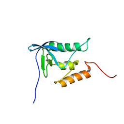
1Y9X
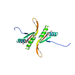 | |
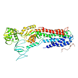
3SQ0
 | | DNA Polymerase(L561A/S565G/Y567A) Ternary Complex with dUpNpp Opposite dA (Mn2+) | | 分子名称: | 2'-DEOXYURIDINE 5'-ALPHA,BETA-IMIDO-TRIPHOSPHATE, 5'-D(*GP*CP*GP*GP*AP*CP*TP*GP*CP*TP*TP*AP*(DOC))-3', 5'-D(*TP*CP*AP*AP*GP*TP*AP*AP*GP*CP*AP*GP*TP*CP*CP*GP*CP*G)-3', ... | | 著者 | Xia, S, Konigsberg, W.H, Wang, J. | | 登録日 | 2011-07-04 | | 公開日 | 2011-10-12 | | 最終更新日 | 2024-02-28 | | 実験手法 | X-RAY DIFFRACTION (2 Å) | | 主引用文献 | Structural Insights into Complete Metal Ion Coordination from Ternary Complexes of B Family RB69 DNA Polymerase.
Biochemistry, 50, 2011
|
|
4ZYN
 | | Crystal Structure of Parkin E3 ubiquitin ligase (linker deletion; delta 86-130) | | 分子名称: | E3 ubiquitin-protein ligase parkin, SULFATE ION, ZINC ION | | 著者 | Lilov, A, Sauve, V, Trempe, J.F, Rodionov, D, Wang, J, Gehring, K. | | 登録日 | 2015-05-21 | | 公開日 | 2015-08-19 | | 最終更新日 | 2023-09-27 | | 実験手法 | X-RAY DIFFRACTION (2.54 Å) | | 主引用文献 | A Ubl/ubiquitin switch in the activation of Parkin.
Embo J., 34, 2015
|
|
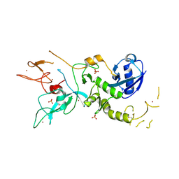
2A5J
 | | Crystal Structure of Human RAB2B | | 分子名称: | GUANOSINE-5'-DIPHOSPHATE, MAGNESIUM ION, Ras-related protein Rab-2B | | 著者 | Dong, A, Wang, J, Shen, Y, Arrowsmith, C.H, Edwards, A.M, Sundstrom, M, Bochkarev, A, Park, H.W, Structural Genomics Consortium (SGC) | | 登録日 | 2005-06-30 | | 公開日 | 2005-07-19 | | 最終更新日 | 2023-08-23 | | 実験手法 | X-RAY DIFFRACTION (1.501 Å) | | 主引用文献 | Crystal structure of human RAB2B
To be Published
|
|
8H99
 | | Crystal structure of E. coli ThrS catalytic domain mutant | | 分子名称: | ADENOSINE-5'-TRIPHOSPHATE, IMIDAZOLE, MAGNESIUM ION, ... | | 著者 | Qiao, H, Xia, M, Wang, J, Fang, P. | | 登録日 | 2022-10-25 | | 公開日 | 2023-02-08 | | 最終更新日 | 2023-10-25 | | 実験手法 | X-RAY DIFFRACTION (1.94 Å) | | 主引用文献 | Tyrosine-targeted covalent inhibition of a tRNA synthetase aided by zinc ion.
Commun Biol, 6, 2023
|
|
8H9C
 | | Crystal structure of chemically modified E. coli ThrS catalytic domain 4 | | 分子名称: | N-(2,3-dihydroxybenzoyl)-4-(4-nitrophenyl)-L-threonine, Threonine--tRNA ligase, ZINC ION | | 著者 | Qiao, H, Xia, M, Wang, J, Fang, P. | | 登録日 | 2022-10-25 | | 公開日 | 2023-02-08 | | 最終更新日 | 2023-10-25 | | 実験手法 | X-RAY DIFFRACTION (2.15 Å) | | 主引用文献 | Tyrosine-targeted covalent inhibition of a tRNA synthetase aided by zinc ion.
Commun Biol, 6, 2023
|
|
8H98
 | | Crystal structure of chemically modified E. coli ThrS catalytic domain 1 | | 分子名称: | N-(2,3-dihydroxybenzoyl)-4-(4-nitrophenyl)-L-threonine, Threonine--tRNA ligase, ZINC ION | | 著者 | Qiao, H, Xia, M, Wang, J, Fang, P. | | 登録日 | 2022-10-25 | | 公開日 | 2023-02-08 | | 最終更新日 | 2023-10-25 | | 実験手法 | X-RAY DIFFRACTION (2.5 Å) | | 主引用文献 | Tyrosine-targeted covalent inhibition of a tRNA synthetase aided by zinc ion.
Commun Biol, 6, 2023
|
|
8H9B
 | | Crystal structure of chemically modified E. coli ThrS catalytic domain 3 | | 分子名称: | N-(2,3-dihydroxybenzoyl)-4-(4-nitrophenyl)-L-threonine, Threonine--tRNA ligase, ZINC ION | | 著者 | Qiao, H, Xia, M, Wang, J, Fang, P. | | 登録日 | 2022-10-25 | | 公開日 | 2023-02-08 | | 最終更新日 | 2023-10-25 | | 実験手法 | X-RAY DIFFRACTION (2.23 Å) | | 主引用文献 | Tyrosine-targeted covalent inhibition of a tRNA synthetase aided by zinc ion.
Commun Biol, 6, 2023
|
|
8H9A
 | | Crystal structure of chemically modified E. coli ThrS catalytic domain 2 | | 分子名称: | N-(2,3-dihydroxybenzoyl)-4-(4-nitrophenyl)-L-threonine, Threonine--tRNA ligase, ZINC ION | | 著者 | Qiao, H, Xia, M, Wang, J, Fang, P. | | 登録日 | 2022-10-25 | | 公開日 | 2023-02-08 | | 最終更新日 | 2023-10-25 | | 実験手法 | X-RAY DIFFRACTION (1.9 Å) | | 主引用文献 | Tyrosine-targeted covalent inhibition of a tRNA synthetase aided by zinc ion.
Commun Biol, 6, 2023
|
|
8WGF
 | | The Crystal Structure of JNK3 from Biortus. | | 分子名称: | MAGNESIUM ION, Mitogen-activated protein kinase 10, PHOSPHOAMINOPHOSPHONIC ACID-ADENYLATE ESTER | | 著者 | Wang, F, Cheng, W, Lv, Z, Ju, C, Wang, J. | | 登録日 | 2023-09-21 | | 公開日 | 2023-11-22 | | 実験手法 | X-RAY DIFFRACTION (1.85 Å) | | 主引用文献 | The Crystal Structure of JNK3 from Biortus.
To Be Published
|
|
8WGQ
 | | The Crystal Structure of L-asparaginase from Biortus. | | 分子名称: | 1,2-ETHANEDIOL, GLYCEROL, L-asparaginase | | 著者 | Wang, F, Cheng, W, Lv, Z, Ju, C, Wang, J. | | 登録日 | 2023-09-22 | | 公開日 | 2023-11-22 | | 実験手法 | X-RAY DIFFRACTION (2.75 Å) | | 主引用文献 | The Crystal Structure of L-asparaginase from Biortus.
To Be Published
|
|
8X70
 | | The Crystal Structure of IFI16 from Biortus. | | 分子名称: | 1,2-ETHANEDIOL, BROMIDE ION, Gamma-interferon-inducible protein 16, ... | | 著者 | Wang, F, Cheng, W, Lv, Z, Meng, Q, Wang, J. | | 登録日 | 2023-11-22 | | 公開日 | 2023-12-27 | | 実験手法 | X-RAY DIFFRACTION (1.7 Å) | | 主引用文献 | The Crystal Structure of IFI16 from Biortus.
To Be Published
|
|
8XOX
 | | The Crystal Structure of FAK2 from Biortus. | | 分子名称: | 1,2-ETHANEDIOL, N-methyl-N-{3-[({2-[(2-oxo-2,3-dihydro-1H-indol-5-yl)amino]-5-(trifluoromethyl)pyrimidin-4-yl}amino)methyl]pyridin-2-yl}methanesulfonamide, Protein-tyrosine kinase 2-beta | | 著者 | Wang, F, Cheng, W, Lv, Z, Ju, C, Wang, J. | | 登録日 | 2024-01-02 | | 公開日 | 2024-01-24 | | 実験手法 | X-RAY DIFFRACTION (1.9 Å) | | 主引用文献 | The Crystal Structure of FAK2 from Biortus.
To Be Published
|
|
8XPN
 | | The Crystal Structure of USP8 from Biortus. | | 分子名称: | 1,2-ETHANEDIOL, DI(HYDROXYETHYL)ETHER, Ubiquitin carboxyl-terminal hydrolase 8, ... | | 著者 | Wang, F, Cheng, W, Yuan, Z, Lin, D, Wang, J. | | 登録日 | 2024-01-04 | | 公開日 | 2024-03-06 | | 実験手法 | X-RAY DIFFRACTION (2.1 Å) | | 主引用文献 | The Crystal Structure of USP8 from Biortus.
To Be Published
|
|
8YGZ
 | | The Crystal Structure of TGF beta R2 kinase domain from Biortus. | | 分子名称: | 1,2-ETHANEDIOL, DI(HYDROXYETHYL)ETHER, TGF-beta receptor type-2 | | 著者 | Wang, F, Cheng, W, Lv, Z, Ju, C, Wang, J. | | 登録日 | 2024-02-27 | | 公開日 | 2024-03-13 | | 実験手法 | X-RAY DIFFRACTION (2.1 Å) | | 主引用文献 | The Crystal Structure of TGF beta R2 kinase domain from Biortus.
To Be Published
|
|
8YHP
 | | Structure of the PGK1 from Biortus. | | 分子名称: | 1,2-ETHANEDIOL, Phosphoglycerate kinase 1 | | 著者 | Wang, F, Cheng, W, Lv, Z, Ju, C, Wang, J. | | 登録日 | 2024-02-28 | | 公開日 | 2024-03-13 | | 実験手法 | X-RAY DIFFRACTION (1.95 Å) | | 主引用文献 | Structure of the PGK1 from Biortus.
To Be Published
|
|
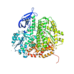
2PQE
 | | Solution structure of proline-free mutant of staphylococcal nuclease | | 分子名称: | Thermonuclease | | 著者 | Shan, L, Tong, Y, Xie, T, Wang, M, Wang, J. | | 登録日 | 2007-05-01 | | 公開日 | 2007-06-19 | | 最終更新日 | 2024-05-22 | | 実験手法 | SOLUTION NMR | | 主引用文献 | Restricted backbone conformational and motional flexibilities of loops containing peptidyl-proline bonds dominate the enzyme activity of staphylococcal nuclease.
Biochemistry, 46, 2007
|
|
4BYG
 | | ATPase crystal structure | | 分子名称: | COPPER EFFLUX ATPASE, MAGNESIUM ION, POLYETHYLENE GLYCOL (N=34), ... | | 著者 | Mattle, D, Drachmann, N.D, Liu, X.Y, Pedersen, B.P, Morth, J.P, Wang, J, Gourdon, P, Nissen, P. | | 登録日 | 2013-07-19 | | 公開日 | 2014-08-06 | | 最終更新日 | 2023-12-20 | | 実験手法 | X-RAY DIFFRACTION (2.85 Å) | | 主引用文献 | Dephosphorylation of Pib-Type Cu(I)-Atpases as Studied by Metallofluoride Complexes
To be Published
|
|
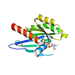
5ZQY
 | | Crystal structure of a poly(ADP-ribose) glycohydrolase | | 分子名称: | MAGNESIUM ION, Poly(ADP-ribose) glycohydrolase ARH3, [(2R,3S,4R,5R)-5-(6-AMINOPURIN-9-YL)-3,4-DIHYDROXY-OXOLAN-2-YL]METHYL [HYDROXY-[[(2R,3S,4R,5S)-3,4,5-TRIHYDROXYOXOLAN-2-YL]METHOXY]PHOSPHORYL] HYDROGEN PHOSPHATE | | 著者 | Wang, M, Yuan, Z, Ma, Y, Wang, J, Liu, X. | | 登録日 | 2018-04-20 | | 公開日 | 2018-08-15 | | 最終更新日 | 2023-11-22 | | 実験手法 | X-RAY DIFFRACTION (1.577 Å) | | 主引用文献 | Structure-function analyses reveal the mechanism of the ARH3-dependent hydrolysis of ADP-ribosylation.
J. Biol. Chem., 293, 2018
|
|


