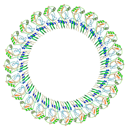
2QYO
 
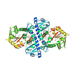 | |
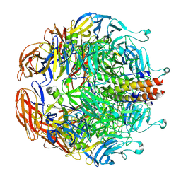
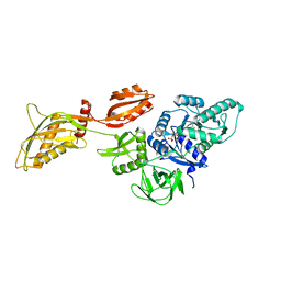
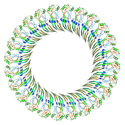
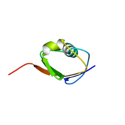
5J6P
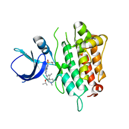
 | | Crystal Structure of Mis18(17-118) from Schizosaccharomyces pombe | | 分子名称: | Kinetochore protein mis18, ZINC ION | | 著者 | Wang, C, Shao, C, Zhang, M, Zhang, X, Zang, J. | | 登録日 | 2016-04-05 | | 公開日 | 2017-11-01 | | 最終更新日 | 2024-03-20 | | 実験手法 | X-RAY DIFFRACTION (2.6 Å) | | 主引用文献 | Crystal Structure of Mis18(17-118) from Schizosaccharomyces pombe
To Be Published
|
|
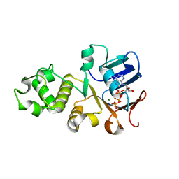
4Y4R
 | | Crystal structure of ribosomal oxygenase NO66 dimer mutant | | 分子名称: | ACETATE ION, Bifunctional lysine-specific demethylase and histidyl-hydroxylase NO66, NICKEL (II) ION | | 著者 | Wang, C, Hang, T, Zang, J. | | 登録日 | 2015-02-11 | | 公開日 | 2015-10-07 | | 最終更新日 | 2024-03-20 | | 実験手法 | X-RAY DIFFRACTION (3.3 Å) | | 主引用文献 | Structure of the JmjC domain-containing protein NO66 complexed with ribosomal protein Rpl8.
Acta Crystallogr.,Sect.D, 71, 2015
|
|
8EB7
 | |
6DKZ
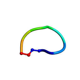 | | Racemic structure of ribifolin, an orbitide from Jatropha ribifolia | | 分子名称: | ribifolin | | 著者 | Wang, C.K, King, G.J, Ramalho, S.D. | | 登録日 | 2018-05-31 | | 公開日 | 2018-11-14 | | 最終更新日 | 2020-01-01 | | 実験手法 | X-RAY DIFFRACTION (0.99 Å) | | 主引用文献 | Synthesis, Racemic X-ray Crystallographic, and Permeability Studies of Bioactive Orbitides from Jatropha Species.
J. Nat. Prod., 81, 2018
|
|
2V9C
 | | X-ray Crystallographic Structure of a Pseudomonas aeruginosa Azoreductase in Complex with Methyl Red. | | 分子名称: | 2-(4-DIMETHYLAMINOPHENYL)DIAZENYLBENZOIC ACID, FLAVIN MONONUCLEOTIDE, FMN-DEPENDENT NADH-AZOREDUCTASE 1, ... | | 著者 | Wang, C.-J, Hagemeier, C, Rahman, N, Lowe, E.D, Noble, M.E.M, Coughtrie, M, Sim, E, Westwood, I.M. | | 登録日 | 2007-08-23 | | 公開日 | 2007-11-13 | | 最終更新日 | 2023-12-13 | | 実験手法 | X-RAY DIFFRACTION (2.18 Å) | | 主引用文献 | Molecular Cloning, Characterisation and Ligand- Bound Structure of an Azoreductase from Pseudomonas Aeruginosa
J.Mol.Biol., 373, 2007
|
|
5IMX
 | |
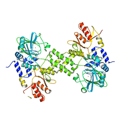
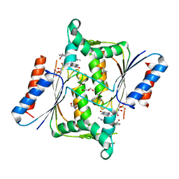
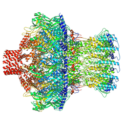
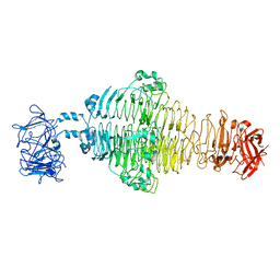
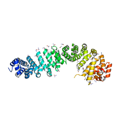
5BOE
 | | Crystal structure of Staphylococcus aureus enolase in complex with PEP | | 分子名称: | Enolase, GLYCEROL, MAGNESIUM ION, ... | | 著者 | Wang, C.L, Wu, Y.F, Han, L, Wu, M.H, Zhang, X, Zang, J.Y. | | 登録日 | 2015-05-27 | | 公開日 | 2015-12-09 | | 最終更新日 | 2023-11-08 | | 実験手法 | X-RAY DIFFRACTION (1.6 Å) | | 主引用文献 | Octameric structure of Staphylococcus aureus enolase in complex with phosphoenolpyruvate
Acta Crystallogr.,Sect.D, 71, 2015
|
|
8DSP
 | |
8EAP
 | |
8EAO
 | |
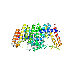
5E8L
 | | Crystal structure of geranylgeranyl pyrophosphate synthase 11 from Arabidopsis thaliana | | 分子名称: | Heterodimeric geranylgeranyl pyrophosphate synthase large subunit 1, chloroplastic | | 著者 | Wang, C, Chen, Q, Fan, D, Li, J, Wang, G, Zhang, P. | | 登録日 | 2015-10-14 | | 公開日 | 2015-11-11 | | 最終更新日 | 2024-03-20 | | 実験手法 | X-RAY DIFFRACTION (2.807 Å) | | 主引用文献 | Structural Analyses of Short-Chain Prenyltransferases Identify an Evolutionarily Conserved GFPPS Clade in Brassicaceae Plants.
Mol Plant, 9, 2016
|
|
5E8K
 | | Crystal structure of polyprenyl pyrophosphate synthase 2 from Arabidopsis thaliana | | 分子名称: | Geranylgeranyl pyrophosphate synthase 10, mitochondrial | | 著者 | Wang, C, Chen, Q, Fan, D, Li, J, Wang, G, Zhang, P. | | 登録日 | 2015-10-14 | | 公開日 | 2015-11-11 | | 最終更新日 | 2024-03-20 | | 実験手法 | X-RAY DIFFRACTION (3.028 Å) | | 主引用文献 | Structural Analyses of Short-Chain Prenyltransferases Identify an Evolutionarily Conserved GFPPS Clade in Brassicaceae Plants.
Mol Plant, 9, 2016
|
|
8DMF
 | | Cryo-EM structure of the ribosome-bound Bacteroides thetaiotaomicron EF-G2 | | 分子名称: | GUANOSINE-5'-TRIPHOSPHATE, MAGNESIUM ION, Tetracycline resistance protein TetQ | | 著者 | Wang, C, Han, W, Groisman, E.A, Liu, J. | | 登録日 | 2022-07-08 | | 公開日 | 2023-01-04 | | 最終更新日 | 2024-06-12 | | 実験手法 | ELECTRON MICROSCOPY (4 Å) | | 主引用文献 | Gut colonization by Bacteroides requires translation by an EF-G paralog lacking GTPase activity.
Embo J., 2022
|
|
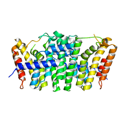
6UK1
 | | Crystal structure of nucleotide-binding domain 2 (NBD2) of the human Cystic Fibrosis Transmembrane Conductance Regulator (CFTR) | | 分子名称: | ADENOSINE-5'-TRIPHOSPHATE, Cystic fibrosis transmembrane conductance regulator, MAGNESIUM ION | | 著者 | Wang, C, Vorobiev, S.M, Vernon, R.M, Khazanov, N, Senderowitz, H, Forman-Kay, J.D, Hunt, J.F. | | 登録日 | 2019-10-03 | | 公開日 | 2020-10-07 | | 最終更新日 | 2023-10-11 | | 実験手法 | X-RAY DIFFRACTION (2.693 Å) | | 主引用文献 | A thermodynamically stabilized form of the second nucleotide binding domain from human CFTR shows a catalytically inactive conformation
To Be Published
|
|
8EAN
 | |
8E4G
 | |
8ET1
 | |
8ET2
 | | CryoEM structure of the GSDMB pore | | 分子名称: | Isoform 1 of Gasdermin-B | | 著者 | Wang, C, Ruan, J. | | 登録日 | 2022-10-15 | | 公開日 | 2023-03-29 | | 最終更新日 | 2024-06-19 | | 実験手法 | ELECTRON MICROSCOPY (4.96 Å) | | 主引用文献 | Structural basis for GSDMB pore formation and its targeting by IpaH7.8.
Nature, 616, 2023
|
|
3JB8
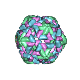 | | Insight into Three-dimensional structure of Maize Chlorotic Mottle Virus Revealed by Single Particle Analysis | | 分子名称: | Coat protein | | 著者 | Wang, C.Y, Zhang, Q.F, Gao, Y.Z, Zhou, X.P, Ji, G, Huang, X.J, Hong, J, Zhang, C.X. | | 登録日 | 2015-08-04 | | 公開日 | 2016-07-13 | | 最終更新日 | 2024-03-20 | | 実験手法 | ELECTRON MICROSCOPY (3.6 Å) | | 主引用文献 | Insight into the three-dimensional structure of maize chlorotic mottle virus revealed by Cryo-EM single particle analysis.
Virology, 485, 2015
|
|
8EFP
 | |
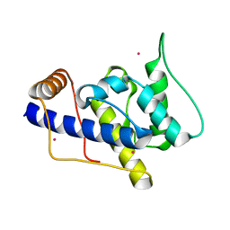
1F0Z
 | | SOLUTION STRUCTURE OF THIS, THE SULFUR CARRIER PROTEIN IN E.COLI THIAMIN BIOSYNTHESIS | | 分子名称: | THIS PROTEIN | | 著者 | Wang, C, Xi, J, Begley, T.P, Nicholson, L.K. | | 登録日 | 2000-05-17 | | 公開日 | 2001-01-10 | | 最終更新日 | 2024-05-22 | | 実験手法 | SOLUTION NMR | | 主引用文献 | Solution structure of ThiS and implications for the evolutionary roots of ubiquitin.
Nat.Struct.Biol., 8, 2001
|
|
4EVA
 | |
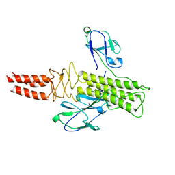
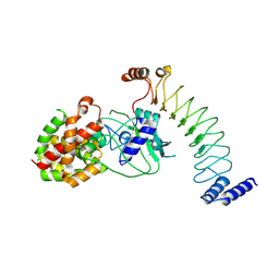
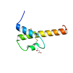
1P5S
 | | STRUCTURE AND FUNCTION OF THE CALPONIN-HOMOLOGY DOMAIN OF AN IQGAP PROTEIN FROM SCHIZOSACCHAROMYCES POMBE | | 分子名称: | MERCURY (II) ION, Ras GTPase-activating-like protein rng2 | | 著者 | Wang, C.H, Balasubramanian, M.K, Dokland, T. | | 登録日 | 2003-04-28 | | 公開日 | 2004-05-11 | | 最終更新日 | 2024-02-14 | | 実験手法 | X-RAY DIFFRACTION (2.22 Å) | | 主引用文献 | Structure, crystal packing and molecular dynamics of the calponin-homology domain of Schizosaccharomyces pombe Rng2.
Acta Crystallogr.,Sect.D, 60, 2004
|
|
4I6O
 | | Crystal structure of chemically synthesized human anaphylatoxin C3a | | 分子名称: | (4S)-2-METHYL-2,4-PENTANEDIOL, Complement C3 | | 著者 | Wang, C.I.A, Ghassemian, A, Collins, B, Lewis, R.J, Alewood, P.F, Durek, T. | | 登録日 | 2012-11-29 | | 公開日 | 2013-02-27 | | 最終更新日 | 2013-03-06 | | 実験手法 | X-RAY DIFFRACTION (2.14 Å) | | 主引用文献 | Efficient chemical synthesis of human complement protein C3a.
Chem.Commun.(Camb.), 49, 2013
|
|


